SIALADENIT
(
sialoadenitis
; Greek sialon saliva + aden gland + -itis) - inflammation of the salivary gland.
In the etiology of S., infection plays an important role. With S., in the ducts of the salivary glands (see), a mixed flora is found, consisting of staphylococci, pneumococci, streptococci, as well as pathogens of actinomycosis, tuberculosis, and syphilis. One of the causes of S. may be mumps and cytomegaly viruses.
Infectious agents enter the salivary gland through the mouth of the excretory duct, sometimes this is facilitated by the entry of a foreign body into the duct (toothbrush villi, apple peels, etc.). as well as lymphogenous or hematogenous route. The emergence of S. is promoted by inf. diseases, surgical interventions, especially on the abdominal organs, stagnation of secretions in the ducts of the salivary gland.
There are acute and chronic S.
Acute sialadenitis on ultrasound
Inflammation of the salivary glands is called sialadenitis.
Acute bacterial sialadenitis develops when the infection is introduced hematogenously or through the excretory ducts, as well as from an open injury. Many viruses (for example, mumps virus, influenza, cytomegalovirus) have an affinity for glandular tissue, including the parenchyma of the salivary glands. In acute sialadenitis, a painful swelling appears in the area of the salivary glands, and the amount of saliva secreted decreases. As a rule, during viral infections several salivary glands are affected. In acute sialadenitis, ultrasound shows an increased size of the affected glands; the echogenicity of the parenchyma is reduced, often heterogeneous due to oval hypoechoic areas; the contour of the gland is clear and slightly convex; blood flow is often increased. It is likely to encounter enlarged regional lymph nodes with increased central blood flow. Acute sialadenitis is not characterized by dilated ducts, increased echogenicity of their walls, or hyperechoic inclusions in the parenchyma of the gland.
| Photo. With viral mumps on ultrasound, the parotid gland (A, B) is enlarged, the parenchyma is hypoechoic, heterogeneous due to small anechoic ovals, the color circulation determines dilated vessels; blood flow is noticeably increased (B). Conclusion: Echo signs of acute mumps. | ||
| Photo. A 5-year-old girl with a temperature of 38.6ºC, a wet cough and painful swelling in the parotid region on the right. On ultrasound, the right parotid gland (A, B) is enlarged, the parenchyma is hypoechoic, heterogeneous due to small anechoic ovals, the color circulation determines dilated vessels; blood flow is noticeably increased. The parotid gland on the left (B) is unchanged. In the general blood test, lymphocytosis, the level of IgM antibodies to the mumps virus is increased. Diagnosis: acute mumps. | ||
| Photo. A, B — On ultrasound, the parotid gland is enlarged, the contour is wavy, the parenchyma is hypoechoic, heterogeneous due to small almost anechoic ovals, probably dilated vessels; enlarged lymph nodes are detected. Conclusion: Echo signs of acute mumps, secondary lymphadenitis. B - Echo signs of acute parotitis in the main gland and accessory lobule. | ||
Reasons for the development of sialadenitis
The first cause of sialadenitis is the penetration of infectious agents of a bacterial, viral or specific nature into the salivary glands, namely:
- oral bacteria;
- weakened immune system;
- angina;
- bacterial infections - staphylococcal, pneumococcal, streptococcal, tuberculosis and syphilis;
- cytomegalovirus infection;
- influenza virus;
- parotitis;
- actinomycosis (fungal infection);
- cat scratch disease, which occurs as a result of cat bites and scratches;
- oncological diseases.
Infection of the gland occurs through the main excretory duct when foreign bodies are introduced into it. Occasionally, infection occurs by hematogenous or lymphogenous route.
The second reason for the development of inflammation of the salivary glands can be a temporary cessation or decrease in salivation during severe infectious diseases, after surgical operations in the abdominal cavity, injuries to the gland, stagnation of secretions in the ducts, as well as in the following diseases:
- tuberculosis;
- syphilis;
- dehydration;
- fever;
- hypercalcemia;
- pathomorphology;
- grape-shaped atrophy of the mucous membrane;
- purulent or serous-purulent exudate inside the duct;
- infiltration by leukocytes.
In addition, with viral damage to the glands, bacterial sialadenitis occurs, and with diseases such as actinomycosis, tuberculosis, syphilis, specific sialadenitis occurs. In this case, normal salivation is disrupted, which prevents the gland from clearing infectious agents - this leads to a worsening of the disease. Therefore, it is very important to begin treatment for sialadenitis as soon as its first symptoms appear.
Chronic sialadenitis on ultrasound
Congenital disorders of the development of ducts and glandular tissue, salivary gum disease, as well as autoimmune diseases contribute to the development of a chronic inflammatory process in the salivary glands. Regardless of the cause, there are three stages of chronic inflammation: initial, clinically pronounced and late. In each stage there are periods of exacerbation and remission. In chronic sialadenitis on ultrasound, the parenchyma is hypoechoic, heterogeneous due to point and linear hyperechoic inclusions, the contour is uneven. During the period of exacerbation of large iron, its echogenicity is significantly reduced. During the period of remission, the size of the gland decreases and its echogenicity slightly increases. The decrease in the echogenicity of the gland is due to swelling of the parenchyma and hyperplasia of the intraglandular lymphoid tissue. Hyperechoic inclusions appear when parenchyma is replaced by fibrous tissue. Due to severe fibrosis in the later stages, the iron decreases in size.
| Photo. On ultrasound, the parotid gland on the right (A, B) is enlarged, the parenchyma is hypoechoic, heterogeneous due to small anechoic ovals and linear hyperechoic inclusions. The parotid gland on the left is unchanged. Conclusion: Echo signs of chronic mumps. | ||
| Photo. On ultrasound, the parotid gland is enlarged, the parenchyma is hypoechoic, heterogeneous due to small anechoic foci, point and linear hyperechoic inclusions; the vessels are dilated and blood flow is increased. Conclusion: Echo signs of chronic mumps. | ||
Sjögren's syndrome is a chronic autoimmune disease, predominantly affecting women over 40 years of age, in which lymphocytes and plasma cells intensively infiltrate and destroy the salivary and lacrimal glands. The main complaints of Sjögren's syndrome include dry mouth and keratoconjunctivitis, as well as arthritis. The disease often affects all salivary glands. In Sjögren's syndrome, ultrasound shows that the salivary glands are enlarged, the parenchyma is heterogeneous due to diffusely scattered multiple hypo- or anechoic oval foci; blood flow is usually increased. It is believed that hypo- and anechoic foci are lymphocytic infiltrates against a background of unchanged parenchyma. In the presence of lesions larger than 2 cm and rapid growth, a biopsy is recommended.
| Photo. A 50-year-old man complains of dry mouth and keratoconjunctivitis. On ultrasound, all salivary glands are enlarged, the parenchyma is hypoechoic, heterogeneous due to small hypoechoic foci and linear hyperechoic inclusions. Conclusion: Echo signs of chronic sialadenitis. Diagnosis: Sjögren's syndrome. | ||
A special form of chronic sialadenitis is chronic sclerosing sialadenitis or Küttner's tumor, when a persistent painful tumor appears in one of the salivary glands. This disease is rare and most often affects the submandibular gland. Microscopy reveals atrophy of glandular tissue, dense fibrosis, and periductal lymphocytic infiltrates. Sialoliths and mucus plugs are often found in altered ducts. It remains unclear whether they are a cause or a consequence of the inflammatory process. Some authors suggest that thick secretion blocks small ducts, which leads to inflammation, fibrosis, and atrophy of glandular tissue. Other authors point to the autoimmune nature of the pathology. With Küttner's tumor, ultrasound reveals diffuse parenchymal damage in the salivary gland: multiple round hypoechoic foci. Less common is a focal lesion: a hypoechoic heterogeneous focus against the background of unchanged parenchyma, which simulates a malignant process. In all doubtful cases, a biopsy is recommended. Chronic sclerosing sialadenitis is an exclusively benign inflammatory disease. Often the affected gland has to be removed; no additional treatment is required.
Symptoms of sialadenitis
The mild form of acute sialadenitis begins with an inflammatory process, which is accompanied by the appearance of edema. However, the general condition of the patient remains unchanged. A small compaction may appear in a separate area of the salivary gland, which can subsequently grow. There is a slight decrease in salivary secretion, but no changes in the oral cavity are observed.
In the moderate form, the patient experiences infiltration, as a result of which pus spreads in the area of the salivary glands. During this period, this area of the oral tissue hurts when touched, swallowing, turning the head and chewing. The pain intensifies in the lower jaw, as well as in the temple and ear. The patient may also experience a feeling of congestion in the ears, and he cannot open his mouth wide as before.
With significant inflammation, the patient may develop a febrile state, accompanied by an increase in body temperature and signs of general malaise. In some cases, acute sialadenitis does not reach the stage of suppuration and necrosis in the gland and is eliminated on its own. But if the patient develops a severe form of sialadenitis, then his health deteriorates sharply, abscesses, fistulas, narrowing of the salivary duct or phlegmon of the parotid and submandibular region may form.
In turn, with the chronic course of sialadenitis, the symptoms of inflammation are not so pronounced. In the area of the affected gland, a slight swelling is detected, which can be slightly painful and replace the internal tissue and excretory ducts of the gland. The patient may complain of a decrease in saliva volume and an unpleasant taste in the mouth. However, over time, the gland changes in size and begins to resemble a tumor. In case of exacerbation of chronic sialadenitis, mucus or pus may leak from the gland, and an abscess may develop on it.
Sialosis (sialoadenosis) of the salivary glands on ultrasound
Sialosis (sialoadenosis) is a non-inflammatory and non-malignant, recurrent, often painless, usually bilateral enlargement of the salivary glands. The causes of sialosis are unclear. Sialosis occurs in patients with endocrine and autoimmune diseases, liver cirrhosis, chronic alcoholism, vitamin deficiencies, etc. With sialosis, ultrasound reveals enlarged hyperechoic salivary glands, but without focal changes and increased blood flow.
| Photo. On ultrasound, the submandibular (A, B) and parotid (C) glands are enlarged, the parenchyma is hyperechoic, poorly transmits ultrasound waves (the posterior contour of the gland is not visible), homogeneous, the blood flow is not changed. Conclusion: Echo signs of sialoadenosis. | ||
Diagnosis of sialadenitis
To identify sialadenitis, specialists use diagnostic methods such as:
- PCR (polymerase chain reaction) analysis is aimed at identifying nucleic acid fragments in biological material;
- microbiological examination of secretions;
- cytological examination of secretions;
- sialometry;
- Ultrasound of the salivary glands;
- sialography;
- sialotomography;
- sialoscintigraphy.
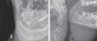
The final diagnosis can only be made by a doctor during examination. To do this, the patient is prescribed an X-ray examination of the affected area in order to exclude or confirm the presence of salivary gland stones.
Inflammation of the lymph nodes in the salivary glands on ultrasound
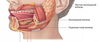
The parotid glands contain a large number of lymph nodes that collect lymph from the upper jaw, lips, mouth, cheeks, outer parts of the nose, upper and lower eyelids, temporal and frontal regions, outer ear and tissues of the parotid-masticatory region. Inflammatory disease in these areas causes a reaction in the lymph nodes of the parotid glands. Inflammation of the intraglandular lymph nodes imitates mumps. Misdiagnosis here is unacceptable, since mumps and lymphadenitis have different management tactics and prognosis.
Important!!! With bacterial lymphadenitis, the infection lymphogenously penetrates into the regional lymph nodes through the “entry gate”. Viral infections hematogenously affect the lymph nodes of several anatomical areas.
Ultrasound with acute serous lymphadenitis within the parotid gland reveals an oval-shaped anechoic formation with clear and slightly wavy contours, often with central blood flow - an inflamed lymph node. Inflammatory edema and infiltration of the tissues surrounding the lymph node (periadenitis) lead to the development of lymphogenous sialadenitis (pseudosialoadenitis). A painful, diffuse swelling appears and the amount of saliva decreases. On ultrasound with lymphogenous sialadenitis, the echogenicity of the parenchyma is reduced, an almost anechoic lymph node is determined; the contour of the lymph node is often not clearly visible, which does not always indicate destruction of its capsule. With a positive outcome, the size of the lymph node decreases, its echogenicity increases, and the contour becomes clearer.
| Photo. Ultrasound of the parotid gland: the echogenicity of the parenchyma is not changed, heterogeneous due to oval-shaped hypoechoic foci with a central scar. Conclusion: Echo signs of reactive lymphadenitis. | ||
Purulent lymphadenitis on ultrasound differs little from acute serous lymphadenitis - an oval-shaped anechoic focus, but as the intensity of the reflected signal increases (GAIN regulator) with purulent melting of the lymph node in the peripheral sections (parietal), point inclusions of medium echogenicity appear, while the central sections the lymph node remains without inclusions. The clarity of the contours and the oval shape indicate the preservation of the capsule of the lymph node during its purulent melting - this is a forming abscess in the salivary gland. An anechoic area of uncertain shape with unclear and uneven contours due to purulent melting of the lymph node and its capsule is adenophlegmon of the salivary gland.
| Photo. On ultrasound, both parotid glands are enlarged, hypoechoic, heterogeneous due to hypo- and anechoic foci. Conclusion: Echo signs of multiple microabscesses. | ||
| Photo. A 2-year-old child with swelling in the parotid region on the right. On ultrasound, the right parotid gland is noticeably enlarged, heterogeneous due to many hypo- and anechoic foci with acoustic enhancement behind and increased blood flow along the periphery. Conclusion: Echo signs of a formed abscess in the parotid gland. | ||
Long-term entry of low-virulent flora into the lymph nodes or inadequate treatment of acute serous lymphadenitis can lead to chronicity of the inflammatory process. An enlarged lymph node in chronic lymphadenitis on ultrasound is hypoechoic, oval in shape with clear contours and (necessarily!) inclusions of high echogenicity in the center. It is the presence of echogenic inclusions in the central parts of the lymph node that confirms the existence of a chronic inflammatory process.
Salivary stone disease on ultrasound
Salivary stones are most often located in the submandibular gland (60-90% of cases) and can be multiple. The parotid glands are affected in 10-20% of cases. Stones can form due to metabolic disorders. Therefore, it is often possible to trace the hereditary nature of the disease. A rather characteristic combination of salivary stone disease and a chronic inflammatory process in the gland.
Sialilithiasis causes partial or complete mechanical obstruction of the salivary duct, which leads to recurrent swelling of the salivary gland during meals and may be complicated by bacterial infection. Sialoliths in the distal submandibular duct can be palpated at the base of the mouth.
Important!!! In preschool children, the role of a calculus can be played by foreign bodies of the submandibular ducts, which enter the ducts through gaping orifices. Such foreign bodies are often toothbrush lint, wood chips from pencils, seed husks, blades of grass, etc.
Calcified stones in the duct of the salivary gland are visible on ultrasound in the form of hyperechoic point or linear inclusions with a pronounced distal acoustic shadow. The duct proximal to the stone is usually dilated.
Only 20% of sialoliths are radiopaque. Uncalcified stones in the salivary gland duct are visible on ultrasound as inclusions of medium or high echogenicity without a distal acoustic shadow. Air bubbles mixed with saliva can mimic stones. An enlarged intraglandular duct indicates the location of the hyperechoic inclusion within the duct, and not in the parenchyma of the gland.
| Photo. Cross section of the parotid gland: 1 - parotid gland, 2 - excretory duct, 3 - stone with an acoustic shadow behind in the distal part of the excretory duct, 4 - masticatory muscle (m. masseter), 5 - ascending arch of the mandible, 6 - buccal muscle ( m. buccinator). | ||
| Photo. A, B — Stones in the excretory duct of the submandibular gland. B - Small stones in the excretory duct of the parotid gland. | ||
| Photo. The parotid gland is enlarged, echogenicity is reduced, somewhat heterogeneous due to small hypoechoic foci, blood flow is increased; the excretory duct is greatly expanded, in the distal section a hyperechoic inclusion with an acoustic shadow behind is determined. Conclusion: dilation of the excretory duct of the parotid gland, echo signs of a calculus in the distal part, secondary parotitis. | ||
Treatment methods for sialadenitis
Basically, the course of treatment for sialadenitis depends on the form of the disease. For mild inflammation, the patient is prescribed physical therapy and antibiotics, which are injected into the salivary gland duct. The patient is also taking pilocarpine hydrochloride. The affected area is treated with dimexide.
In cases of moderate sialadenitis, the patient is given intramuscular antibacterial agents and novocaine blockades are performed in the area of the gland. Also during this period, therapy is prescribed; for purulent inflammation, surgical opening of purulent foci is used. If the causative agent of the disease is a virus or fungus, then the patient takes appropriate antiviral drugs.
In case of severe sialadenitis and its complications, the patient may be prescribed surgery. If the structure of the salivary gland duct is disturbed, its bougienage can be performed. If the patient has been diagnosed with salivary stones or other foreign bodies, he is prescribed lithotripsy, lithoextraction, sialendoscopy and other techniques. In some particularly severe cases, it is necessary to perform surgical intervention, which is accompanied by opening the capsule of the gland and duct with removal of the contents or complete removal of the affected gland with the duct. Moderate and severe forms of sialadenitis must be treated in the infectious diseases department of a hospital.
In the chronic form of sialadenitis, in addition to drug therapy, the patient is recommended to undergo a course of physiotherapy, namely:
- salivary gland massage;
- UHF;
- galvanization;
- electrophoresis;
- fluctuarization.
Also, in parallel, the patient is prescribed anti-inflammatory drugs and drugs that stimulate metabolism. Good results are achieved with novocaine blockade of the salivary gland. However, regardless of the form of development of this disease, its treatment is quite long, since, in addition to complications, concomitant diseases are taken into account.
Salivary gland cysts on ultrasound
Simple cysts are uncommon in the salivary glands. They can be congenital or acquired. Some acquired cysts develop due to obstruction of the salivary ducts in the presence of tumor, stones, or inflammation. Clinically they appear as a painless tumor. On ultrasound, simple cysts are defined as avascular anechoic lesions with a clear contour and posterior acoustic enhancement.
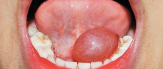
Retention cysts of the sublingual glands are located under the tongue on one side of the frenulum of the tongue: sometimes the cyst displaces the frenulum, but never moves to the opposite side. On palpation, a soft, elastic, painless swelling of a round or oval shape with a smooth, slightly wavy surface is determined. Being covered with a thinned mucous membrane, retention cysts have a characteristic cyanotic or bluish tint. On ultrasound, a small retention cyst (up to 2.0 cm in size) is visible as an anechoic formation, on one side of the frenulum and fibrous septum of the tongue; the outer and anterior contour are not clearly defined, since in these places the cysts are adjacent to the lower jaw. Retention cysts grow slowly. Sometimes they penetrate through the mylohyoid muscle into the submandibular region. If the cyst is large, the wall can be damaged when eating solid food (baking bags, crackers, candies, etc.). If the cyst empties completely, it cannot be detected on ultrasound. The cyst begins to grow again as fluid accumulates in it. In long-term large retention cysts there is often a suspension of medium echogenicity.
| Photo. A — Retention cyst of the sublingual gland. B — Ultrasound shows a simple cyst of the parotid gland. B — Ultrasound shows a complex cyst of the parotid gland. Such cysts require additional research. | ||
Forecast and prevention of sialadenitis
In most cases, acute sialadenitis heals within two weeks. However, the patient should remember that if not treated in a timely manner, the disease can cause irreversible changes in the salivary gland and even necrosis. Prevention of sialadenitis consists, first of all, of maintaining oral hygiene, strengthening the immune system, as well as timely treatment of infectious diseases and eliminating existing chronic foci of infection. If treatment for acute inflammation of the salivary glands is started on time, the disease can be easily cured and the prognosis will be favorable. As for chronic sialadenitis, complete recovery usually does not occur. In this case, it is important to prevent exacerbations of its course and the transition of the disease to severe forms.
Benign tumors of the salivary glands on ultrasound
The most common benign neoplasms of the major salivary glands are polymorphic adenomas and Warthin tumors (adenolymphoma, cystadeniolymphoma). They grow slowly and may be asymptomatic. It is not possible to make a final diagnosis based on ultrasound alone.
Polymorphic adenomas are most common in the parotid gland (60%-90%) in people in the fourth and fifth decades of life, but can occur at any age. Polymorphic adenoma (pleomorphic adenoma, mixed tumor) is a benign tumor with a well-defined fibrous capsule, has slow expansive growth, and is not prone to malignancy, metastasis and relapse. One of the previously existing names for this tumor, fibrochondromyxoepithelioma, indicates that this tumor develops from two germ layers (meso- and ectoderm). Polymorphic adenoma is very diverse and can be represented by both homogeneous dense masses of a grayish color, and mushy masses, areas of mucus with a soft gelatinous consistency, cartilaginous tissues with islands of ossification. Polymorphic adenoma of the salivary glands on ultrasound looks like a round or oval formation of reduced or low echogenicity with a clear, even or coarsely wavy contour. The echogenicity of polymorphic adenoma can be from slightly reduced to low and almost anechoic. The echostructure of the tumor can be homogeneous or contain many pinpoint inclusions of high echogenicity, randomly distributed throughout the tumor, or have large zones with inclusions of medium echogenicity. Blood flow in polymorphic adenoma is often poor or absent, but may be abundant.
| Photo. Polymorphic adenomas of the parotid gland on ultrasound: A - Hypoechoic focus with a wavy contour, almost homogeneous echostructure, blood flow in the center and along the periphery of the focus is increased. B — Hypoechoic focus with a wavy contour, heterogeneous due to hyperechoic foci. B — Hypoechoic focus of lobular structure, heterogeneous due to hyperechoic inclusions. | ||
Important!!! After unsuccessful surgery, polymorphic adenomas can recur multifocally. Untreated pleomorphic adenomas can undergo malignant changes after decades.
Infantile hemangiomas are the most common parotid tumor in infants. Like infantile hemangiomas of other areas, it is benign, appears soon after birth, grows quickly, and spontaneously regresses after 18 months. Surgery is usually not required. On ultrasound, in the projection of the parotid gland, a lobulated hypoechoic formation with a clear contour and extreme vascularization is found.
Warthin's tumor (adenolymphoma, cystadeniolymphoma) accounts for 5-10% of all benign neoplasms of the salivary glands and occurs more often in men in the fifth and sixth decades of life. In 10-60% of cases, Warthin tumors can occur at two or more points, grow and appear at different times. Warthin's tumors are benign; extremely rarely, the epithelial component can undergo malignant transformation. Adenolymphomas consist of a bilayered eosinophilic epithelium and lymphoid stroma. The epithelium forms various glandular and papillary structures: solid, cystic, lobular, papillary. Warthin's tumors on ultrasound are oval, hypoechoic lesions with a clear contour, often containing multiple anechoic areas. Warthin tumors are often hypervascularized but may also contain only short segments of vessels.
| Photo. Warthin tumors on ultrasound. | ||
Cystic lymphangioma occurs from dilated lymphatic vessels when drainage is impaired. Lymphangiomas are not true tumors, but malformations of the lymphatic system. Lymphangioma of the parotid gland is manifested by the presence of an extensive painless formation of soft elastic consistency without clear contours. The color of the skin over the lymphangioma remains normal. Lymphangioma of the parotid gland can extend to adjacent areas (temporal, buccal and submandibular), as well as to the superolateral parts of the neck. Lymphangiomas increase with colds. In this case, the lymphangioma becomes denser and even painful. Inflammation and even suppuration of lymphangioma is often observed, which is accompanied by an increase in volume, thickening, pain that sharply increases with palpation, a change in the color of the skin over the lymphangioma, a local and general increase in temperature. Cystic lymphangioma on ultrasound is visible as an anechoic cavity formation of irregular shape with clear contours, easily changing with pressure. In the lymphangioma cavity, septa are usually visible. The walls of the cysts are relatively thick, up to 1-3 mm, and may contain blood vessels, nerves, and adipose tissue. An inflamed lymphangioma on ultrasound is characterized by greater resistance of the cavity to compression and a convexity of the outer contour, the septa become thicker and more noticeable, and multiple point inclusions appear in the cystic fluid. With hemorrhage, the lymphangioma changes from anechoic to hypoechoic.
| Photo. Cystic lymphangioma. | ||
The parotid gland contains its own veins and transit venous lines - the superficial temporal vein, the mandibular vein, the maxillary vein, therefore, of the hemangiodysplasias in the parotid glands, venous dysplasia or isolated phleboectasis are most common. Venous dysplasia can spread beyond its borders. Large venous nodes located in the subcutaneous tissue and skin create a characteristic clinical picture. Deep venous dysplasia manifests itself as a painless swelling of soft elastic consistency with unclear contours. A characteristic clinical sign of venous dysplasia is an increase in the size of this formation when the child strains or cries, as well as when the head is tilted or in a horizontal position. In a calm state, venous dysplasia on ultrasound is represented by an area of reduced echogenicity of an indeterminate shape with uneven and unclear contours. When performing the Valsalva maneuver, the characteristics of the zone of change change dramatically: its echogenicity decreases, its contours become clearer, and its shape becomes more rounded. This technique allows you to confidently differentiate venous dysplasia from all other diseases of the parotid gland.