The salivary glands are paired organs whose role is to secrete saliva. The liquid contains a set of enzymes and is involved in the processes of food digestion, protein synthesis, and removal of metabolites from the body.
Diseases of the salivary glands lead to digestive disorders and severe pain during meals, and increase the likelihood of developing caries. To solve the problem and restore proper flow of saliva, bougienage of the salivary gland duct is performed. Find out when the procedure is necessary and how it is performed.
Indications for use
Bougienage of the ducts of the salivary glands is a procedure aimed at artificially widening narrow areas. Indications for its implementation are:
- strictures formed as a result of sialadenitis of any origin;
- sialolithiasis, or salivary stone disease.
These pathological conditions have the same symptoms, caused by impaired outflow of salivary fluid. You should consult a doctor if the following problems occur:
- feeling of fullness in the mouth during and immediately after eating,
- unpleasant sensations of varying intensity caused by food intake,
- acute pain when eating spicy and sour foods.
Symptoms of impaired saliva flow cannot be ignored. If the liquid released during eating cannot move properly in the ducts for a long time, the salivary glands become denser and stones form in them. If left untreated, the pathological process becomes chronic.
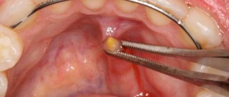
Removing stones from the salivary glands
Stones in the salivary glands can occur both in the parenchyma of the gland itself and in its ducts, which leads to disruption of the outflow of saliva into the oral cavity. In most cases, stones occur in the submandibular salivary gland, which is located in the floor of the mouth. Less commonly, this condition affects the parotid or sublingual salivary glands. In some cases, multiple salivary gland stones may be present.
Salivary gland stones form when chemicals found in saliva accumulate in a duct or gland. In most cases, these stones are composed of calcium salts. The exact cause of salivary gland stones is unknown. But there are known factors that contribute to a decrease in saliva secretion and or its thickening, which increases the risk of salivary gland stones. These factors include dehydration, poor diet, and the use of certain medications, including antihistamines, hypertension medications, psychotropic medications, and bladder medications. Trauma to the salivary glands may also contribute to the risk of salivary gland stones.
The risk of salivary gland stones is also increased in patients with endocrine and metabolic disorders, such as gout, hyperparathyroidism, urolithiasis and hypervitaminosis D, as well as diabetes mellitus. Most often, sialolithiasis - salivary gland stones - occurs at the age of 25-40 years. In general, more than half of patients with dental diseases go to the doctor with this particular ailment.
Salivary gland stones usually do not cause any symptoms, however, when they reach a certain size, when the stone blocks the duct of the salivary gland, stagnation of saliva occurs in the ducts and parenchyma of the gland, which is manifested by pain and swelling of the gland. In addition, when an infection occurs, suppuration of the salivary gland may occur.
There is also the appearance of pain in the area of the affected salivary gland during meals. An unpleasant taste appears in the mouth. With acute blockage of the salivary gland duct, acute pain occurs. When the stone is localized in the duct of the submandibular salivary gland, the pain may intensify during swallowing movements and radiate to the ear. The attachment of an infection to a salivary gland blocked by a stone is manifested by malaise, fever and constant dull pain.
In the diagnosis of salivary gland stones, in addition to palpation examination, instrumental research methods are used, such as ultrasound of the salivary glands, CT, radiocontrast study - sialography and sialoscintigraphy - radioisotope study of the salivary glands. In many cases, a simple ultrasound examination is sufficient to identify a stone in the salivary gland.
Currently, treatment of salivary gland stones can be either conservative or surgical. The latter is resorted to when drug therapy is ineffective.
Currently, among the invasive methods for removing salivary gland stones, the following are used:
- bougienage of the salivary ducts
- crushing of salivary gland stones (lithotripsy)
- endoscopic methods of stone removal - sialendoscopy
- open surgery to remove a stone in the salivary gland and
- removal of the entire salivary gland
When a stone is detected in the salivary gland, the goal of treatment is its removal. In some cases, a salivary gland stone, if it is in the duct, can be removed with a gentle massage. In other cases, they resort to the above invasive methods. Currently, minimally invasive interventions based on endoscopic techniques are increasingly used in maxillofacial surgery. When removing salivary gland stones, sialoendoscopy is performed. For this manipulation, special endoscopic instruments are used, which include a thin endoscope equipped with a high-quality camera and light source. Under local anesthesia, such an endoscope is carefully inserted into the mouth of the salivary gland, and the doctor sees the entire course of the intervention on a monitor screen with multiple magnification. Once the tip of the probe is in the duct and the surgeon sees the stone, using special instruments it is either grasped and removed, or if it is difficult to remove, it is destroyed. After such manipulation, antibiotics are prescribed to prevent infectious complications.
Another innovative way to remove salivary gland stones is by crushing, as is the case with kidney stones. Shock wave lithotripsy allows you to crush the stone into several small fragments without any incisions. Then these fragments either come out on their own using conservative measures or are removed endoscopically.
In some cases, traditional opening of the mouth of the salivary gland duct may be used when the stone is close to the exit.
Most minimally invasive procedures for removing salivary gland stones are performed on an outpatient basis under local anesthesia.
Removal of the salivary gland (extirpation) is used for recurrent salivary gland stones or irreversible structural changes in the tissue of the salivary gland. This procedure is usually performed under general anesthesia in a hospital setting.
In modern foreign clinics, minimally invasive and innovative treatment methods that are effective and safe are actively used to remove salivary gland stones. Clinics in Western Europe, the USA, Israel, India, Turkey and South Korea today employ highly qualified maxillofacial surgeons who have extensive experience in performing minimally invasive interventions in the treatment of salivary gland stones.
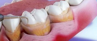
Features of the event
Bougienage of the ducts of the salivary glands does not require complex preparation. 2-3 hours before the procedure, the patient needs to refrain from eating. To widen the duct, the doctor uses conical probes of different sizes. During the procedure, the dentist:
- selects a probe of suitable diameter,
- introduces it into the duct to the maximum depth,
- Leaves for 10-15 minutes and removes.
Bougienage is carried out daily. Each time, a larger diameter instrument is used to gradually and without injury expand the gland duct.
Types and functions of salivary glands
Salivary glands are paired organs that differ in location:
- large parotid - located on the side of the lower jaw, under the left and right ears. They produce saliva containing high concentrations of potassium and sodium chlorides;
- large submandibular - adjacent to the lower jaw, have an excretory duct in the area of the lingual frenulum, produce saliva with a low acid index;
- large sublingual - located in the oral cavity under the mucous membrane, secrete alkaline saliva containing protein saturated with mucin (high molecular weight glycoproteins).
The minor salivary glands are located on the mucous membrane of the cheeks, lips, and tongue.
The role of the salivary glands is to secrete a fluid that contains special enzymes and is involved in the digestive process. They also perform the following functions:
- exocrine - production of proteins, fluids;
- endocrine - produce biologically active substances;
- excretory - participation in the removal of metabolic products from the body.
Bougienage during examination of the salivary gland duct
Examination of patients with diseases of the salivary glands requires certain skills from the dentist, since often the first symptoms of this disease are inflammatory processes in the oral cavity.
After collecting anamnesis, visual and palpation examination, the following may be prescribed:
- sialometry using the Andreeva method - after making a bougienage into the duct of the salivary gland, a cannula (special tube) is inserted through which saliva is collected for quantitative and qualitative research;
- sialography (contrast x-ray diagnostics) - 1 - 2 ml of iodolipol or other iodine-containing substance, heated to a temperature of 37 - 40 degrees, is injected into the ducts of the salivary glands using a blunt needle. The mouth of the duct is first bougiened with a conical probe. An x-ray is taken immediately;
- pantomosialography (contrast x-ray diagnostics of several glands at once) - this procedure helps to determine the ongoing hidden inflammatory process of paired salivary glands, narrowing or widening of the ducts;
- digital (digital) subtraction sialography - performed with the introduction of a contrast agent.
Sialolithiasis (salivary gland stones) - symptoms and treatment
Sialolithiasis, or salivary stone disease (Greek: sialon saliva + lithos stone) is a disease characterized by the formation of stones in the salivary gland and its chronic inflammation.
Normally, a person has three pairs of salivary glands: sublingual, submandibular and parotid. There are also small glands in the oral cavity, such as the palatine, buccal and lingual glands. Their task is to produce and secrete saliva. The submandibular gland most often undergoes the process of stone formation in the gland ducts and subsequent inflammation (89 - 95% of all cases), then in descending order is the parotid gland and extremely rarely the sublingual gland; sometimes stones can form in the minor salivary glands [3].
The disease is often detected at the age of 20-45 years - calculi (stones), which began to grow in childhood, by this age reach sizes that impede the flow of saliva and cause complaints. Sialolithiasis is more common in men than in women [11]. Salivary stone disease is rarely observed in children [1].
The formation of saliva is the initial stage of food digestion. Saliva contains enzymes, antibacterial substances and dissolved minerals. The exact reasons for the formation of stones in the salivary glands are unknown. Only factors that favor the development of pathology are identified:
- Mechanical effects on the salivary glands in the area of the excretory duct , for example, due to injuries from teeth and crowns. A change in the lumen of the gland duct due to injury disrupts the outflow of saliva, which contributes to the formation of stones.
- Inflammation , in which microflora accumulates in the gland and pus appears. The disorder appears due to the presence of an inflammatory process in nearby tissues or the entry of pathogenic microorganisms into the gland duct. If the cause persists, then over time the stones increase due to deterioration in the outflow of saliva, metabolic disorders and exacerbation of the process. The sublingual and submandibular glands are susceptible to inflammation.
- Narrowings and kinks of the salivary glands and ducts.
- Impaired calcium metabolism due to bad habits, taking medications, poor quality drinking water, systemic diseases (for example, problems with the thyroid gland).
- Lack or complete absence of vitamins - most often a deficiency of vitamin A leads to the disease.
- Increased blood clotting.
- Entry of a foreign body into the gland duct. Bacteria actively multiply around it, forming a stone. This could be a solid fragment of food, a bone, a stone remaining in the grain, a fish scale, etc.
Mostly one stone is formed, but sometimes multiple stones are also found. It is extremely rare for several glands to be affected at the same time. The weight of stones can vary from fractions of a gram to several tens of grams. Stones come in various shapes: oblong, round or irregular; in their center there are often foreign bodies. Salivary stone mainly consists of inorganic salts - phosphates and calcium carbonates. With sialolithiasis, chronic inflammation and disruption of tissue nutrition are observed inside the gland tissues, which leads to the formation of connective tissue surrounding the lobules of the gland and its dilated ducts.
The development of sialolithiasis is also promoted by the use of anticholinergic drugs. Taking these medications inhibits the secretion of saliva, which leads to the accumulation of various food debris in the oral cavity. At the same time, the risk of them entering the gland duct increases and the number of bacteria in the oral cavity increases, which contributes to the formation of stones. These reasons increase with a decrease in water consumption, but studies have not been conducted to identify the relationship between these factors.
Treatment using bougienage of the salivary gland duct
The salivary glands can be susceptible to the following diseases:
- Stricture (narrowing) of the salivary gland duct - occurs due to tissue scarring due to frequently recurring inflammatory processes or traumatic damage to the gland. Bougienage is performed by inserting a thin conical probe (bougie) into the duct for 15 minutes. The next day, the procedure is repeated, slightly increasing the diameter of the inserted probe. The number of sessions can vary from 15 to 30.
- Sialadenitis is an inflammation of the salivary gland resulting from its infection. Causes functional disruption of the glands. It is treated with medication; instillation of antibiotics into the duct of the salivary gland is also prescribed, which is carried out after bougienage.
- Also, with the help of bougienage, the integrity of the parotid gland duct is restored. The procedure protects the end of the damaged duct from becoming overgrown. Bougienage should be carried out daily with the requirement that a larger diameter probe must enter the created cavity.
Prevention of diseases of the salivary glands includes, first of all, strengthening the immune system, timely detection and treatment of foci of infection, and proper level of oral hygiene.
How is an ultrasound of the salivary glands done?
This examination is one of the most comfortable and efficient for the patient. The general cycle of the salivary gland ultrasound procedure follows the following algorithm:
The ultrasound procedure of the salivary glands lasts 20-30 minutes, without causing pain or discomfort to the patient.