Vestibuloplasty in children is an operation performed to deepen the vestibule of the oral cavity.
The vestibule of the mouth is the space between the cheeks and lips in front and the teeth and gums in the back. At the SM-Doctor clinic, plastic surgery of the oral vestibule in children is performed for the following indications:
- speech therapy problems
- preparation for orthodontic treatment
- inflammatory-destructive processes in the periodontium
- gum recession (raising of its edges and exposure of tooth roots)
- cosmetic defect
Symptoms
The vestibule of the oral cavity is a gap of soft tissue that is located between the lip or cheeks and the elements of the dentition.
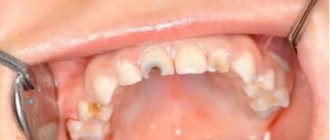
A variant of the norm is the depth of the vestibule from 5 to 10 mm. If this indicator does not reach 5 mm when measured from the end of the gum to the stationary area of the mucous membrane, dentists diagnose a shallow vestibule of the oral cavity.
An anomaly in the size of the vestibule can be identified by the following signs:
- excessive narrowing or complete absence of the attachment zone of the mucous membrane;
- tension of the gum tissue in the area of the periodontal junction;
- inflammation and bleeding of the gums;
- increased sensitivity of incisors;
- exposure of the necks and roots of bone organs in the area of attachment of ligaments;
- dentofacial deformities;
- the presence of a short frenulum;
- violation of diction.
With a decrease in the size of the upper vestibule, incomplete closure of the lips, their partial immobility, as well as malocclusion and a reduced size of the upper jaw compared to the lower jaw may be observed.
Vestibuloplasty according to Clark
This technique is the simplest of all known. As a rule, the Clark operation is performed on large areas. Most often, vestibuloplasty using this method is done on the upper jaw.
The intervention algorithm looks like this:
- anesthesia is administered;
- the mucous cavity between the gums and the moving part of the jaw is dissected;
- the mucous membrane of the lips peels off;
- muscles and tendons move deeper;
- single muscle fibers are removed;
- the mucous flap is sutured in the area of the periosteum in the depths of the vestibule of the mouth;
- the wound is covered with a protective film.
The approximate healing time for the operated area is about 14 days.
Causes
A small vestibule of the oral cavity can be the result of both surgical intervention and congenital pathology.
Acquired pathology is a consequence of the following points:
- surgery to correct nonunion of the upper lip;
- surgical interventions to eliminate the consequences of burns, mechanical trauma to soft tissues, and removal of tumors.
The cause of a congenital reduction in the size of the vestibule is most often heredity and the presence of certain pathologies in the development of the dentofacial apparatus.
What is a crooked jaw and methods for straightening it.
We will tell you here about how long the swelling lasts after compactosteotomy.
At this address https://orto-info.ru/zubocheliustnye-anomalii/zubov/polozheniya/etiologiya-razvitiya-i-metodyi-korrektsii-protruzii.html we will discuss why tooth protrusion occurs after braces.
The main advantages of using laser in vestibuloplasty
Other advantages of using laser in vestibuloplasty include:
- the cut is made as accurately as possible;
- it is possible to avoid swelling of the mucous membrane;
- no bleeding;
- microcirculation of the vascular walls is significantly reduced;
- tissues regenerate faster;
- bactericidal effect available;
- pathogenic microflora decreases;
- it is possible to avoid the appearance of scars.
In most Moscow clinics, vestibuloplasty can be performed using any known technique. You can find out more detailed information and also familiarize yourself with the cost of the procedure on our website.
Consequences
The depth of the vestibule and the attachment of the gums play an important role in the process of protecting the marginal periodontium from external influences.
In case of insufficient attachment or complete absence of attached gum tissue, the following problems may arise:
- trauma to the marginal periodontium while eating food;
- increased muscle tone of the chin;
- impaired blood supply to gum tissue;
- formation of pathological bite;
- partial decrease in lip mobility;
- slower growth of the upper jaw row;
- loosening of teeth;
- inflammatory diseases of the gums, their atrophy;
- development of periodontitis.
Indications for surgery
Vestibuloplasty is indicated in the following cases:
- absolute absence of gum attachment;
- a symptom of tension, the signs of which are pallor and displacement of the gingival margin when the lip is pulled back;
- lack of attached gum zone – distance less than 1 mm;
- signs of inflammation of the gum tissue;
- preparation for orthodontic therapy - installation of a structure to correct the bite will not bring the required effect with a small vestibule due to the fact that the alveolar processes of the incisors will return to their original position due to gingival tension;
- the need for further prosthetics;
- elimination of recession or atrophy of gum tissue.
The small vestibule of the oral cavity is often diagnosed not only in adults, but also in childhood. In this case, during the mixed dentition, observation of the child by the dentist is indicated. The operation is permissible after complete eruption of all teeth.
Course of action
Before proceeding with surgery, the dentist conducts a thorough examination of the patient’s oral cavity using certain instruments and equipment.
This allows you to identify associated problems and determine the most suitable vestibuloplasty technique among the many existing ones.
Before the operation, the dentist also professionally removes mineralized plaque from the front surface of the dentition.
During preparation for surgery, the patient is required to comply with the following rules:
- refusal to take painkillers and other medications not prescribed by the dentist;
- refraining for 6 hours before surgery from consuming foods that can cause mechanical injury to the gum tissue.
Vestibuloplasty is performed with preliminary anesthesia of the treated area. Anesthesia can be either local or general, at the request of the patient.
There are many options for performing surgery to change the size of the vestibule, which can be divided into several large groups.
Open method
Open vestibuloplasty techniques are based on changing the depth of the vestibule in such a way that a wound is formed on the surface of the lip and alveolar process, which takes about two weeks to heal.
The key disadvantage of such methods of surgical intervention is the formation of scars on the soft tissue, which in the future can contribute to the re-development of the pathology.
The open method is carried out as follows:
- a dissection of the mucous membrane of the lower lip is performed using an incision in the sector of the front teeth;
- an apron-shaped flap is peeled off, the base of which is a transitional fold in the area of the incisors;
- soft tissues are displaced to the prescribed depth, which helps to increase the depth of the vestibule;
- the detached flap of tissue is placed in the area of the alveolar process of the lower jaw, after which it is fixed with suture material;
- the wound formed on the mucous membrane is sutured and healed due to secondary intention.
Closed
Plastic surgery using a closed surgical technique involves closing the wound surface formed after enlargement of the vestibule using local soft tissue.
The essence of the method is that soft tissue is cut off through a small vertical incision.
The mucous membrane is practically not damaged. Thanks to this, the recovery process proceeds more quickly.
Dentists note a significant drawback that is inherent in closed operations - the possibility of relapse of the disease. According to statistics, three years after the operation, the size of the vestibule is again reduced by almost half.
Patchwork
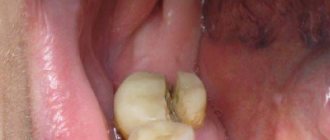
The key indication for flapplasty is the exposure of the necks and roots of the teeth due to the strong tension of the soft tissue, which over time can lead to inflammation of the gum tissue and loosening of the teeth.
During vestibuloplasty, several horizontal and vertical incisions are made, which allow the edge of the gum tissue to be excised to form a flap.
After this, the separated flap is placed in the intended place in the dentition and fixed with suture materials.
Use of the plate
Vestibuloplasty using special plates is practically no different from the methods described above, however, its peculiarity is the use of a forming plate during the final stage.
This vestibular structure is applied to the wound surface after applying the flap and fixing it with sutures. The duration of its use is about two months.
Possible causes of crowded teeth in the lower jaw and ways to eliminate the anomaly.
In this publication we will talk about the reasons for the development of a small lower jaw in a child.
Follow the link https://orto-info.ru/zubocheliustnye-anomalii/ryadov/zuboalveolyarnogo-ukorocheniya.html if you are interested in the operation of dentoalveolar shortening.
Plastic surgery of the frenulum of the lips and tongue
The frenulum is a connective tissue cord present on the mucous membrane of the oral cavity. There are frenulums of the upper and lower lip and tongue.
If the frenulum develops incorrectly, various functional disorders develop.
- with a short frenulum of the tongue in children, there is a violation of the pronunciation of individual sounds; by the age of 15-18, this can lead to exposure of the necks of the teeth on the oral side.
“How to determine that a child has a short frenulum?” There are the following signs of a short frenulum of the tongue:
- child's speech delay
- The frenulum does not start from the middle of the tongue, as is normal, but is woven into its tip
- if you ask a child to reach his nose with his tongue, the tip of the tongue bends downwards
- If you ask a child to reach his nose with his tongue, it splits
- if you ask a child to reach his nose with his tongue, then ischemia (blanching) of the frenulum is visible
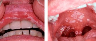
- with a short frenulum of the lip (most often observed on the upper lip), there is a gap between the central teeth (diastema)
Signs of a short frenulum of the lip:
- blanching of the frenulum when the lip is pulled back
- presence of diastema
- low weaving of the frenulum (directly into the gingival margin).
With age, a short frenulum of the lip can lead to exposure of the necks of the teeth.
The short frenulum of the tongue can be stretched with the help of speech therapy exercises, but with severe pathology, surgery is necessary - frenulotomy (dissection of the frenulum).
Diastema can be corrected with the help of orthodontic structures, however, if the cause, in the form of a short frenulum, is not eliminated, over time the teeth will return to their original position.
The most effective treatment for short frenulum of the lips and tongue is surgical excision. Excision of a short frenulum is a fairly simple operation, it takes little time and healing also takes place in a short time.
Other treatments
Among the above methods, there are some variations, the choice of a specific one is made by the dentist after examining the patient’s oral cavity.
Edlan-Meicher method
This method is most often used when it is necessary to eliminate a small vestibule on the lower jaw due to its high efficiency.
After anesthesia of the operated area, an incision is made into the mucous membrane along the bend of the bone arch. Next, the mucous membrane and periosteum are peeled off, as well as the submucosal tissue moves to the lateral and anterior sections of the vestibule.
For fixation, sutures are placed on the mucous membrane, after which the wound is covered with a special bandage. The patient's recovery time is about two weeks.
The performance of vestibuloplasty according to Meyhar can be seen in the video.
Schmidt method
The Schmidt technique differs from the previous version of the operation only in that exfoliation of the periosteum tissue is not performed.
The soft and muscle tissues in this sector are also incised along the periosteum. The resulting flap edge is placed deep into the formed vestibule and then fixed.
This method of vestibuloplasty is equally effective for treating both the lower and upper jaw.
Glickman method
Plastic surgery using the Glickman method can be performed both on a limited sector of the oral cavity and on a larger area.
After administering anesthesia, the surgeon dissects the mucous membrane in the area of its attachment to the lip, then detaches the soft tissue and forms a depression.
After this, the detached section of the mucous membrane is attached to the resulting depression.
Clark's method
Plastic surgery of the small vestibule using the Clark method is performed for pathology of the upper jaw row.
After administering the anesthetic drug, a section is made at the border of the junction of the gingival margin and the moving section of the mucous tissue of the vestibule.
Using scissors, the mucous surface of the upper lip is peeled off. The soft tissue of the vestibule is dissected as close as possible to the periosteum and parallel to the curvature of the bone surface. The depth of the cut should not exceed 15 mm.
The previously detached section of the mucous membrane of the lip is placed in the vestibule formed as a result of tissue dissection, and then secured with sutures.
At the end of the procedure, the wound is covered with an iodoform swab.
Tunnel vestibuloplasty
The least traumatic method of plastic surgery, which is used to correct the depth of the vestibule of both jaws, is tunnel vestibuloplasty.
In certain areas of the oral cavity, two horizontal incisions are made along the premolars, as well as one vertical in parallel with the frenulum. After this, the mucosal flap is shifted inside the formed vestibule and secured.
The wound surface during this procedure is significantly reduced compared to other surgical techniques. The duration of the rehabilitation period practically does not exceed 10 days.
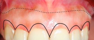
The concept of the depth of the vestibule of the oral cavity
The depth of the vestibule of the oral cavity is the length of the gap between the border of the transition of the mobile mucosa to the fixed one and the end of the gum. This area can have varying depths from 5 mm to 1 cm (and even more) and, depending on the distance, there are deep, medium and shallow vestibule. If the vestibule of the oral cavity is shallow, there is a risk of marginal periodontal disease. In this case, with a high degree of probability, so-called periodontal pockets can form - this is a depression between the gum and tooth. The problem arises if the pocket depth becomes more than 3 mm. The reason for their occurrence can be a normal conversation, the process of brushing teeth, the process of eating food. This occurs due to increased mobility of the papillae and retraction of the free end of the gum, which in turn causes periodontal damage. The tissues become inflamed and periodontal pockets form. In order to eliminate this problem, it is necessary to perform an operation to deepen the vestibule of the oral cavity. Operations of this kind are called vestibuloplasty. Their essence is the redistribution of soft tissues in the oral cavity in order to deepen the vestibule. There are various methods of vestibuloplasty. Among them, there are methods with an open surface of wounds and methods that involve covering wounds on the alveolar process and lip. Both have various advantages and disadvantages. The undoubted advantage of open methods is the simplicity of the operation. Disadvantages include deterioration of the trophism of the alveolar process and bone resorption. The disadvantage of closed methods can be the occasional relapse of the small vestibule. Technique of the operation. During the operation, a horizontal incision is first made at the border of the attached gum and the free mucosa of the alveolar process within up to 6 teeth. From the ends of the first incision, two more – now vertical – incisions, each 10 mm long, are made. Thus, a trapezoidal flap is formed along the contours of the incisions. The incisions are made so that the base of the trapezoid faces the lip. Then the flap is placed in the now deeper vestibule and fixed with catgut. After the operation, about 11 mm of exposed bone remains, which epithelializes and scars. Technique for performing surgery with a closed wound surface. The mucous membrane of the vestibule is dissected from one canine to another. The cut passes 5 mm below the transitional masonry and has the shape of an arc. Then a second similar incision is made 5 mm away from the end of the red border. This is followed by a third incision in the middle of the lip at an angle of 70 degrees. It connects the two previous cuts. The resulting flaps peel off. If the vestibule area is less than 4 mm, the periosteum is fenestrated and the chin muscles are moved downwards. Together with the muscles, the periosteum also moves by about 1.5 cm. Next, plastic surgery of the mucous membrane is performed - for this, opposing flaps are used. This operation is used if the vestibule is not deep enough within the entire frontal region of the lower jaw. There are cases when the small vestibule occupies the area of the lower incisors in the presence of a short frenulum of the lower lip. The operation begins with making a main incision on the crest of the frenulum from the place of its attachment to the end of the vermilion border (the incision should end 5 mm from the vermilion border). Then two more cuts follow. The first of them is made on the alveolar process (the angle is 70 degrees, the length of the incision is up to 3 cm). The second is in the lip area at the same angle and the same length as the first. The resulting flaps are peeled off, moved and fixed with sutures. After the operation is completed, a forming plate is applied to the patient’s teeth and worn for two weeks.
Elimination of isolated gum recessions. Recession is the process in which the root of a tooth is exposed. As a rule, this occurs from the vestibular surface. The cause of this problem can be a short frenulum of the tongue or lips, tooth dystopia, improper dental care (traumatic brushing) as well as inflammation of the gums, which can appear due to the formation of microbial plaque.
Gum recession is divided into 4 types: 1. Recession of the marginal part of the gums, passing into the mucogingival junction. It is not accompanied by loss of alveolar bone tissue. 2. Recession reaches the mucongingival junction or extends below. In this case, it may or may not be accompanied by loss of alveolar bone tissue. 3. Recession extends beyond the mucogingival junction. Accompanied by some non-critical loss of interdental bone tissue and gums. The end of the gum is located above the end of the bone. 4. Recession extends beyond the mucongingival junction. Loss of interdental bone tissue and gums reaches significant proportions. The margins are at the level of the apical border of the recession.
There are 2 types of elimination of isolated gum recession: 1. The cause of recession is eliminated by loosening the tension of the short frenulum of the lip or tongue and normalizing the position of the dystopic tooth, eliminating dental plaque from under the gums, making good fillings and dentures. 2. The tooth root is closed surgically. All operations of this kind are divided into two types: flap and free grafts.
Particular attention should be paid to the so-called patchwork operations. Flap operations are operations in which flaps are used to eliminate periodontal pockets, correct damage to the gum margin and to restore damaged tissue.
The flaps can be full, that is, consisting of periosteum, connective tissue and epithelium, and split, that is, consisting of epithelium and connective tissue.
In addition, simple and positional flaps are also separated. At the end of the operation, the first of them is placed in its old place, while the second is moved to a new position.
A simple flap is used to eliminate periodontal pockets and, to some extent, bone pockets. Such operations are carried out in cases of periodontitis with present bone pockets deeper than 0.5 cm, as well as in cases of periodontal pockets with fibrous changes and thinning of the gums, and resorption of the alveolar bone of the vertical type with mobility of teeth I and II degrees.
First, the area where the surgery will be performed is anesthetized. Then two vertical incisions are made from the end of the gum to the transitional fold so that the gingival papillae are not touched, and two horizontal incisions are made along the edges of the gums on the oral and vestibular sides.
The gum edge is cut using scissors, the section width is approximately 2 mm. Granulation tissue is removed. Then the tooth root is polished, and the edges of the alveolar bone and the inner surface of the mucoperiosteal flap are processed. After all this, the flap is moved into place, and sutures are placed in each segment between the teeth and on the vertical incisions.
The advantages of this type of operation are free access to bone pockets, the ability to treat them under visual control, as well as wound healing by primary intention. Disadvantages include exposure of the dental necks and lowering of the alveolar process.
There are patch surgeries that use agents that activate regenerative processes in marginal periodontal tissues. This type is called gingivosteoplasty. After completing the main stages of the operation, a drug that activates restorative osteogenesis is injected into the pre-planned bed.
Bone grafts, tissue grafts, bioplastics, and bioceramics are used for the same purpose. Various membranes are used as a barrier to the proliferation of the oral epithelium.
This is due to the fact that proliferation of the oral epithelium of the tooth root during wound healing begins to occur earlier than the process of proliferation of bone tissue cells and periodontal ligament starts, which does not lead to complete adhesion of the gum and tooth.
Membranes must meet certain stringent requirements. Among them are compatibility with a biological organism. In addition, they must provide protection against cell penetration and tissue germination.
There must be sufficient space between the membrane, the tooth root and the wall of the bone pocket to restore periodontal tissue. Its important property is the ability to fuse with the gum to strengthen the position of the flap.
Membranes are either absorbable or non-absorbable. Resorbable membranes are divided into natural ones, made from components taken from animals and humans, and synthetic polymers.
Non-absorbable membranes have a number of disadvantages: – They must be removed, which requires an additional operation – Tissues interacting with such a membrane can fester – The removal process itself does not contribute to 100% tissue restoration.
Absorbable membranes also have their own characteristics. On the one hand, repeated surgery is not required, on the other hand, it is difficult to control the time of their resorption.
Rehabilitation
The duration and severity of the rehabilitation process depends not only on the dentist’s manipulations, but also on the patient’s compliance with the following recommendations:
- at the end of the procedure, use a cold compress to relieve swelling from the operated area;
- Avoid eating too hard, spicy or hot foods for two weeks after surgery;
- reduce the amount of dairy products consumed, as they contribute to the formation of persistent plaque;
- use soft-bristled toothbrushes for daily hygiene;
- rinse the mouth with special anti-inflammatory and antiseptic drugs;
- after five days after the operation, begin performing myogymnastic exercises recommended by a specialist;
- visit the dentist on the appointed days to monitor the recovery process.
Vestibuloplasty price
It is difficult to immediately name the full cost of the plastic surgery procedure, since the volume of surgical intervention, the need for additional medical procedures, and the amount of suture material are unknown. The final cost can be found out at your doctor's appointment. Prices for vestibuloplasty at Aladen do not differ from the average in Minsk.
Contraindications to vestibuloplasty Surgery may not always be performed. There are a number of relative and absolute obstacles to vestibuloplasty.
Contraindications
- a large number of teeth with caries and its complications,
- inflammatory processes in the bones,
- pathologies of the central nervous system,
- hematological diseases,
- tumors
- collagenoses.
Relative contraindications include caries, pulpitis or osteomyelitis. This procedure can be started after treatment of carious teeth and osteomyelitis.
Malignant neoplasm is an absolute obstacle to vestibuloplasty.
If you have other chronic diseases, consultation with an appropriate specialist before surgery is advisable.