Indications
Indications for removal of the submandibular salivary gland are:
- chronic sialadenitis (inflammation);
- benign or malignant tumors;
- multiple cystosis;
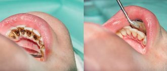
- salivary stone disease;
- severe injury to the gland;
- complete blockage of the salivary ducts with the impossibility of restoring their patency.
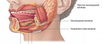
Depending on the diagnosis, various tests are prescribed before the operation - ultrasound, computed tomography or magnetic resonance imaging, chest x-ray. In the case of a tumor, a biopsy is also necessary to determine the nature of the formation. Salivary stone disease is examined using contrast sialography.
Chronic interstitial sialadenitis
The disease causes inflammatory processes.
In 90% of cases, the salivary glands in children are affected. In chronic interstitial sialadenitis, narrowing, dilatation and deformation of all ducts are observed. Treatment necessarily involves the use of antibiotics that have a wide spectrum of action.
In case of purulent inflammation, the gland is treated until the admixture of pus disappears from the salivary secretions.
At the discretion of the attending physician, ointment compresses may be prescribed.
With effective therapy, pain and swelling are quickly eliminated, and salivation is significantly enhanced.
Preparation for the operation and features of its implementation
Preoperative preparation includes:
- laboratory tests;
- abolition of blood-thinning medications (at least a week before the procedure);
- changing your diet three days before surgery - excluding alcohol, spicy and rough foods, hot drinks.
- Smoking is also prohibited before surgery.
Removal of the submandibular salivary gland is a complex and specific operation, since its anatomical location near the nerve and choroid plexuses, muscles and other organs is associated with risks of serious complications. Such a procedure can only be performed by experienced specialists - dental surgeons or maxillofacial surgeons.
In the case of a malignant tumor, especially when it grows beyond the gland, the organ is removed along with the affected tissue of nearby muscles and lymph nodes. In other cases, only gland tissue is removed.
Surgery to remove the submandibular salivary gland can be performed in several ways:
- The traditional method is surgical excision of tissue with removal of the pathological gland. This type of operation does not require specific equipment, which explains its lower cost and speed of implementation. Postoperative wounds are quite large; the time for suture removal varies from 1 to 3 weeks.
- A microsurgical endoscopic operation that does not require large tissue intersections - all actions are carried out using manipulators, and observation is carried out using a probe that transmits an image to the screen. The great advantage of this type of operation is the targeted effect, due to which in case of salivary stone disease, benign tumors and cystosis of the gland, there is no need for its complete removal - only a small area of it is excised. After the operation, small marks remain, the stitches are removed after a few days. The disadvantage of this method is its high cost.
Material and methods
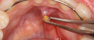
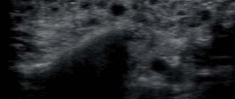
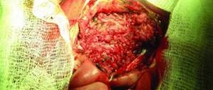
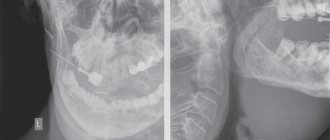
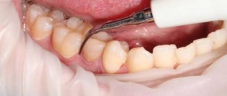
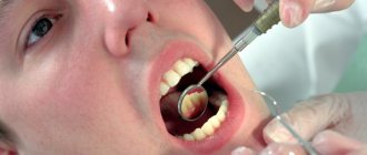
In the Department of Surgical Dentistry of the Central Research Institute of Dentistry and Maxillofacial Surgery (TsNIIS and Maxillofacial Surgery) in 2013-2015. 43 patients with stones in the duct of the submandibular gland were observed. The clinical examination included: anamnesis, examination, palpation, radiography of the floor of the mouth, orthopantomography (Fig. 1) and ultrasound examination to identify the exact location of stones, their number and determine the degree of fibrous changes in the submandibular S.J. After the diagnosis was made and minor changes were identified in the tissues of the submandibular gland, surgical intervention was performed using the following technique: a salivary bougie was injected into the duct intraorally under local anesthesia (Fig. 2 et seq.); then the mucosa was cut with a scalpel near the duct of the submandibular SG, then the duct of the submandibular SG was bluntly and sharply isolated (Fig. 3) along its entire length with visualization of the lingual nerve and lingual artery; the localization of the stone was determined by palpation and visually; the duct was fixed on a holder distal to the stone, it was cut with a scalpel along the duct above the stone (Fig. 4) and the stone was removed (Fig. 5, 6); the wound in the area of the floor of the mouth was medicinally treated with a solution of 0.05% chlorhexidine, close sutures were placed, and drainage was installed in the wound. The patient was prescribed a salivary diet and drug therapy: decongestants, painkillers and hemostatic drugs. A repeat examination and dressing was carried out the next day, the drainage was removed and the wound was treated with medication. In the postoperative period, there were no signs of inflammation in the surgical area. If fibrous changes are detected in the tissues of the submandibular gland and its function is impaired, it is advisable to carry out a radical intervention - removal of the submandibular gland along with the stone.
Fig. 1. X-ray examination (orthopantomography). 1 - stone.
Rice. 2. Bougienage of the submandibular SG duct. 1 - salivary bougie; 2 - the mouth of the duct of the submandibular salivary glands.
Rice. 3. Isolation of the submandibular SG duct using a blunt method. 1 — “Mosquito” type clamp; 2 - salivary bougie; 3 - duct of the submandibular fluid.
Rice. 4. Dissection of the mucous membrane of the submandibular duct above the calculus. 1 — scalpel; 2 - salivary bougie; 3 - mucous duct of the submandibular gland.
Rice. 5. Removal of stone from the duct of the submandibular fluid. 1 - needle holder; 2 - stone.
Rice. 6. Concrete removed from the duct of the submandibular fluid. 1 - stone.
Performing surgery to remove the salivary gland
With the traditional method of surgery, the patient assumes a lying position with his head thrown back and to the side. The operation involves local infiltration anesthesia. Several parallel incisions are made in the submandibular area, the tissue is lifted, and the capsule of the salivary gland is discovered, which is opened, and the gland tissue is extremely carefully removed and removed. When a malignant tumor grows together with nearby tissues, the latter are also removed. After this, stitches are applied.
During endoscopic surgery, both local and general anesthesia can be used. Through small punctures in the tissue, manipulators and a video probe are inserted, which projects all the actions performed on the screen. When the manipulators reach the gland, small incisions are made with their help, through which pathological tissue is removed. The endoscopes are then removed and sutures are placed at the puncture sites.
Postoperative care of the surgical site includes:
- hygiene - no water or dirt should get into the wounds;
- special diet - the diet will consist of semi-liquid warm food, no hot or cold drinks, no alcohol;
- smoking ban;
- special treatment - it is recommended to regularly treat the seams with antiseptics, and the oral cavity after each meal should be rinsed with a mixture of antiseptic and water.
If all the recommendations of the attending physician are followed, complete wound healing occurs within a few months.
Results and discussion
In 26 patients, the stones were localized in the middle third of the duct of the submandibular SG, in 17 - in the distal parts of the duct. In all patients, stones were removed using the method described above; in no case was removal of damage to blood vessels or nerves observed. The wounds healed by primary intention without signs of inflammation. Here is a clinical example. The patient had stones removed from the submandibular SG duct using the method described above. Patient P.
appealed to the surgical department of the Central Scientific Research Institute and Maxillofacial Surgery with complaints of swelling in the submandibular region on the right, worsening with food intake.
During a clinical examination, bimanual palpation of the submandibular region revealed a painful round-shaped formation of elastic consistency along the duct of the submandibular fluid on the right. When collecting anamnesis, it was established that patient P.
undergone therapeutic treatment without surgical intervention. However, after some time the symptoms returned.
An X-ray examination of the floor of the mouth revealed stones in the anterior, middle and posterior third of the submandibular duct with a diameter of 0.2 or 0.8 mm.
Removal of stones from the duct of the submandibular gland is a complex surgical procedure that requires a dental surgeon to have good knowledge of the anatomy and topography of the tissues and organs of the oral cavity. In order to prevent possible complications (injury of the lingual nerve and lingual artery) during surgery, it is necessary to isolate the duct of the submandibular SG along its entire length to the calculus, which will allow visualizing the relationship of the lingual nerve and lingual artery and avoiding their damage, especially when removing calculi in the distal part of the duct .
Recovery period
Activities during the early recovery period are assigned to a medical professional. The doctor checks the level of functioning of the facial muscles, treats the operated area and the place with a drainage device, and prepares a dietary diet for the patient.
It is recommended to start following a proper diet immediately after discharge from the hospital. Food is served ground, semi-liquid, neither hot nor cold (optimally at room temperature). Spicy, salty, and sour foods are excluded from the diet.
Among the permitted products:
- vegetable puree: from potatoes, carrots, zucchini;
- pureed soup from broth cooked with lean meat (chicken or turkey);
- semi-liquid porridge (wheat, oatmeal).
After eating, rinse the mouth with warm water or herbal infusion (chamomile, calendula, oak bark).
After surgery, the person operated on can lead a normal lifestyle.
To exclude recurrence of the primary disease and complications, it is enough to adhere to the following simple rules:
- keeping the postoperative area clean;
- cleansing the wound surface with hydrogen peroxide twice a day or antibacterial ointment (as prescribed by a doctor);
- Cleaning the operated area with mild soap and water after removing the suture.
Symptoms such as fever, general malaise, swelling, pain, bleeding, nausea and vomiting syndrome, feeling of shortness of breath, discomfort in the sternum should be a reason to consult a doctor.