Stones in the salivary gland (salivolithiasis) are a pathology that is also known as “sialolithiasis”. This is an inflammatory process in which stones form in the salivary glands.
As the stones grow, the risk of formation of an abscess or phlegmon (purulent inflammation, which, unlike an abscess, has no clear boundaries) increases. We will tell you in the article what symptoms accompany the disease and how to treat it.
Characteristic features of pathology
The size of the stones can vary - from a few millimeters to several centimeters
A person has three pairs of salivary glands, located respectively in the earlobe, under the jaw and tongue.
Under the influence of unfavorable factors, stones, or salivolitis, can form in them.
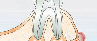
Salivary gland stones are mineral compounds that block the ducts of the salivary glands. Typically, this pathology affects people aged 20 to 45 years.
In most cases (95%), salivolitis is localized in the submandibular glands.
In the parotid glands, such formations are observed in only 8% of cases. They are most rarely formed in the sublingual salivary glands.
Stones can be small or large. Small stones are easily washed out of the ducts with saliva. Larger ones remain in the gland, clogging its lumen.
These formations are taken from organic substances and minerals: amino acids, ductal epithelium, sodium, iron, chlorine.
As for external characteristics, salivary gland stones have an uneven surface and a yellowish color. Their size can vary - from a few millimeters to several centimeters. The weight of the stones varies from 3 to 30 g.
Note! The larger the salivolitis reaches, the more difficult it is for the patient to open the jaws, speak, and chew food.
Small stones are easily washed out of the ducts with saliva, while larger ones remain in the gland, clogging its lumen
Symptoms
The appearance of stones in the salivary glands is accompanied by the following symptoms:
- Inflammation of the salivary gland
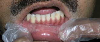
swelling of the face in the areas where the salivary glands are located where salivolitis is formed (parotid space, neck, jaw);
- discomfort, pain when trying to open the mouth, as well as when swallowing;
- acute attacks of pain in the affected area;
- tension and pain in the ears, jaw, cheeks;
- drying out of the membranes of the oral cavity;
- increased eosinophils in the blood;
- production of mucus mixed with pus instead of saliva;
- the appearance of an unpleasant taste in the mouth;
- recurrent headaches;
- redness of the facial skin;
- loss of appetite;
- prostration;
- slight increase in temperature.
Important! Without treatment, an abscess or phlegmon gradually forms. There is also a possibility of piercing the salivary gland with the sharp edges of the calculus and the latter exiting to the soft tissues.
Reasons for the development of stones in the salivary gland
Stones in the salivary glands are formed as a result of unfavorable factors, which include:
- Metabolic disorders in the body can lead to the formation of stones in the salivary gland
inflammatory or infectious processes in the area of the salivary glands (cysts, granulomas);
- injuries to the area of passage of the salivary glands and ducts caused by bruise, blow, tooth decay;
- metabolic disorders in the body, especially with problems with calcium metabolism;
- penetration of a foreign body into the duct;
- smoking;
- the presence of malignant or benign neoplasms;
- dysfunction of salivation;
- vitamin A deficiency;
- diabetes;
- gout;
- taking certain medications (blood pressure medications, diuretics, antihistamines);
- increased blood clotting.
Under the influence of the described reasons, stagnation of the secretion in the salivary glands occurs, which causes the formation of a sediment in the form of salts. The latter, uniting into a single whole, form salivolites.
Salivary Stone Removal with Home Remedies
Home remedies to get rid of salivary stones include:
- Citrus lollipops
. Lemon or orange slices will increase the flow of saliva, which will help dislodge the stone. A person may also try sugarless gum or hard, sour candy. - Drink more fluids.
Regular fluid intake helps keep your mouth hydrated and increase saliva flow. - Soft massage
. Massaging the affected area may relieve pain and the stone may pass through the salivary duct. - Medications
. Some medications, such as ibuprofen and acetaminophen, can reduce pain and swelling. - Ice cubes
. Sucking on something cold, such as an ice cube, reduces pain and swelling caused by salivary stones.
Diagnosis of pathology
Diagnosis of sialolithiasis is carried out using the following methods:
- After diagnostic measures, an appropriate course of treatment is prescribed
Palpation of the inflamed area;
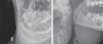
- X-ray to identify foreign formations in the salivary glands. If necessary, the procedure is carried out with the introduction of a contrast agent into the vein;
- Examination of the salivary canal using a probe. This procedure is prescribed only if the disease is not at the acute stage;
- Sialography. The technique allows you to determine the location of the stone and obtain data on the condition of the duct;
- Cytogram of gland secretion. The procedure allows you to identify the inflammatory process occurring in the gland.
After diagnostic measures, an appropriate course of treatment is prescribed, taking into account the general condition of the patient and the degree of development of the pathology.
Diagnosis of salivary stone disease
- Complete blood count (leukocytosis, increased erythrocyte sedimentation rate).
- Saliva cytology.
Ultrasound of the salivary glands.- Probing the salivary gland duct.
- X-ray of the gland and floor of the oral cavity, sialography.
Differential diagnosis:
- Sialadenitis.
- Stenosis of the salivary gland duct after injury.
- Neoplasm of the salivary gland.
Forms of the disease
Salivary stone disease can occur in acute and chronic forms.
Acute sialolithiasis is characterized by sudden development. This form is characterized by severe acute pain and fever. In this case, complications often appear in the form of the formation of phlegmon or an abscess.
If the disease becomes chronic, the inflammatory process disappears, but slight swelling remains. When the pathology reaches this stage, mandatory surgical intervention is required.
Treatment of salivary stone disease
A salivary diet is indicated; surgical removal of stones from the ducts; If the disease recurs, the issue of removing the affected gland is decided. Prescribed only after confirmation of the diagnosis by a medical specialist.
Essential drugs
There are contraindications. Specialist consultation is required.
- Canephron N (antispasmodic, anti-inflammatory, antimicrobial agent). Dosage regimen: orally, 50 drops 3 times a day. The course of treatment is 4 weeks.
- Potassium iodide (mucolytic, antiseptic). Dosage regimen: orally, in the form of a 3% solution, a tablespoon 3 times a day. The course of treatment is 4 weeks.
- Loratadine (antihistamine, antiallergic drug). Dosage regimen: orally, at a dose of 10 mg 1 time/day.
Treatment approaches
The choice of the optimal treatment method depends on what stage the pathological process is at. When diagnosing the initial stage of sialolithiasis, surgical intervention is not required: if the stone is small in size, then over time it can come out on its own.
As an auxiliary technique, the patient is recommended to eat properly, give up too much solid food, as well as smoking and alcohol.
It is also recommended to massage the affected areas, spend more time in the fresh air, play sports, follow a salivary diet, and generally lead a healthy lifestyle.
Radical intervention
Surgical intervention is required if the stones are large and prevent the patient from speaking, chewing, swallowing, or if the disease has become chronic.
Depending on where the stone is located, it can be accessed in different ways:
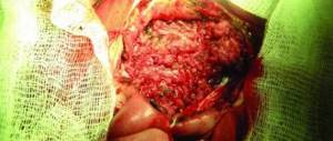
- For stones in the submandibular area, an incision is made in the neck. Pathological tissue is usually removed along with the salivary gland. In advanced cases, removal of the affected lymph nodes is also required;
- If there are stones in the parotid gland, external access is performed;
- For stones in the sublingual salivary gland, a cystectomy is performed.
If, as a result of the formation of stones, an abscess has formed, then it is opened and conditions are created for the safe outflow of purulent contents.
If a relapse occurs even after radical intervention, surgery is performed to remove the salivary gland.
After surgery, the patient should eat only liquid food.
It is easier to remove a stone that is located close to the mouth of the duct. Under such conditions, a specialist can remove it with tweezers or by gradual squeezing.
A low-traumatic way to eliminate salivolitis is lithotripsy - crushing with ultrasound.
Sialoscopy of the salivary glands is an event involving endoscopic removal of stones.
The procedure is an alternative to surgery and has several advantages:
- the likelihood of damage to ducts, glands, vessels and nerves is minimal;
- The rehabilitation period is not too long.
Sialoscopy allows you to remove stones even from the most difficult to reach areas.
Bougienage is a technique, the essence of which is to insert a probe into the duct of the salivary gland in order to expand it. The procedure can be repeated up to 15-30 times, with each procedure increasing the size of the probe.
Removal of parotid stones
The cost of dental procedures varies and depends on the complexity of their implementation:
- the price of surgical intervention ranges from 3,000 to 10,000 rubles;
- bougienage of the ducts of the salivary glands costs about 400-700 rubles per procedure;
- Sialoscopy costs about 15,000-20,000 rubles.
Treatment of sialolithiasis with medications
If there are stones in the salivary glands, medications are prescribed to enhance saliva production.
If a specialist determines that the pathological process is at the initial stage of development, the patient is prescribed medications that can be used to correct the condition.
If there are stones in the salivary glands, the following is prescribed:
- drugs to enhance saliva production;
- topical antibacterial agents;
- anti-inflammatory drugs that reduce the severity of pain;
- carrying out physiotherapeutic procedures (dry heat, massage, compresses).
Traditional methods
Alternative treatment methods can also be used, but only as an addition to the main treatment - conservative or surgical.
Among the most effective means:
- Using cranberry puree. It is enough to mash the washed berries and place the mixture in the oral cavity and leave for a few minutes. This simple method activates the salivary glands;
- Rinsing the mouth with soda. You should prepare a weak solution of soda (a teaspoon per glass of warm water) and rinse your mouth with it;
- Rinsing the mouth with a combined decoction of medicinal herbs, which includes chamomile, sage, mint.
Important! Treatment of sialolithiasis must be carried out under the supervision of a physician. Independent attempts to get rid of this phenomenon can provoke the release of purulent contents of the abscess, which is fraught with its penetration into the blood and tissues.
Preparing for surgery
Preliminary preparation is of great importance, since if it is not followed, surgical intervention may be ineffective. The preparation procedure consists of collecting anamnesis, as well as the need to carry out the following diagnostic procedures:
- X-ray of the salivary gland;
- CT or MRI of the salivary gland;
- Ultrasound examination of the salivary gland;
- Sialography (this is a procedure during which X-ray contrast agents are injected into the salivary duct to help see the presence of salivary stones in the ducts and its location. After this, an X-ray is taken.)
The removal procedure, depending on the location of the damaged salivary gland, can be carried out both on an outpatient basis and in a hospital. The submandibular and parotid ducts are subject to outpatient treatment. In a hospital setting, the intraglandular parts of the parotid gland are operated on.
Complications of pathology
Salivary stone disease is fraught with the following consequences:
- soft tissue abscess;
- phlegmon;
- passage of stone into the duct;
- vascular injury during surgery;
- impaired sensitivity of the tongue due to injury to the lingular nerve, which is possible during surgery.
In order to prevent life-threatening consequences, in case of manifestations of salivary stone disease, it is necessary to contact a specialist in a timely manner.
Diagnostics
The diagnosis and treatment of the disease is carried out by oral and maxillofacial surgeon . Our clinic carries out a thorough diagnosis, which allows not only to determine the presence of the disease, but also to determine its stage, the location of the stone, and to obtain a complete picture for drawing up a subsequent treatment plan. Diagnostics consists of several stages:
- examination at the dentist's appointment with a surgeon, collection of the patient's medical history and complaints;
- instrumental examination: ultrasound, MRI, CT, contrast sialography.
After receiving all the data, suitable treatment is prescribed.
Prevention
To prevent the development of pathology, it is necessary:
- follow the rules of oral care;
- promptly treat diseases that spread to any organs or systems of the body;
- stop smoking, alcohol;
- consume enough vitamins;
- promptly eliminate congenital ductal anomalies.
Salivary stone disease is characterized by the formation of mineral compounds in the salivary glands. Its danger lies in the possibility of the formation of an abscess and phlegmon, the purulent contents of which can spill into the surrounding tissues. Treatment of the pathology is carried out either conservatively or surgically.
When to see a doctor
Saliva stones can sometimes cause infections or abscesses, so people who are unable to remove the stones themselves should see a doctor. If signs of infection or abscess are observed, seek medical attention. The doctor usually treats the infection with antibiotics.
The doctor will examine your mouth to identify painful areas and the size and shape of the stones. Sometimes an x-ray or CT scan is needed to determine the number of stones and their exact location.
The doctor may use an ultrasound machine to help break the stone into smaller pieces and make it easier to remove. Stones that are large or located deep in the salivary gland are more difficult to remove. In these cases, the doctor may require sialoendoscopy
. This procedure involves using an endoscope to widen the affected salivary duct, allowing the stone to pass through it. Because this procedure can be uncomfortable, your doctor will use a local anesthetic to numb your mouth.


