Mucous membrane - what is it and where? Construction, types, diseases
The mucous membrane is the characteristic lining of some organs. This is part of the ducts and cavernous internal organs. It performs many functions in the body and has different structures depending on its location. What is the mucous membrane and is it really so important for the proper functioning of every body?
Mucous membranes are of great importance for many internal systems. But primarily for those who introduce external elements into the body, that is, for the digestive, respiratory, urinary and reproductive systems. The mucous membranes perform many protective and protective functions, protecting internal organs from the harmful effects of external factors.
Oral cavity examination methods
Methods for examining the oral cavity come down, first of all, to a thorough examination using directional (preferably shadowless) lighting and special instruments (see Dental Instruments) - a spatula, wide hooks for retracting the cheeks, lips, tongue and stomatol. mirrors for inspecting hard-to-reach areas. Sometimes, to identify luminescent compounds in the oral mucosa (see Luminescence), an inspection of the oral cavity is performed under UV light. During the examination, pay attention to the presence of bad breath. With the help of palpation, the mobility, density, consistency and soreness of various parts of the mucous membrane and patol are determined. formations.
To study the organs surrounding the oral cavity, various X-ray diagnostic methods are used (see). Research methods such as ultrasonic echolocation (see Ultrasound diagnostics) and thermography (see) are also used. According to indications, Citol is produced. examination of swabs and prints from pathologically changed areas of the mucous membrane (see Cytological examination), as well as the study of the microflora of the oral cavity. According to strict indications, a biopsy is performed (see).
In some cases, there is a need to conduct immunological and biochemical studies, as well as to determine various types of sensitivity: tactile, pain, temperature, taste (see Esthesiometry).
See also Patient examination, dental examination.
What mucous membrane?
The mucous membrane is also called the mucosa. This is a kind of special membrane that covers the ducts in the body of every person and the so-called cavernous internal organs, that is, those that are filled with blood.
Each mucous membrane consists of two main layers:
- outer layer formed by the epithelium of the mucous membrane,
- the inner layer, which is called plaque itself.
The inner layer of the mucosa is the connection between the mucosa and its substrate. This substrate is nothing more than the substrate of the organ (internal system) in which this mucous membrane is located. This part fits tightly to the submucosa. Dental plaque is of great importance because it contains blood and lymph vessels, glands, smooth muscles and nerves. This is a dental plaque that stabilizes and firmly fixes the mucous membrane on a given substrate.
Differences in the structure of individual mucous membranes depend on their location in the body. Other functions of the mucous membrane also appear here. It plays different roles in the digestive system, another in the respiratory system, and another in the urinary or reproductive system. To understand its importance and function in the human body, it is necessary to distinguish the location of a specific mucous membrane, and this occurs in the most important internal organs of a person.
About
Presentation. Introduction to the oral cavity. presentation for a biology lesson (8th grade) on the topic
Slide 1
Introduction to the oral cavity Prepared by: biology teacher of the highest qualification category Andreeva E.S. Municipal educational institution secondary school No. 2 of Maloyaroslavets named after A.N. Radishchev
Slide 2
Purpose: to become familiar with the structure, diseases and prevention of diseases of the oral cavity.
Slide 3
Let's remember what we pay our attention to first of all when we look at a person? Of course, a smile and white, straight teeth attract people. People with a beautiful smile are charming, conducive to communication, and they are, as a rule, more successful in life.
Slide 4
Prevention of diseases of the oral cavity and face is a priority in modern dentistry. Currently, dental disease is especially common among children: 96% suffer from caries, 70% from periodontal disease and dental anomalies.
Slide 5
Dental anomalies are deviations from the norm in the shape and size of the jaws. Normally, the jaws have a semicircular shape, but under the influence of various reasons they can acquire an irregular shape (for example, trapezoid), are large in size, or, conversely, remain underdeveloped and small.
Slide 6
How to prevent the development of dental anomalies? You cannot suck your finger (In this case, the upper jaw is “pushed forward); you should not bite your lip (in this case, the lower jaw takes the shape of a trapezoid and there is not enough space for the teeth); you cannot place your tongue between the teeth (in this case, the teeth will not close and a gap will form between them); You cannot sit at a table with your head propped up with your hand or fist (in this case, the lower jaw may move to the side).
Slide 7
The bite is the relationship of the upper and lower teeth when they are closed. Special orthodontists treat malocclusion. Open bite Deep bite Crossbite
Slide 8
Types of occlusion Normal straight Normal biprognathic Normal progenic
Slide 9
Permanent teeth form a person's permanent bite. The main signs of a correct bite: Correct shape of the dental arches; The position of the upper jaw relative to the lower jaw; Coincidence of the midline passing between the central teeth of the upper jaw with the line of the lower jaw and with the midline of the face; Overlapping 1/3 of the crowns of the lower front teeth with the upper front teeth.
Slide 10
Bite disorders
Slide 11
Adolescents with permanent dentition and adults undergo complex hardware treatment. To carry out such treatment, doctors use a brace system, which is a non-removable device for the treatment of malocclusion. The bracket system consists of brackets and an arch. Braces are special locks that are fixed to the teeth using a composite material. Each tooth has its own bracket.
Slide 12
The arch has a certain shape to which it always strives (shape memory), is attached to each brace, and thus pulls the teeth into the correct position. Previously, metal braces and arches were mainly used, but now ceramic braces and Teflon arches are becoming increasingly common, because have a more aesthetic appearance and sufficient strength.
Slide 13
Oral hygiene products A toothbrush cleans teeth from plaque. People have been using toothbrushes for a very long time. Ancient people made toothbrushes from wooden sticks, splitting them at the end, resulting in special “brooms.” Then toothbrushes began to be made from animal bristles, such as pig bristles.
Slide 14
Basic criteria for choosing (purchasing) a toothbrush and rules for caring for it. It is necessary to choose a brush with softer bristles; The length of the bristles should be equal to the width of two or three teeth; Before and after brushing your teeth, you should thoroughly wash your brush; The brush must be stored in an individual glass with the bristles facing up; The brush is a personal hygiene product and cannot be passed on to others; Do not use the brush for purposes other than brushing your teeth; The toothbrush must be changed every 1.5-2 months, because... The bristles wear out.
Slide 15
Toothpaste removes soft plaque. Toothpastes are divided into adults and children. They differ from each other in different ingredients.
Slide 16
In addition, toothpastes are divided into hygienic, which do not have a therapeutic and prophylactic effect, but only cleanse teeth of plaque, and therapeutic and prophylactic, which not only clean tooth surfaces well from plaque, but also have an anti-caries and/or anti-inflammatory effect, and They can also whiten teeth and reduce sensitivity.
Slide 17
Teeth brushing technique. Teeth are brushed with the jaws open. First, the teeth of the upper jaw are cleaned. Cleaning begins from the outer surface of the teeth.
Slide 18
The brush is positioned at an angle of 45 to the tooth surface. On each section consisting of two teeth, 10 movements are performed in the direction from the gum to the cutting edge of the tooth. The inner surface of the teeth is cleaned in the same way.
Slide 19
When brushing the front teeth, the brush is directed perpendicular to the cutting edges and brushed towards the front. Then they clean the chewing surface of the small and large molars, making 10 “scraping”, “sweeping” movements in each area.
Slide 20
Next, brush the teeth of the lower jaw in the same order. Complete the cleaning by massaging the gums of the upper and lower jaw. The procedure is carried out with the teeth closed. To do this, use a brush in a circular motion in the area of the teeth and gums. The brush is washed and placed in a glass with the bristles facing up.
Slide 21
Mouthwash has a disinfecting effect, removes food debris from the surface of the teeth, nourishes the teeth, and freshens breath. After brushing your teeth, you need to rinse your mouth with water or a special mouthwash; they come in different colors and contain many nutrients that are beneficial for enamel.
Slide 22
An advertisement for Dirol chewing gum states that it cleans teeth after eating, keeps them white, restores the acid-base balance and protects teeth from diseases. This chewing gum contains dyes and preservatives that have a negative effect on the body. In addition, chewing gum causes fillings to break down and fall out.
Slide 23
Dental floss – floss – removes plaque and food debris from the spaces between teeth. Since it is not possible to completely remove plaque from the interdental spaces with a brush, cleaning the dental surfaces with floss is a necessary procedure. It is recommended to use floss after every brushing of your teeth and after every meal.
Slide 24
Rules for using floss: 30-40 cm of floss is pulled out of the cassette; Most of the floss is wound on the middle finger of the left hand, the rest is wound on the middle finger of the right hand so that the gap between the fingers is approximately 10 cm;
Slide 25
The floss is carefully inserted between the gum and the tooth and tightly adheres to its surface; The surface of the tooth is cleaned with movements back and forth and towards the cutting edge of the tooth; The procedure is repeated on all teeth.
Slide 26
Diseases of teeth and tissues surrounding teeth. Caries (from the Latin Caries - rotting) is a disease of the hard tissues of the tooth, which develop under the influence of the vital activity of microorganisms and leads to tooth destruction.
Slide 27
Destruction of tooth tissue occurs as a result of exposure to organic acids produced by microorganisms. The ideal food for these microorganisms is sugar. They process carbohydrates to produce energy and release organic acids, which destroy tooth tissue.
Slide 28
With the constant accumulation of dental plaque, a focus of demineralization (reduced mineralization) of the enamel appears - caries in the chalk spot stage.
Slide 29
Further exposure to acids leads to the destruction of enamel and then dentin. A hole forms in the tooth - a carious cavity. At first it is very small and is located in the enamel - this is superficial caries.
Slide 30
Over time, the hole in the tooth constantly increases. As caries deepens, it damages not only the enamel, but also the dentin—medium caries develops. Pain appears from sweet, sour or cold foods.
Slide 31
Since dentin is softer than enamel, it is destroyed much faster, so the carious cavity deepens and deep caries develops. In this case, the tooth hurts more often and more severely.
Slide 32
Tooth destruction by caries leads to the following consequences: Difficulty chewing; development of various chronic diseases; early tooth extraction; bad breath; speech disorder.
Slide 33
Pulpitis is inflammation of the pulp. In acute pulpitis, the leading symptom is paroxysmal pain, which occurs more often in the evening or at night. Treatment is carried out by removing the pulp from the tooth root with subsequent filling of the canal and carious cavity.
Slide 34
Periodontitis is an inflammatory process in the periodontium (periodontium is the space between the root of the tooth and the socket filled with ligaments, vessels and nerves). An inflammatory focus, a granuloma, forms at the root of the tooth. Periodontitis is treated by removing decayed pulp from the root of the tooth.
Slide 35
Gingivitis is an inflammation of the gums, which is characterized by bleeding gums. If you consult a doctor promptly, gingivitis can be cured without further unpleasant consequences.
Slide 36
Periodontitis is inflammation of the periodontium (a complex of tissues that hold the tooth in the bone, including: the root, the tooth socket, the periodontium and the gum). As a result, the tooth begins to loosen until it falls out.
Slide 37
The simplest rules for maintaining dental health It is better to drink sweet foods with unsweetened tea or milk, or you can simply rinse your mouth with water; After breakfast, lunch or dinner, it is good to eat an apple, carrot or other hard vegetables and fruits, as they help cleanse the teeth of food debris and also contain vitamins and mineral salts; You should definitely brush your teeth with toothpaste in the morning after breakfast and in the evening after dinner, then harmful microorganisms will remain hungry; Visit a dentist twice a year.
Slide 38
Practical work Progress of work Sit at the table, place a mirror in front of you. Examine the oral cavity. Study the appearance of the teeth and the order in which they are located in the jaw. Find and examine the incisors, canines, small and large molars. Determine which teeth are missing. Wash the small pocket mirror thoroughly, insert it into the mouth and use the two-mirror system to examine the outside and inside of the dental crowns. Determine which teeth are damaged and which are filled.
Slide 39
Fill out the table “My Teeth” (presence of teeth – number 1, filled teeth – asterisk, removed teeth – “-”).
Slide 40
dentofacial system Left side Right side incisors incisors canines canines small molars small molars large molars large molars total upper lower
Slide 41
Make a general conclusion about the condition of your teeth. Give recommendations to improve their condition.
Slide 42
Be healthy!
Oral mucosa
The oral mucosa is part of the mucous membrane of the entire gastrointestinal tract. Each section of the digestive tract has this characteristic lining. Its name already pinpoints its location. In the mouth, this type of mucous membrane is formed by taste buds and salivary glands.
The mucous membrane of the pharynx performs such functions as:
- protective functions of the oral cavity, which consist in preventing the passage of most microorganisms through the epithelium and ensuring the “passage” of harmful substances due to the saliva formed in the oral cavity and the lysozyme contained in it,
- ability to distinguish between temperatures (from very hot to icy),
- the ability to feel pain due to the presence of sensory receptors,
- ability to feel pressure
- the ability to distinguish different tastes thanks to taste buds,
- absorption of many substances, known as the resorptive function of the oral mucosa,
- renewal of the epithelium due to its specific structure.
The oral mucosa is covered with flat stratified epithelium, keratinized or non-keratinized. This type of epithelium provides a balance between the formation of new cells and the removal of old ones. This is why medicine places so much emphasis on the ability to renew the oral mucosa.
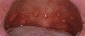
Poor oral hygiene or irritants (such as physical, thermal or mechanical) can cause oral inflammation. This causes inflammation, ulceration, and viral and fungal infections. Very often, such unfavorable changes occur during puberty, pregnancy or menopause.
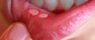
Inflammatory changes in the mucous membrane also occur with certain infectious diseases, vitamin deficiencies or ailments associated with gastrointestinal diseases. Many factors negatively affect the oral mucosa. One of the most common causes is aphthous stomatitis, the presence of bacteria, celiac disease (that is, gluten intolerance), allergies to certain foods, or serious diseases such as cancer.
But very often the causes of inflammation of the mucous membrane are quite ordinary, for example, biting the cheek, excessive brushing of teeth, or eating too sour or spicy foods.
About
Causes of oral tumors
The origin of benign tumors has not been fully studied, but today there are several possible causes or predisposing factors that influence the formation of formation:
- bad habits - smoking, alcohol abuse;
- oral cavity trauma (including tooth extraction, permanent mechanical trauma to the mucous membrane from sharp edges of teeth or fillings, dental structures);
- viral infection - may cause the formation of some forms of benign tumors;
- disruption of tissue differentiation during intrauterine development of the fetus (as a rule, tumors formed for this reason manifest themselves already in childhood - within 1 year of the child’s life).
Stomach mucosa
The gastric mucosa contains cells responsible for the production of:
- digestive enzymes
- hydrochloric acid,
- mucus that protects the stomach from acids.
This type of mucous membrane is covered with single-layer columnar epithelium, and its role in the functioning of the entire digestive system is truly enormous. First of all, the gastric mucosa is involved in the processing and digestion of food.
Why is it such an important part of the digestive system? Well, thanks to such activities and supporting the digestive system, food is better (healthier) absorbed by the body. This also makes it possible to produce the so-called intrinsic factor, whose task is to bind vitamin B12. This allows food to be properly absorbed in the digestive tract.
If the lining of the stomach is damaged in any way, inflammation of the lining of the stomach occurs, called mucositis. Doctors often call this condition gastritis or runny nose. Then the gastric mucosa is hyperemic and irritated.
Unfortunately, in most cases, catarrh is the result of infectious agents and, above all, the presence of Helicobacter pylori in the stomach. But also toxic factors such as bile, alcohol or certain medications cause inflammation of the gastric mucosa. How to recognize that the mucous membrane is damaged and inflamed? Well, there are a few classic symptoms that, once diagnosed, resolve quickly and do not cause too much disruption to the mucous membrane.
These include frequently occurring ones:
- heartburn ,
- nausea,
- vomit,
- epigastric pain ,
- too much satiety
- bleeding from the digestive system.
In 80% of cases, inflammation of the gastric mucosa is detected and treated correctly (under the supervision of a specialist). In such cases, home remedies alone should not be used. This, unfortunately, raises the risk of developing cancer, which is a serious disease and requires long-term treatment.
Vascular tumors of the oral cavity
- Hemangiomas. Classified into the following types:
- capillary - formations from red to bluish in color, protruding above the mucous membrane (can be of different sizes);
- cavernous - formations with clear boundaries 1-2 cm in diameter, have a bluish color, most often formed on the mucous membrane of the cheeks;
- capillary-cavernous;
- mixed.
- Lymphangiomas. The cause is a violation of embryogenesis, so diagnosis is most often carried out in early childhood. They have the appearance of a limited tumor. Lymphangiomas are classified into the following forms:
- cavernous;
- cystic;
- capillary-cavernous;
- cystic-cavernous.
They are characterized by blanching and reduction in size when pressed; injury to these formations can cause bleeding.
These neoplasms are characterized by a tendency to inflammation: it can often be triggered by trauma to the oral mucosa and exacerbation of chronic inflammatory processes in the upper respiratory tract. The tumor is prone to rapid growth, but its distinctive feature is its spread over the surface without penetrating deep into the mucous membrane. Lymph nodes may also be enlarged. In the absence of inflammation, the formation is not filled with lymphatic fluid, however, experienced dentists diagnose the disease at this stage.
Uterine mucosa
The lining of the uterus is called the endometrium. This is the tissue that covers the inside of the uterus. Its role is of great importance for every woman, because it is the place where the embryo is implanted after fertilization of the egg.
The endometrium varies depending on the age and phase of each woman's cycle, and its thickness varies and varies depending on the different stages of the menstrual cycle. At the first stage up to 9 mm, at the second - a little thicker. This is a very important mucous membrane for every female. What characterizes him? This layer peels off during each menstruation. And it should never exceed a thickness of 15 mm, and for postmenopausal women - 12 mm. Endometrial hyperplasia can lead to cancer. This is why it is so important to systematically examine the uterine mucosa.
About
Diagnosis of oral tumors
Benign tumors of the oral cavity are diagnosed through a visual examination by a specialist, and cases of diagnosis are not uncommon during a planned, preventative visit. Also, in the process of treating other diseases of the teeth and oral cavity, a diagnosis can be established, but an accurate determination of the shape and type of tumor is possible only after laboratory analysis - histological examination of tissues. It can be carried out either after complete removal of the tumor, or through a biopsy - partial sampling of material.
To determine the depth of tumor growth, an ultrasound diagnostic method is used - this makes it possible to assess the condition of the soft tissues.
The condition of bone tissue is assessed using radiography.
In case of fibromatosis of the gums, the doctor prescribes an orthopantomogram - a panoramic image to diagnose the destruction of the alveolar process.
Angiography is used to assess the condition of tumors of a vascular nature.
Operations
Small-scale surgical interventions in the oral cavity are performed, as a rule, by dental surgeons, most often on an outpatient basis. Before any operation in the oral cavity, the oral cavity is sanitized (see).
The most common are tooth extraction operations (see Tooth extraction), as well as interventions associated with diseases of the periodontal tissues and odontogenic processes. They are usually performed under local (in the lower jaw, mainly conduction) anesthesia (see Local anesthesia, maxillofacial area). Opening of gingival abscesses and purulent foci during periostitis is carried out using incisions to the bone followed by drainage.
Incisions for cysts (see Dental cyst), benign neoplasms, and pathologically altered areas of the mucous membrane are made within unchanged tissues, followed by suturing with catgut. Wound suturing for oral injuries is performed tightly. Features of surgical treatment of wounds for wounds, including gunshots, affecting the oral cavity - see Face, Jaws.
More extensive interventions in the oral cavity are performed in an inpatient setting under local anesthesia with premedication or general anesthesia. Plastic surgery is performed in the presence of congenital malformations (cleft lip and palate, “double lip”, shortened frenulum of the upper lip and tongue, etc.), as well as for the consequences of injuries and diseases (scars, defects).
In cases of cicatricial deformities in the area of the corners of the mouth, to eliminate the narrowing of the oral fissure, the so-called. microstomy, the edges of the oral fissure with their end-to-end scar change are dissected and epithelialized, everting the mucous membrane from the cheek (Evdokimov’s method). If the strip of the red border is preserved, it is cut off with a through incision, keeping it in the form of a bridge between the upper and lower lips, and then, after dissecting the scars and tissues, the cheeks are pulled up to the required level, where they are secured with sutures, thus forming a new corner of the mouth (Vasiliev’s method).
It is very rare to resort to plastic surgery for an excessively wide mouth gap, the so-called. macrostomy, resulting from a unilateral transverse cleft of the face of a congenital nature (see Face, malformations).
In the postoperative period, careful hygienic care of the oral cavity is necessary (excessive rinsing of the mouth, rinsing), as well as the administration of liquid or softened food, introducing it through a sippy cup if chewing is impossible.