Benign and malignant tumors arise in the ear under the influence of external and internal causes. They are diagnosed at the Yusupov Hospital using modern equipment from the world's leading manufacturers. Otolaryngologists provide conservative treatment using medications that are highly effective and have minimal side effects.
Candidates and doctors of medical sciences, doctors of the highest category make a decision on the need and choice of surgical intervention method at a meeting of the Expert Council. Surgeons use the latest instruments and perform operations under the control of a microscope and computer technology. The medical staff is attentive to the wishes of patients.
Polyp in the ear
Benign polyps of the ear include polyps. These are neoplasms that arise as a result of the proliferation of granulation tissue. The polyp may be located in the external auditory canal or middle ear. Neoplasms localized in the ears can spread to other parts of the skull.
Most often, a polyp is a complication of a chronic inflammatory process in the ear. At the site of chronic inflammation of the mucous membrane, a gradual proliferation of tissue occurs, replacing normal tissue with connective tissue. When the pathological process is localized in the middle ear, the formation can remain invisible for a long time to conventional otoscopy. As the polyp grows, it “falls out” into the external auditory canal through the perforation of the eardrum.
A polyp in the ear is manifested by the following symptoms:
- Suppuration, sometimes mixed with blood (stopping the flow of pus can be caused by blockage of the ear canal by a polyp);
- Itching, noise and pain in the ear;
- A feeling of constriction, the presence of a foreign body in the ear cavity;
- Decreased or loss of hearing;
- Headaches.
In the absence of adequate treatment, a polyp caused by an infection in the ear often becomes the cause of chronic otitis media, supports the inflammatory process and prevents the penetration of drugs to the site of infection. The growth of the polyp leads to blockage of the ear canal and deafness. Under certain conditions, there is a risk of its degeneration into a malignant tumor.
For polyps of small size, in some cases, otolaryngologists carry out conservative treatment with creams containing glucocorticoids and antibacterial drops. If the disease is fungal in nature, antifungal drugs are used. The main treatment for a polyp in the ear is surgery.
The polyp is cut off on an outpatient basis with a special loop or using another instrument: a curette, an ear conchotome. Radical surgery is performed in a hospital. The operation is performed if the fistula is localized in the semicircular canal. An alternative treatment option is laser removal of polyps. A modern method of treating a polyp in the ear is OTO NUS therapy in combination with endoural LILI (exposure to the pathological focus with low-frequency ultrasound through various medicinal solutions).
Homeopathic treatment
All of these remedies are used for symptomatic treatment . The cause is not eliminated with such drugs, but nevertheless, symptomatic drugs not only relieve symptoms, but also increase immune defense and improve the balance of all body functions.
These remedies are recommended to be used to alleviate the condition until you consult a doctor.
For toothaches, the following remedies are most effective: Aconite alternately with Chamomilla, Antimonium crudum, Belladonna, Bryonia, Calcarea carbonica, China, Clematis ( Clematis), Coffea, Ignatia.
They can be combined with warm herbal rinses or medications to relieve inflammation.
Note! The effect is temporary - most drugs have to be used every 2-3 hours.
For increased tooth sensitivity - Argentum nitricum, Causticum, Colkhicum.
This group of remedies is effective in cases where the teeth are not affected by the disease, but the pain in them still bothers the person . Such pains are aggravated by eating sweet, cold or hot foods.
If pain occurs when the jaw is clenched and radiates to the ear - Ammonium carbonicum, Lachesis.
Arnica helps with jaw injuries
Reduces discomfort and pain, the feeling of pulsation in the wound goes away. In parallel with the administration, it is necessary to care for the wound using antiseptic rules.
Pain along the nerve is relieved by Arsenicum album, Bismuthum, Glonoinum, Hypericum, Kreosotum, Plantago.
At the same time, it is recommended to apply cold to the sore spot (if it is pinched). In case of inflammation, it is impossible to further cool or heat the nerve.
Relieves swelling, pain, anti-inflammatory effect - Hekla lava, Hepar sulfur, Kali phosphoricum.
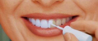
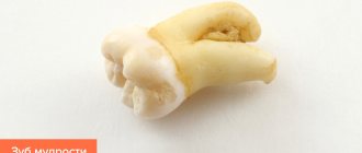
Photo 2. Before starting treatment, you should definitely visit a doctor. Source: Flickr (Denmark Dental)
Glomus tumor of the middle ear
Tympanic paraganglioma (glomus tumor of the middle ear) develops from glomus bodies, which are located on the medial wall or roof of the tympanic cavity, jugular - on the bulb of the jugular vein. Paraganglioma is a benign neoplasm, but mature forms of the tumor have infiltrating and locally destructive growth.
Due to the impossibility of total removal, a glomus tumor of the middle ear can pathologically spread to vital structures of the body (brain stem, internal carotid artery). It can destroy the walls of the temporal bone pyramid, penetrate the posterior cranial fossa and cause compression of the medulla oblongata. Glomus cells often affect vessels over a considerable distance, leading to various complications with a fatal outcome. Patients complain of a “pulsating” noise in the ear. During an objective examination behind the eardrum, the doctor sees a pulsating red mass. As the tumor grows, the following symptoms occur:
- Hearing impairment;
- Facial asymmetry;
- Dysphonia (speech disorder);
- Dysphagia (swallowing disorder).
The Yusupov Hospital has accumulated extensive experience in diagnosing and treating patients with glomus tumor of the middle ear. Otolaryngologists determine the degree of invasion of the glomus tumor of the middle ear into adjacent structures using computed and magnetic resonance imaging of the temporal bones with contrast, angiography and retrograde jugulography. Doctors make the final diagnosis based on the results of histological examination.
If the glomus tumor of the middle ear is widespread, angiography is mandatory. The study is necessary to confirm the vascular nature of the neoplasm, determine its size, location and sources of blood supply. This plays a role in the possibility of embolization, a minimally invasive procedure that is an alternative to surgery. The procedure is aimed at preventing blood supply to the damaged area, which helps to reduce the size of the tumor and achieve a good effect with further surgical removal of the identified tumor. Treatment consists of surgical removal of the glomus tumor of the middle ear. Total surgery is performed in the presence of a glomus tumor that does not spread beyond the middle ear. For subtotal (incomplete) removal of the tumor, and depending on the patient’s age, radiation therapy or stereotactic radiotherapy (gamma knife) is used.
Benign tumors of the ear
Benign tumors of the middle ear include hemangioma and various neurogenic neoplasms. Hemangiomas of the middle ear are manifested by the following symptoms:
- Decreased hearing;
- Ear congestion;
- Feeling of noise.
Often the first symptom of the disease is a slowly occurring paralysis of the facial muscles on the side where the hemangioma is located. For middle ear hemangioma, otolaryngologists usually perform abdominal surgery or widely remove the mastoid process.
Chemodectoma of the middle ear develops from glomus bodies, which are normally located at the bottom of the tympanic cavity, on the dome of the bulb of the internal jugular vein and in the temporal bone. They differ in structure from glomus bodies, which are located in other areas. Depending on the histological structure and the ratio of cell accumulations, there are 3 types of glomus tumors: adenoid-like, alveolar and angioma-like. According to the clinical course, limited and widespread forms of chemodectoma are distinguished.
Chemodectomas are observed at different ages and can be multiple in both ears. Sometimes neoplasms have a malignant course from the very beginning, despite the benign structure of the chemodectoma.
Chemodectomas, which are located in the tympanic cavity, in the initial period of the disease cause hearing loss and pulsating noise in the ear. At this time, the neoplasm appears through the eardrum. Then the tumor protrudes it and causes hyperemia (redness). Gradually, the neoplasm penetrates the external auditory canal and looks like a polyp. When you try to remove it, bleeding occurs. Sometimes patients report ear pain.
Chemodectomas that originate in the jugular vein bulb first destroy the dome of the jugular fossa and spread into the tympanic cavity. As the tumor increases and bone destruction occurs, symptoms of damage to the VII-XII pairs of cranial nerves develop. Patients are bothered by noise in the ear, and otoscopic changes occur. Chemodectomas can grow into the cranial cavity.
Diagnosis of chemodectoma is carried out using radiography of the jugular fossa, pyramid of the temporal bone, attic-antral region, mastoid process. X-ray examination includes radiography of the temporal bone in three main projections and tomography in direct and lateral projections.
Chemodect treatment is surgical. Small tumors that do not destroy the eardrum are removed or exposed to ultra-low temperatures. Tumors that have spread to the external auditory canal, mastoid process, or antrum are also subject to surgical treatment. Otolaryngologists perform operations of varying scope - from tympanotomy to extended radical surgical interventions on the ear. Sometimes cryotherapy is used. For tumors that destroy the pyramid and spread into the cranial cavity, external gamma irradiation is performed, which often causes growth arrest or a decrease in chemodectoma.
Preventive measures
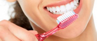
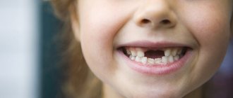
Since jaw pain is most often caused by dental caries, then, first of all, in order to avoid unpleasant sensations, it is necessary to take proper care of the oral cavity:
- brush your teeth 2 times a day;
- use dental floss;
- rinse the mouth after eating;
- do not get carried away with sweet carbonated drinks (if you drink, it is better through a straw);
- less candy;
- do not combine too cold and hot dishes;
- visit the dentist for a preventive examination 1-2 times a year at least;
- If symptoms or signs of tooth decay appear, seek treatment immediately.
One rule applies to the prevention of neuralgia: do not overcool, avoid drafts .
Note! To prevent inflammatory diseases of the jaw, you should monitor the cleanliness of your mouth, the health of your nose and paranasal sinuses, and constantly strengthen your immunity with proper nutrition, exercise, and walks in the fresh air.
Osteoma in the ear
Osteoma in the ear (exostosis, osteophyte) develops mainly from the compact layer of the posterior wall of the bony part of the external auditory canal. Much less often, neoplasms are found on the lower and upper walls of this section. Endophytic osteomas penetrate into the mastoid process. Osteoma is a benign tumor that grows rather slowly.
Osteoma has the appearance of a round formation, which is covered with a skin layer, very dense when palpated with a Vojacek probe. It is treated surgically. The operation is performed after the tumor has grown to medium size. In this case, removing the tumor is technically as convenient as possible. If the tumor is small, there is a risk of not completely removing the pathological tissue. If the osteoma is large, it is possible to capture a significant part of the healthy bone tissue during surgery. This will cause a large bone defect.
What to do if both jaw and ear hurt at the same time
When acute pain occurs from the jaw joint to the ear, it is worth reducing the impact on the affected area.
Following these simple recommendations will help with this:
- try not to open your mouth wide, thus reducing the load on the joint;
- It is worth eating food in small portions, and it should not be very hot or very cold;
- The food consumed should not be difficult to chew, preferably soft; if the pain syndrome is severe, the food is preferably liquid;
- avoid pressure and mechanical impact on the painful area;
- exclude the possibility of hypothermia.
These precautions can reduce the number and force of chewing movements and reduce the load on the jaws.
Instructions on how to help yourself or a loved one while waiting for a doctor’s appointment:
| Main complaints | What to do to alleviate the condition |
| Chewing pain, stiffness, clicking or catching of the jaw | take a pain reliever; reduce or eliminate the load on the jaw. |
| Pain syndrome in the jaw and ear | Taking a pain reliever |
| Edema and swelling of the temporomandibular joint | Short-term use of cold |
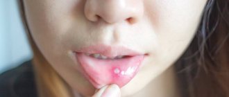
| Dental pain | taking painkillers; rinsing with antiseptic drugs; herbal rinses with an antiseptic and soothing effect. |
| A significant increase in body temperature, the appearance of symptoms of viral mumps in the child | use of antipyretic drugs; fastest house call. |
Prolonged pain that does not go away is a reason for examination
If you simultaneously complain that your jaw and ear hurt, you need to see a doctor as soon as possible. A dentist or ENT doctor is suitable for the initial visit; they will help differentiate the cause of the pain and, if necessary, refer you to other specialists.
Every person knows himself better than any doctor. Therefore, when the first signs of illness appear, you can roughly guess what may be causing your poor health.
Lipoma and atheroma behind the ear
The area of skin around the ear contains a huge number of sebaceous glands. For this reason, lipomas and atheromas often form behind the ear. Lipomas that form behind the ear grow slowly and are often not cancerous. They are a soft-elastic formation with a smooth surface, surrounded by a capsule. The lipoma in the photo looks like a wen.
Atheroma is a cavity formation filled with sebum. Formed due to blockage of the sebaceous glands. Atheromas occur for the following reasons:
- Disorders of fat or carbohydrate metabolism;
- Genetic predisposition to increased oily skin;
- Hormonal imbalances and diseases of the endocrine system;
- Hyperhidrosis is a disease associated with excessive sweating;
- Failure to comply with personal hygiene rules.
Atheroma is a rounded formation protruding above the surface of the skin, which can reach up to 4.5 cm in diameter. When the tumor becomes infected or inflammatory reactions occur, the following symptoms occur:
- Pain behind the ear;
- Redness of the skin;
- Burning and itching;
- Fluctuation is a symptom that indicates the presence of fluid in a cavity formation.
When pressure is applied to the walls of the atheroma or they are damaged, the viscous mass contained inside comes out to the surface of the skin. It has a white color and an unpleasant odor. When atheroma suppurates, the contents have a green-yellow tint. Lipomas and atheromas behind the ear are removed surgically. Modern treatment methods are used - laser or radio wave removal.
Salivary gland adenoma is a benign formation that appears in the glandular epithelium of the salivary glands. The salivary glands are parotid, submandibular, and sublingual. The most common occurrence of tumors is on the parotid gland. If the components of such a tumor are benign, then it is an adenoma of the parotid salivary gland.
Adenoma behind the ear
A benign tumor, a parotid adenoma, often develops in the parotid region. The structure of the neoplasm resembles the salivary gland itself. The cause of the development of benign tumors of the salivary glands is the formation of altered glandular epithelium.
The neoplasm is enclosed in a capsule, has a soft-elastic consistency, and is not fused to the skin and surrounding tissues. The skin above the adenoma behind the ear is not changed. It is treated surgically. In order to undergo examination and treatment of benign tumors of the ear and parotid region, call the contact center of the Yusupov Hospital.
Comments
I can’t understand, my tooth hurts and that’s why it radiates into my ear. Or, on the contrary, the ear hurts and it hurts the tooth. Which doctor is better to go to?
Nathan (02.11.2019 at 16:37) Reply to comment
- Dear Nathan! After reading our article, you should have realized that pathology can be caused by completely different reasons. Only a doctor after examination can determine the true one. In your case, it is recommended to first visit two specialists: a dentist and an otolaryngologist. If these doctors do not find anything, then further examination by an orthopedist or neurologist may be required.
Editorial staff of the portal UltraSmile.ru (09.11.2019 at 09:14) Reply to comment
Ear and tooth hurt. I did an independent examination in front of the mirror, all the teeth in my mouth were intact, there were no holes. I haven’t gotten my ticket to the ENT specialist yet; in our city it’s a big problem to get to see this specialist for free. To avoid pain, I take pills, my grandmother also advises warming my cheek, supposedly this helps. Is it possible to do this?
AlisaV. (11/26/2019 at 12:19 pm) Reply to comment
- Dear Alisa! If you have not noticed any pathologies externally, this does not mean that you do not have dental problems. For example, it is almost impossible for the patient to notice a cyst in the early stages on his own. Therefore, it is still recommended that you go to the dentist. As for warming up the cheek, this is strictly forbidden, because... if the inflammatory process is to blame, then the infection, when warmed up, can spread through the bloodstream to the surrounding tissues, and your condition will only worsen.
Editorial staff of the portal UltraSmile.ru (01.12.2019 at 09:21) Reply to comment