Many people ask a completely logical question when they find blue veins under the tongue: what can this mean and does it indicate the development of any disease? We hasten to reassure you that in most cases this phenomenon is considered absolutely normal, due to the peculiarities of blood circulation, vessels located close to the surface and a number of other factors caused by physiological processes.
However, some diseases and borderline conditions can provoke the appearance of blue veins under the tongue, so it is important to learn to distinguish normality from pathology.
We will tell you how to do this further.
Anatomical structure of the tongue
To better understand the reasons for the appearance of blue veins, one should turn to the anatomical features of the structure and functioning of the human tongue.
It is a muscular organ covered with a mucous membrane. The tongue has many nerve endings, thanks to which the necessary signals are sent to the central nervous system.
The tongue consists of two main parts - the body, which can be easily seen with the help of a mirror, and the root, located near the pharynx. They are separated from each other by a V-shaped terminal groove. The surface of the body is rough due to the presence of multiple papillae on it, which are responsible for taste, temperature and pain perception.
In the middle of the lower part of the organ, the mucous membrane forms a frenulum , which connects it to the mucous membrane of the oral cavity. On the sides there are fringed folds, converging to the tip. It is between them and the frenulum that the elements of the circulatory system are visible.
The lingual artery is responsible for the blood supply to the lingual tissues; with its help, oxygen-enriched blood is supplied to the organ. This is how the tongue is nourished. Venous outflow is provided by a complex of lingual vessels connecting to the internal jugular vein. It is the change in their color that most often causes concern among people.
Types of benign tumors of the tongue
Papilloma
Focal skin growth caused by activation of the human papillomavirus in the body. The virus is transmitted when the tongue comes into contact with the mucous membrane or skin of an infected person. In the rarest cases, transmission of the virus occurs through household contact. The virus can remain dormant for a long time and appear when the immune system is weakened.
Externally, papillomas look like small pink, flesh-colored or white papillae of various shapes. Their size usually does not exceed two centimeters. You can detect these tumors yourself. They are usually localized on the back, surface and tip of the tongue. These small tumors pose a serious threat to human health because they are very often damaged and susceptible to malignancy.
Adenoma
The occurrence of this tumor is caused by the proliferation of glandular epithelium. As a rule, neoplasms are single. The following types of tongue adenoma are distinguished:
- polymorphic;
- basal cell;
- canalicular;
- sebaceous;
- adenolymphomas;
- monoform.
The pathology is usually accompanied by the formation of polyps on the tip of the tongue.
Botriomyxoma
A benign tumor of the tongue, the occurrence of which is caused by traumatic injuries - burns, cuts, injections. A couple of months after the injury, a red tumor-like formation with a lobulated or smooth surface may appear in its place. Botriomyxoma has a dense elastic consistency, sits on a stalk, and with minor damage begins to bleed.
Fibroma
A neoplasm that is formed from connective tissues covered with mucous membrane. Usually appears as small nodules, but can also look like branched polyps. The main causes of the disease are traumatic and inflammatory processes in the oral cavity. Most often, a tumor forms against the background of advanced inflammatory dental diseases - stomatitis, glossitis, periodontitis, gingivitis.
Retention cyst
The tumor occurs due to problems with the outflow of secretion from the gland, which causes blockage of the duct. Because of this, the secretion gradually accumulates in the gland, stretches it and fills it with new contents. This type of tumor is removed surgically.
Lipoma
A tumor that is predominantly localized in the back of the tongue. A lipoma forms in the submucosal layer of the tongue and has a soft and elastic consistency. Often the process of lipoma formation occurs without symptoms, so diagnosing it at an early stage is extremely difficult.
Myoma
A tumor in the tongue that occurs due to the growth of muscle tissue. Myoma has a fairly dense consistency, is covered with a mucous membrane, and small papillary outgrowths can form on it. This tumor usually reaches a centimeter in size.
Neurofibroma
A tumor develops from the sheaths of peripheral nerves. Doctors associate its formation with a defect of the facial or trigeminal nerve. This tumor is extremely rare.
Hemangioma
This is a vascular neoplasm that occurs in the tissues of blood vessels. The tumor is benign - it grows quite slowly and does not penetrate other organs. However, hemangioma is completely unpredictable, as it can suddenly increase in size and begin to interfere with the functioning of the tongue. Depending on the structure of the tumor, it can be of two types: simple (looks like a tangle of capillaries located on the mucous membrane of the tongue) and cavernous (a tangle of large vessels under the mucous membrane of the tongue).
Lymphangioma
The tumor usually grows in the walls of the lymphatic vessels, which then leads to a significant increase in the size of the tongue. Tumors often form on the tip of the tongue and its surface.
Struma of the tongue
A congenital pathology that occurs due to a violation of the location of fragments of the thyroid gland. It looks like a small organic node. After surgical removal of the tumor, the prognosis for the patient is favorable.
Why do blue veins appear under the tongue?
Since ancient times, changes in structure, color, and the formation of plaque on the surface of the tongue have been considered evidence of disturbances in the functioning of internal organs . That is, it acted as a kind of identifier of a person’s health status.
Bluish vessels, as already noted, are not always a pathological sign, but in some cases they, like a litmus test, make it possible to promptly suspect any disorders in individual organs and systems.
If the appearance of the sublingual vascular pattern changes, you should consult a doctor who will give an expert assessment of what is happening.
It’s worth noting right away that the presence of any disease is not limited to just increased coloration of the tongue; it should be considered, rather, as an additional sign. Let's consider a number of reasons that provoke such a clinical manifestation.
Nutrient deficiency
A lack of B vitamins can affect the appearance of not only blood vessels, but the entire oral cavity. In particular, when there is insufficient intake of vitamin B2, known as riboflavin, similar symptoms appear in the body.
In addition, a person becomes susceptible to a number of diseases of the oral cavity and throat, such as stomatitis, pharyngitis, tonsillitis , he regularly develops cracks on his lips that do not heal for a long time, the color of the tongue surface also changes, darkens and acquires a purple tint.
This condition is corrected by additional intake of vitamins in tablet form and selection of an appropriate diet.
Respiratory diseases
Insufficient saturation of the circulatory system with oxygen due to various pathologies of the respiratory system leads to increased coloration of the lingual vessels , even black. For any of the diseases of the lungs and bronchi, the main symptoms are characterized by lack of air, coughing with or without sputum, and the presence of wheezing, which can be heard when listening to the chest with a phonendoscope.
High cholesterol
Disturbed lipid metabolism and the appearance of fatty plaques significantly disrupt the natural blood flow and nutrition of organs. For this reason, the sublingual space may have an excessively dark color.
Circulatory disorders
In most cases, it is problems with the circulatory system that provoke visual darkening of the vascular pattern. In particular, varicose veins in the sublingual area can appear as a result of:
- increased intravascular pressure as a result of heart failure due to atrial fibrillation and flutter and valve defects;
- hereditary predisposition, characterized by some thinning of individual walls, as a result of which nodes and bloating appear;
- hemorrhoids - a pathological condition accompanied by a violation of the outflow of blood with the formation of nodes localized in the rectal area. You can learn more about varicose veins of the rectum from our article;
- decrease in the elasticity of vascular walls due to age-related changes.
Causes of tongue cancer
In modern medicine, it is believed that the release of carcinogens during the combustion of tobacco is the main factor that influences the occurrence and development of tongue cancer. When combined with alcohol, carcinogens from tobacco smoke increase the likelihood of developing cancer among alcoholics and smokers.
Also, the development of tongue cancer can be influenced by components that are classified as occupational hazards, such as perchlorethylene, asbestos, heavy metal salts, and petroleum distillation products.
The formation of this tumor can be influenced by mechanical trauma to the mucous membrane. This type of injury can occur from sharp tooth edges, poorly fitting dentures, frequent tongue biting, poorly processed fillings, or a broken tooth.
According to research, a connection has been established between chronic persistent infection and tongue cancer, which is caused by the herpes simplex virus, HIV, or HPV. Patients who receive immunosuppressive drugs for a long time are also predisposed to tongue cancer.
If the carcinogenic factor continues to act on the organ, then changes in the mucosa will lead to oncology. Today, the following factors are classified as precancerous conditions of the tongue:
- papillomas;
- chronic tongue ulcer;
- hyperkeratic forms of lupus erythematosus;
- leukoplakia of the tongue;
- hairy leukoplakia of the tongue;
- Bowen's disease;
- lichen planus;
- systemic lupus erythematosus.
Long-term exposure to these trigger factors leads to changes in the DNA structure of tongue cells with the development of hyperplasia and dysplasia of the tongue mucosa. Most benign tumors of the tongue are susceptible to transformation into malignant tumors due to trauma to the oral cavity.
Painful sensations
Swollen blue veins that appear under the tongue indicate the development of varicose veins. Due to anatomical features, the vessels themselves cannot hurt due to the absence of nerves in them.
Due to the course of varicose veins , they expand and put pressure on the nerve endings located in the immediate vicinity, causing pain in the sublingual area. It is these sensations that non-medically savvy people confuse with pain in the vessel.
In addition, late stages of the development of the disease provoke violations of the integrity of the integument due to the formation of ulcers, which will definitely cause discomfort.
promptly seeking qualified help , you can get complications from varicose veins in the form of thrombophlebitis. Blockage of the lumen of the sublingual vessel by a thrombus will inevitably make itself felt not only by visual changes, but also by physical pain.
If, during dental surgery, when a tooth was pulled out, blue veins with characteristic swelling were noticed under the tongue, you should consult the doctor who performed the extraction. He will be able to examine the picture in detail, draw appropriate conclusions and, if necessary, refer you to a specialized specialist.
Read more about massage for varicose veins in our material.
Symptoms at different stages
At an early stage, tongue cancer, like other malignant tumors, is treatable. The difficulty in identifying the disease at an early stage is that the signs and symptoms of tongue cancer at an early stage do not bother the patient and therefore go unnoticed. The main signs of tongue cancer are:
- Unpleasant smell. If you have healthy, unproblematic teeth and despite proper care, an unpleasant odor appears in the mouth at the initial stage of tongue cancer, which is one of the first warning signs of the presence of malignant tumors.
- Discomfort. With the onset of tumor development, discomfort begins in the affected area. A person may experience tingling, burning, or numbness in the tongue area.
- Visual changes. At the initial stage of tongue cancer, symptoms include small neoplasms in the form of ulcers, papillomas, erosions and spots. When you press on them, you may feel a tightness.
- Migrating pain. At the initial stage of the disease, a person feels pain inconsistently, the pain can migrate and radiate to nearby locations, gums or cheeks.
In the later stages of the disease, the symptoms of tongue cancer are as follows:
- Increased salivation;
- Trouble swallowing;
- Bleeding of the tongue;
- The appearance of speech defects;
- Difficulty speaking;
- Loss of appetite;
- Severe pain;
- Loss of body weight;
- Significant increase in the volume of the tongue;
- Feeling chronically tired
- Enlargement of the postauricular and submandibular nodes.
For the third stage of development of tongue root cancer, the symptoms are as follows:
- There is a disturbance in the digestive system of the body, loss of sensitivity, immobility of the tongue, and difficulty in eating;
- I am worried about severe pain, it is localized in the frontal sinus and temporal area;
- Malignant tumors disintegrate, which causes an even greater increase in saliva production and a strong unpleasant odor from the oral cavity.
In advanced situations, such as stage 4 tongue cancer, a very aggressive course of the disease occurs, rapid invasive tumor growth, metastases from tongue cancer spread to the lymph nodes, which are located nearby and gradually reach the bones, liver, brain and lungs.
Since there is difficulty in diagnosing tongue cancer at the initial stage, when the disease is detected in an advanced stage, patients wonder how long they can live at stage 4 of tongue cancer. Modern medicine gives a disappointing prognosis for patients with stage 4 tongue cancer and the chance of recovery is about 30%. Few people know what tongue cancer looks like, so at the first sign of discomfort you should seek advice from specialists.
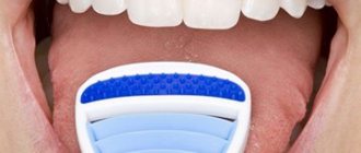
How to diagnose
If you suspect the development of varicose veins, the only correct decision would be to consult a specialized doctor - a phlebologist. He will conduct a visual examination and collect anamnesis, after which he will prescribe additional studies to obtain comprehensive information on a particular case.
Ultrasound examination of the affected areas is an accessible and informative method of diagnosis for varicose veins. With its help, qualitative indicators of blood outflow are checked , allowing one to determine the presence or absence of blood clots that pose a real threat to health. Sometimes radiography is prescribed using a special contrast agent.
Despite the fact that women are more susceptible to developing varicose veins, its manifestations in the sublingual area occur equally often in both sexes.
We wrote in detail about the methods of diagnosis and treatment of varicocele on the left testicle in our article.
Diagnosis of tongue cancer
According to statistics, the success of treatment for tongue cancer depends on timely and high-quality diagnosis. If there are first symptoms of discomfort in the tongue area, the patient should consult a dentist. During the examination, the doctor examines the oral cavity using mirrors, palpates the affected area and, as a result, determines the medical history. If there is a suspicion of oncology, the dentist will refer you to an oncologist for further diagnosis.
In the process of diagnosing tongue cancer, the following methods are used:
- bacterioscopy. The method consists of examining a smear, which is necessary to identify pathogens that cause a number of diseases in the oral cavity;
- biopsy. The method consists of studying biomaterials that are taken from the affected area to determine the presence of metastases;
- computed tomography or MRI. Using this method, you can determine the presence of metastases in the brain;
- radiography. This method is used to determine whether there are changes in the bone structure.
- ultrasound diagnostics. Using this method, the size of the endophytic tumor is determined.
If there are primary signs of tongue cancer, you should consult a specialist, since diagnosing the disease in the early stages increases the chances of recovery. High-quality diagnostics using modern equipment can be performed at the Yusupov Hospital.
How to treat
Modern methods of treating varicose veins can be divided into medicinal and surgical. The former are used to slow down and stop the course of the disease in the initial stages, from initial manifestations to stage 2 disease.
Surgery is a panacea for advanced disease. However, it is the latter that has the most pronounced result and allows you to get rid of not only the cosmetic problem, but also the possible development of complications caused by this pathology.
The choice of the necessary treatment method is the prerogative of the attending physician alone, who can correctly assess the patient’s condition.
Conservative therapy involves taking medications with various pharmacological effects, namely venotonics, anticoagulants, antiplatelet drugs . Some of them come in various forms and are used both internally and topically.
Blue swollen veins that appear under the tongue, photos of which can often be seen on the Internet, can be treated surgically or by injecting a sclerosing substance into the venous cavity, causing it to stick together.
Veins are removed by microphlebectomy or laser coagulation. Any of these techniques is aimed at restoring proper blood circulation. We described in our material how varicocele manifests itself and is treated in adolescents.
Tongue cancer
Patients most often complain of unusual sensations or pain in the gums, tongue, throat, and cheeks. Tongue cancer is most often localized on the lateral surfaces (up to 70% of cases). Much less often the root (about 20%) and lower surface (about 10%) of the tongue are affected. Patients consult a doctor with complaints of a non-healing ulcer (usually about 1 cm in diameter). However, sometimes tumors can reach large sizes (4 cm or more). In later stages, pain, itching, and burning appear. With cancer of the floor of the mouth, patients often consult a doctor when the tumor reaches a large size, the disintegration of the tumor, bad breath, and bleeding are noted. With such processes, almost 50% of patients have signs of regional and distant metastasis by the time they contact a specialized institution. They may also be concerned about swelling or ulcers in the mouth, loosening and loss of teeth, and bleeding of the oral mucosa. Later, complaints include difficulty opening the mouth, difficulty or impossibility of eating, bad breath and excess saliva, swelling of the neck and face, and weight loss. Upon examination and palpation of the oral mucosa, a changed area is detected. It differs from the rest of the mucous membrane in appearance and density. Essentially, we see part of the tumor tissue that takes on different shapes and shapes, and the rest of the tumor tissue is located under the mucous membrane. It can be a dense, painless plaque of gray or pink color, with a finely lumpy surface. It protrudes slightly above the level of the mucosa and has clear boundaries. You can see a dense, painless, gray-pink node with clear boundaries. It protrudes significantly above the level of the unchanged mucosa. Its surface is medium or coarsely lumpy. The cancerous node has a wide and dense base. An irregularly shaped ulcer can be observed, with a lumpy bottom and uneven raised edges. Its color varies from dark red to dark gray. On palpation, the ulcer is moderately painful and dense. There is pronounced tumor infiltration around the ulcer. Cancer of the oral mucosa can also manifest itself in the form of an infiltrate with unclear boundaries, covered with unchanged mucous membrane. The infiltrate most often has a dense consistency and is painful. Oral cancer spreads quickly, destroying surrounding tissues - muscles, skin, bones. Tumor recurrences are possible after radical operations. With regional metastasis, enlarged lymph nodes are palpated on the lateral surface of the neck, usually dense, painless, and inactive. The clinical course of cancerous tumors of the oral cavity can be divided into three phases or periods: initial, developed and neglected. Initial period . At this time, patients most often note unusual sensations in the area of the pathological focus. When examining the oral cavity, various changes can be detected: thickening of the mucous membrane, thickening of tissues, superficial ulcers, papillary neoplasms, white spots, etc. During this period, it is necessary to carefully examine the organs of the oral cavity, since an analysis of observations shows that in almost 10% of cases, at the first visits to the doctor, local lesions on the mucous membrane were not identified. Pain, which usually forces you to see a doctor, occurs during the initial period of cancer development in approximately 25% of cases. However, in more than 50% of cases, pain is associated with sore throat, dental disease, etc. This is especially often observed in cancer localized in the posterior half of the oral cavity and the alveolar edge of the jaw. Doctors' attention is often misdirected. In the initial period of development of oral cancer, it is advisable to distinguish three anatomical forms: a) ulcerative; b) knotty; c) papillary. The ulcerative form is observed most often; in approximately 50% of patients, the size of the ulcer increases slowly, in others quickly. Conservative treatment, as a rule, does not reduce the ulcer. This can be said about the following two forms. The nodular form is manifested by compaction in the mucous membrane with whitish spots around or hardening in the tissues. In the latter case, the mucous membrane above the hardening may be unchanged. Seals usually have clear boundaries and develop faster than with the ulcerative form. The papillary form is characterized by the presence of dense growths above the mucous membrane. They develop quickly and are often covered with an intact mucous membrane.
Developed period. At this time, numerous symptoms appear. First of all, almost all patients are bothered by pain of varying intensity, although sometimes, even with large tumors, pain may be absent. The pain becomes excruciating, is local in nature, or radiates to one or another area of the head, often to the corresponding ear or temporal region. In many patients, salivation increases as a result of irritation of the mucous membrane by tumor decay products. A typical symptom is bad breath, which is a concomitant of tumor decay and infection. In the developed period of cancer of the oral mucosa, we distinguish two anatomical forms:
- exophytic form (papillary - a mushroom-shaped tumor with plaque-like or papillary outgrowths; ulcerative - the presence of an ulcer with a marginal ridge of active tumor growth, despite the increase in its size, it still remains superficial, and the tumor ridge seems to delimit the process) and
- endophytic form (ulcerative-infiltrative - an ulcer on a massive tumor infiltrate. Ulcers often take the form of deep fissures; the infiltrative form is characterized by diffuse damage to the organ. The mucous membrane of these neoplasms does not ulcerate).
Dividing cancer of the oral mucosa into anatomical forms aims to clarify the nature of tumor growth and determine the type of treatment. Clinical experience shows that endophytic forms of tumors, characterized by diffuse growth, have a more malignant course than exophytic forms with a limited type of growth. Period of neglect . Cancer of the oral mucosa, spreading rapidly, destroys surrounding tissue and should be classified as one of those tumors that we consider exclusively aggressive and malignant. It should be noted that in general, cancer of the mucous membrane of the posterior half of the oral cavity is more malignant than the anterior one; it is also much more difficult to treat cancer of the organs of the posterior half of the oral cavity. Tongue cancer most often develops in the middle third of its lateral surface (62-70%) and at the root. Much less often it occurs on the lower surface of the tongue, sometimes on the dorsal surface (7%) and the tip of the tongue (3%). Cancer of the root of the tongue is observed, according to various sources, in 20 - 40% of cases. More often it is squamous cell carcinoma of various differentiation. Malignant tumors emanating from the minor salivary glands develop in the tongue in approximately 1.5-3% of cases. Malignant lymphomas sometimes occur in the back of the tongue. Cancer of the floor of the mouth accounts for 20% of all squamous cell carcinomas of the oral cavity, of which about 3% are adenocarcinomas of the minor salivary glands. Often the floor of the mouth is infiltrated with secondary malignant tumors of the tongue, gums, lower jaw, and submandibular salivary glands. Patients seeking medical help in the early stages is rare. More often we have to deal with tumor processes when they are accompanied by a secondary infection and pain appears. Often, when you first visit a doctor, the spread of the tumor to the lower jaw and muscles of the floor of the mouth is determined. During this period, approximately one third of patients experience regional metastases. Cancer of the buccal mucosa - the histological picture in this case is the same as for cancer of the tongue and floor of the mouth. However, malignant tumors of the minor salivary glands are less common. Often the mucous membrane of the cheek is infiltrated by a tumor secondary to the tonsils, lips and skin. Regional metastases are rarely observed when patients first visit a doctor, with the exception of tumors localized in the retromolar region and with their spread to the tonsils and arches. Cancer of the mucous membrane of the palate - on the hard palate, malignant tumors emanating from the minor salivary glands (adenocystic carcinoma - cylindroma, adenocarcinoma) often develop. Mixed tumors (polymorphic adenomas) are observed somewhat less frequently here, the differential diagnosis of which is very often difficult even for histologists. Squamous cell carcinoma of the hard palate rarely develops. In the soft palate, neoplasms arising from the minor salivary glands are rarely observed and the vast majority of tumors are squamous cell carcinoma. This morphological feature of tumors of the hard and soft palate significantly affects their clinical course. Squamous cell carcinoma of the hard palate ulcerates quite quickly, causing discomfort or pain. Patients usually consult a doctor when the tumors are still small. Neoplasms arising from the minor salivary glands remain encapsulated for a long time, sometimes reaching significant sizes. In such patients, the first and main complaint is the presence of a tumor on the hard palate. As the size of the tumor increases, its pressure on the mucous membrane increases and an area of ulceration appears, then infection occurs, and pain occurs. It should be borne in mind that adenocarcinomas and mixed tumors of the hard palate in the initial period of development are similar for a long time and mainly retain a tendency to encapsulated growth. The adenocarcinoma then grows and destroys the underlying bone structures. Cancer of the mucous membrane of the alveolar edge of the lower and upper jaws
These neoplasms almost always have the structure of squamous cell carcinoma. They manifest themselves quite early, as the teeth are involved in the process and toothache occurs. Often these pains are treated and even tooth extraction is performed. Unwise tooth extraction promotes the spread of cancer into the tooth socket and then into the bone. In the initial period, the swelling is usually local and bleeds when touched. Infiltration of the underlying bone tissue (alveolar edge of the lower or upper jaw) occurs after several months and should be considered as a late manifestation of the disease. The extent of tumor spread to the bone is determined by x-ray, but it must be borne in mind that chronic dental diseases also cause a picture of demineralization of bone tissue. Depending on the location, gum cancer also spreads to the mucous membrane of the cheek, palate or floor of the mouth. Regional metastasis is observed early and is diagnosed in approximately a third of patients. Malignant tumors arising from the minor salivary glands are rare.
How dangerous?
It is impossible to give a definite answer to this question due to various reasons for the manifestation of such symptoms.
If the structure of the vessel is not disturbed, but it is blue or black in color, this may be a normal variant. Venous blood is darker in color than arterial blood due to its saturation with carbon dioxide. Additional blueness is given by the vascular wall, mucous membrane and other tissues that can distort the real shade of blood.
Another situation that also does not require treatment and is considered completely natural is the aging of the body. , a natural thinning of the vascular walls occurs , resulting in dark blue veins appearing under the tongue.
A change in not only the color, but also the structure of the sublingual venous lines, the formation of nodules, multiple or single, the appearance of painful sensations - all this is a reason to contact a specialist to carry out a differential diagnosis and establish the root cause of the manifestation of such symptoms. Read more about varicocele in a 14-year-old teenager in our material.
Treatment of tongue cancer
A combination of methods is used to treat tongue cancer. The tongue is an important organ in the human body that is involved in speech and swallowing, therefore the choice of treatment method largely depends on the impact that the effect of this method can have on these processes. Also, when choosing a treatment method for tongue cancer, doctors take into account how it may affect the patient’s appearance.
Basic treatment methods for tongue cancer
In modern medicine, the following types of treatment for tongue cancer are distinguished:
- Surgery. Today, surgery is the most common treatment for tongue cancer in the early stages, leading to recovery in 80% of cases. However, there is a category of patients who, for certain reasons, cannot undergo surgery or the operation can negatively affect body functions, such as swallowing and speech. Surgical treatment of tongue cancer involves radical removal of the tumor, which includes complete glossectomy or partial hemiglossectomy of the tongue. If the cancer grows into the bone structures and soft tissues of the floor of the oral cavity, surgical intervention is accompanied by resection of the jaw bone and affected tissues. If necessary, doctors use an orthostomy and apply plastic surgery methods to restore lost structures of the maxillofacial part.
- Radiotherapy. Modern medicine has proven that the results of radiotherapy are similar to those of surgery. Clinical studies have shown that radiotherapy is effective as a single treatment or in combination with surgery, with less severe complications. Recovery is achieved in about 80% of patients.
- Radiotherapy in combination with surgery. This combination is usually prescribed in cases of large tumors. But it can also be used to treat patients with a small area of normal tissue that remains after surgery, as well as if cancer grows into the edge of the removed tissue. Also, for leukoplakia of the tongue, complex treatment is prescribed.
- Palliative radiation and chemotherapy are given to patients who have distant metastases of tongue cancer.
Removal of lymph nodes
When treating tongue root cancer in the early stages, doctors decide to perform surgery or radiotherapy on the lymph nodes. If the lymph nodes are not treated, the cancer will spread to the neck through the lymphatic system. Untreated cancer leads to recurrence of the disease. It is very important to determine the presence of a tumor in the lymph nodes of the neck. Currently, this is effectively achieved through surgical removal of the lymph nodes.
For cancerous tumors in the neck, lymph nodes are removed, which is called radical dissection. If cancer cells are found in the lymph nodes, the patient is prescribed radiotherapy to the neck. Otherwise, no further therapy is carried out.
Diagnosis and treatment of various types of tumors are carried out by specialists from the Oncology Center of the Yusupov Hospital. For this purpose, the clinic has modern high-tech equipment, and also employs leading oncologists and chemotherapists with many years of experience.
Do I need to follow a diet?
If any of the reasons listed above have been medically established as causing the appearance of a cosmetic defect, it would not be a bad idea to reconsider your own gastronomic preferences.
Each of these conditions requires at least a slight adjustment of the diet , especially if a person prefers to eat “junk” food. The adjustment is as follows:
- in increasing the volume of consumption of products of plant origin - vegetables, fruits, herbs in fresh, baked form, as well as in salads;
- excluding excessively fatty meat, spicy, salty and fried foods from the menu. When cooking food, you should choose baking, boiling or steaming;
- in regular consumption of sea fish and seafood, as the main source of saturated acids;
- in choosing pasta made from durum wheat or vegetables as a side dish for meat or fish dishes, depending on individual preferences;
- You can’t give up cereals; they are a source of vitamins and microelements. The most useful are buckwheat, oatmeal, and pearl barley;
- You should use olive or any other vegetable oil as a salad dressing. As an exception, it is allowed to consume homemade mayonnaise once a week for this purpose in limited quantities - no more than 30 ml.
Treatment methods at home
The basis of home therapy is following the doctor’s recommendations and maintaining a healthy lifestyle. Quitting smoking, drinking alcoholic beverages, and observing basic dietary restrictions are the main requirements in the therapy process.
If varicose veins are diagnosed in the sublingual area in the initial stages, it is advisable to use various decoctions and infusions of herbal components that have anti-inflammatory and restorative effects as additional measures. Let's look at the best recipes:
- Mix a glass of water at room temperature with a tablespoon of natural apple cider vinegar. Divide the contents into 2 doses and consume immediately after meals;
- Divide the sour apple into 6 slices, fill them with 500 ml of boiling water, leave for 4 hours. Then mash the fruits, add 3 teaspoons of honey and take the resulting liquid three times a day, 150 ml before meals;
- nutmeg-alcohol tincture has also proven itself as an effective remedy in the fight against diseases of the circulatory system. Mix 250 grams of crushed nuts with a bottle of vodka and leave for 20 days in a cool place, protected from direct sunlight. Take 20 drops half an hour before meals.
Under no circumstances should you rely on traditional medicine alone and refuse drug therapy prescribed by your doctor. This can aggravate the course of the disease and provoke complications.
Preventive actions
The following tips will help reduce the chances of pathological changes in the sublingual area:
- nutrition should be regular and balanced;
- oral hygiene measures should be carried out 2 times a day, morning and evening;
- Regular medical examinations by highly specialized doctors will allow you to identify the slightest changes at the initial stages, when they are still being successfully treated. Such events should be held at least once a year;
- if there is a hereditary predisposition to varicose veins, constant moderate physical activity should be maintained;
- Sanitation of the oral cavity at the dentist will avoid the proliferation of pathogenic microflora in it, which becomes the impetus for the formation of pathology.
As you know, it is much easier to prevent the development of a disease than to cure it later, so you should not neglect these simple rules.
Classification of tongue cancer by types and forms
In the oral cavity, the tongue and the mucous membrane of the floor of the mouth are most often affected. Most often, the tumor develops from squamous epithelium, which is why it is called squamous cell carcinoma of the tongue
. Squamous cell carcinoma is prone to metastasis and affects the submandibular lymph nodes. Occurs in 90% of all tongue tumors. Another histological type is adenocarcinoma, which tends to affect the salivary glands.
- Based on localization,
there are three types: cancer of the body of the tongue, cancer of the root of the tongue and the lower part of the organ. - The shape of
a tongue tumor can be infiltrative (with thick seals inside the organ), papillary (with a slowly growing papilloma on a thin or thick stalk) or ulcerative (in the form of bleeding wounds).