Causes and risk factors
The causes of the development of malignant neoplasms of the oral cavity can be divided into local and general risk factors.
Common factors include age, a history of various hazards (exposure to radiation, etc.), and hereditary predisposition.
Local factors are local factors affecting the oral cavity. These include chewing nasvay (tobacco- and drug-containing mixtures), smoking, the habit of drinking scalding hot drinks, chronic traumatization of the mucous membrane (tooth fragments, deformed dentures), as well as the presence of precancerous diseases. A separate risk factor for detecting the disease in later stages is the lack of annual dental examinations.
It is the neglect of preventive visits to the doctor that prevents the diagnosis of cancer at early, treatable stages, detection and treatment.
Methods to prevent and reduce the likelihood of developing oral cancer
The cost of cancer treatment abroad is quite high, so many people choose to take preventive measures and maintain their health to avoid cancer. Effective ways to prevent the development of oral cancer, as well as to combat possible relapse after successful treatment, include:
- complete cessation of alcohol and smoking, as well as the use of any tobacco products;
- following the rules of a healthy diet, saturating the diet with fresh vegetables and fruits, as well as olive oil and fish.
In addition, each person must independently and regularly:
- carry out an examination of the oral cavity (at least once a month). This will allow changes to be identified at an early stage. If any suspicious pathologies are detected, you must immediately contact a specialist (for example, a dentist or otolaryngologist);
- Visit the dentist regularly - a healthy mouth contributes to the overall health of the body.
Localization
Oral cancer is usually classified by location. This is due to the fact that the area under discussion includes a large number of anatomical formations with significant diversity.
When choosing treatment tactics and the type of operation, the position of the tumor in the mouth plays a significant role. Different parts of the oral cavity are innervated differently, have different blood supply, and have different functional significance, so the prospects for treating absolutely identical tumors located in different places can differ significantly.
Based on location, oral cancer is usually divided into:
- Cheek cancer
- Floor of mouth cancer
- Tongue cancer
- Cancer in the alveolar ridge area
- Palate cancer
- Gum cancer
Cancer of the buccal mucosa
Cancer of the buccal mucosa ranks second in frequency (after cancer of the tongue) in the structure of oral cancer. Local factors, chemical and physical agents that cause chronic trauma to the mucous membrane have a significant influence on the increase in risk. To a greater extent than with cancer of other areas, such a predisposing factor as chronic traumatization by dentures and sharp edges of damaged teeth is relevant.
Floor of mouth cancer
This type of tumor accounts for 10-15% of all oral cancers. The floor of the mouth is formed by the structures between the tongue and the hyoid bone. The mucous membrane lining the bottom of the mouth has a developed submucosa, consisting of loose connective tissue and fiber. This area is richly supplied with blood. All this creates favorable conditions for tumor growth, spread and metastasis.
Tongue cancer
Tongue cancer is the most common type of oral cancer. The tongue is a mobile organ with a large number of nerve endings (receptors). Thanks to this, patients, as a rule, pay attention to the tumor that has arisen and have the opportunity to seek help in a timely manner. A developed network of blood and lymphatic vessels contributes to early tumor metastasis, primarily to peripheral lymph nodes.
Cancer in the alveolar ridge area
Cancer in this area develops either from mucosal cells or from the epithelial islets of Malasse. Epithelial islands of Malasse are the remains of epithelial cells in the thickness of the periodontium. Normally, these cells do not manifest themselves in any way, but under unfavorable conditions they can become a source of tumor. A distinctive feature of these tumors is the relatively early onset of symptoms, the teeth in the tumor growth area are exposed to it, and the patient begins to complain of pain.
Palate cancer
Palate cancer is rare. The hard and soft palates are separated, therefore the histological types of tumors of the soft and hard palate are different. Cylindromas and adenocarcinomas are more typical of the hard palate; the soft palate is more susceptible to squamous cell carcinoma.
Causes
The exact causes of tumor development are unknown. It is generally accepted that genetic changes in the body contribute to uncontrolled cell growth. There are known risk factors that increase the likelihood of developing cancer pathologies. The most aggressive factors in the development of oral cancer include:
- Smoking, chewing tobacco
Cancer of the oral mucosa and red border of the lips develops more often in the place where the cigarette comes into contact with the lips.
- Alcohol abuse
A common factor that weakens the body, leading to the development of many types of oncopathologies of the digestive tract.
- HPV
Human papillomavirus is a family of viruses that combine several types. In general, they are all capable of causing cancer, but among them there are the most aggressive ones, for example, type 16.
- Presence of other head and neck tumors
They can metastasize, which then turn into cancer of the oral mucosa and other types.
There are other, less common, causes of the development of cancer pathologies:
- exposure to ultraviolet rays;
- gastroesophageal reflux disease;
- radiation or chemotherapy for the treatment of head and neck tumors; metastases can be considered an additional factor;
- aggressive factors, for example, in production;
- chronic trauma, for example, incorrectly installed dentures, teeth due to malocclusion, bad habits, sharp edges of teeth, but this impact must continue for years.
Studies have shown that following a diet high in fruits and vegetables, especially fresh ones, can reduce the likelihood of developing cancer.
Stages of oral cancer
They show how far the tumor has spread and whether the condition is serious. Practice shows that more often the problem is identified in the later stages, when the symptoms can no longer be ignored.
- Stage zero
– precancerous diseases, which, if left untreated, can become malignant, that is, become malignant and turn into tumors. - The first stage
is a localized tumor that affects only one area and does not spread to other tissues. - The second stage
is regional cancer, which begins to affect nearby tissues. - The third stage
is metastases, which can affect the lungs or liver.
In the absence of timely diagnosis and treatment, oncopathologies can begin in one part of the oral cavity and then spread to other tissues, even organs. The prognosis will depend on the stage at which the diagnosis was made and what treatment was carried out.
Early symptoms
In the early stages, there are no specific symptoms, and those that arise are often attributed to other dental diseases: stomatitis, ulcerative lesions of the mucous membrane, etc. But regular visits to the dentist and the “Oncological Alertness” program allow you to diagnose problems in a timely manner, especially in patients at risk . An early symptom may be the appearance of spots, thickening of the mucous tissue. If such symptoms appear, you should seek medical help. As the cancer process progresses, other symptoms appear:
- Various types of pain, itching, burning in the mouth.
- Numbness.
- The formation of a dense lump on the mucous membrane, this may indicate cancer of the mucous membrane of the floor of the mouth.
- Bleeding gums, bruising.
- Mobility of teeth.
- Persistent, difficult to treat ulcers, spots on the mucous membrane.
- Difficulty opening the mouth, chewing, swallowing, and moving the tongue, including due to pain.
- Decreased performance.
- Severe fatigue.
- Weight loss.
Symptoms of precancerous conditions
The earlier the problem was identified, the better the prognosis. Therefore, it is so important to prevent the development of oncological pathologies and to identify the problem at the stage of precancerous conditions. These include:
- Dysplasia
This is a general term used in various branches of medicine, characterized by the abnormal development of cells in tissues or organs. Outwardly, this looks like a thickening of tissues, a change in their color, etc.
- Leukoplakia
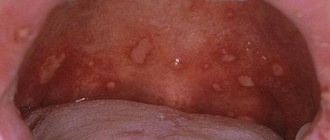
This is the appearance of white, gray spots dispersed throughout the oral cavity.
- Erythroplakia
It is characterized by the appearance of flat, small areas protruding on the surface of the mucous membrane, which are red in color and may bleed due to accidental injury, including after brushing teeth or eating hard food.
- Erythroleukoplakia
This is a combination of the above diseases, that is, the appearance of white, red spots on the mucous membrane.
In this case, the prognosis is the most unfavorable. It is possible to differentiate precancerous and cancerous conditions only with the help of a biopsy - a histological examination of a tissue area. There are cases where oral cancer pathologies developed without precancerous conditions. Therefore, visiting the dentist every 6 months for a preventive examination is the best strategy for preventing and early diagnosing oral cancer. Text: Yulia Lapushkina.
Symptoms of Oral Cancer
Manifestations of the disease depend on the nature of the tumor and the location. Typically, the affected area is either a local ulceration or an induration (bulge, nodule). The patient feels pain and discomfort in the corresponding area. Warning signs include the following:
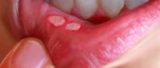
- A long-term, non-healing mouth ulcer. The oral cavity is rich in blood vessels, the mucosal epithelium is well supplied with blood, and normally, in the event of injury, rapid healing occurs. Any damage to the oral mucosa heals faster than a similar injury to the skin of the arm or leg. Therefore, if there is an ulcer in the mouth and for unknown reasons it does not heal for a long time, you should seek medical help.
- Lumps, “balls” under the mucous membrane, regardless of the area of the mouth where they arose. A compaction under the mucosa can be a symptom of both a non-oncological pathology (for example, a cyst of one of the small glands) and a sign of cancer. Only a specialist can distinguish one from the other. The seal, which initially appeared as a cyst, may subsequently become malignant if left untreated.
- Impaired swallowing, chewing, discomfort at the root of the tongue - all this is a reason to suspect cancer in the corresponding area.
- Toothache and bleeding in a specific area of the gum may be signs of cancer. And, although similar symptoms are caused by much less formidable ailments, consultation with a dentist is necessary. Thus, any violation of the anatomical integrity or functionality of the organs of this zone carries a potential threat, and this is a reason to see a doctor.
What are the symptoms of oral cancer?
The list of symptoms characteristic of oral cancer includes:
- inflamed wounds in the mouth that do not heal for a long time;
- the appearance of seals that do not disappear over time;
- unexplained weakness of the gums and teeth (after tooth extraction, the wound may not heal for a long time);
- problems when wearing dentures;
- numbness of part of the lip or tongue;
- spots (white or red) in any area of the mouth, as well as on the lips;
- hoarseness, hoarseness of voice;
- pain or difficulty swallowing;
- bleeding of unknown origin;
- aching pain radiating to the ear, throat, jaw or tongue;
- weight loss that does not depend on changes in diet and exercise.
Please note that some of these signs, such as a sore throat or earache, may indicate other conditions. However, if you notice any of these symptoms, especially if they do not go away or you have more than one, contact your dentist or ENT doctor as soon as possible.
Stages of the disease
Stage I is characterized by the presence of a tumor up to 1-2 cm in diameter, not extending beyond the affected area (cheek, gums, palate, floor of the mouth), limited to the mucous membrane. Metastases are not detected in regional lymph nodes.
Stage II - a lesion of the same or larger diameter, which does not extend beyond any one part of the oral cavity, but extends into the submucosal layer. There are single metastases in regional lymph nodes.
Stage III - the tumor grows into the underlying tissues, but not deeper than the periosteum of the jaw, or has spread to adjacent parts of the oral cavity. In regional lymph nodes there are multiple metastases measuring up to 2 cm in diameter.
Stage IV - the lesion spreads to several parts of the oral cavity and deeply infiltrates the underlying tissues, in the regional lymph nodes there are immobile or disintegrating metastases, and the presence of distant metastases is also characteristic.
Classification by stages is periodically subject to revision; you can find a division of stages into subtypes - A and B. Currently, classification by stages is used less and less, the TNM classification is more relevant. Its principle is that this coding indicates the characteristics of the tumor itself, the condition of the closest (regional) lymph nodes, and the presence or absence of distant metastases.
In the diagnosis, the oncologist indicates the histological type of cancer, since different types of cells differ in growth rates, tendency to metastasize, and sensitivity to treatment. All types of classifications serve one purpose - to correctly assess the extent of the spread of the disease, the degree of damage and develop the right tactics of assistance.
Book a consultation 24 hours a day
+7+7+78
Treatment
The following factors are important for optimal treatment:
- Timely appeal. The factor of advanced disease plays a huge role. Even now, when doctors have learned to successfully fight the disease even in later stages, the final prognosis in most cases is determined by how quickly the patient turned to an oncologist.
- High-quality diagnostics. There are many research methods that allow you to more accurately establish a diagnosis. In oncology, ultrasound, MRI, CT, PET-CT, as well as histological studies are widely used.
- Good contact between the patient and the treating doctor. The doctor’s ability to explain to the patient the essence of what is happening, support him and correctly organize joint work, as well as the patient’s ability to be disciplined, trust the specialist and not delay in carrying out the prescribed studies or treatment procedures.
For oral cancer, surgical treatment, chemotherapy, and radiation therapy (including brachytherapy) are used.
Surgery
The operation is a classic treatment tactic in oncology in general and in oral oncology as well. The difficulty of using oral surgery is that in some cases it is technically difficult to remove the tumor without damaging vital anatomical structures.
The doctor takes into account the following important points:
- The mouth area has a rich blood supply and is at risk of active bleeding.
- The organs and tissues of the mouth area are mobile, and the possibility of this displacement relative to each other determines such important functions as speech, swallowing, etc.
- The anatomical formations of this zone are small in size and closely adjacent to each other. In this area there are few ballast, functionally insignificant tissues; for every centimeter there are the most important vessels, nerves, and organs. This complicates the operation process. The lack of sufficient tissue makes it more difficult to close the defect created after tumor removal.
As in the case of radical surgical treatment of tumors in other areas, during operations on the oral cavity the following conditions are sought to be met:
- Removal of the tumor must occur within healthy tissues; the cut does not pass at the border of healthy and diseased tissue, but along healthy tissue, that is, the tumor is excised as if with a reserve. When the organs are close together, this poses a problem.
- When excision of a tumor, the importance of further restoration of the integrity of the integument is taken into account, as well as, if possible, preservation of function.
- When excising the tumor, in most cases, peripheral lymph nodes are also removed. This is because the lymph nodes closest to the tumor are a trap for metastases and can often contain tumor cells.
In classical oncology, surgical treatment was the method of choice; operations were used, as a rule, immediately after diagnosis. Currently, the approach has changed a little and has become more differentiated. Sometimes surgery is preceded by chemotherapy and/or radiation therapy.
Chemotherapy
A feature of tumor cells is rapid growth and division. This is what chemotherapy is based on. Special substances that are toxic to cells and inhibit their growth are introduced into the body. Chemotherapy affects the entire body, but most cells are insensitive to the substance, and the chemotherapy is harmful to tumor cells.
Chemotherapy is used as an addition to surgery, before or after it, as an independent method, and in combination with radiation therapy.
The appropriateness of chemotherapy is determined by the histological type of tumor. The type of drug is selected taking into account the sensitivity of a particular neoplasm to various types of relevant drugs.
The disadvantage of chemotherapy is its side effects. The body has a number of healthy cells that are frequently renewed and, accordingly, quickly divide. These are cells of the gastrointestinal mucosa, blood cells and others. Chemotherapy drugs affect them. The blood formula changes, the cells lining the stomach and intestines die. The result is nausea, stool upset, weakness, and decreased immunity. These phenomena are temporary and, as a rule, surmountable; however, the use of chemotherapy requires constant medical supervision.
Radiation therapy
The method is based on the damaging effect of X-ray radiation on cells, primarily on actively dividing cells, such as the cells of malignant tumors. Radiation therapy can be used as an independent method of treatment, or in combination with surgical methods and chemotherapy. Before the start of the course, markings are made to determine the direction of impact and ensure maximum impact on the tumor itself and minimal impact on surrounding tissues.
Brachytherapy is a subtype of modern radiation therapy, when a radiation source is introduced directly into the affected area. Brachytherapy allows you to treat the tumor with maximum doses of radiation and reduce the damaging effects on healthy tissue.
Prognosis and survival
The prognosis of the disease depends on the stage, localization, and timely provision of assistance. In the early stages, with adequate treatment, the disease can almost always be overcome. When contacting a doctor was not timely, the prognosis is somewhat worse, and the percentage of cured patients is lower. In assessing the prognosis, the presence or absence of distant metastases is important. Despite enormous progress in oncology, distant metastases sharply reduce 5-year survival; this is a significant problem for a specialist. In addition, the prognosis depends on the histological type of the tumor, because The growth rate of different types of tumors can vary significantly. The age of the patient, the location of the tumor, the presence of concomitant diseases in the patient, the chosen treatment tactics - all these factors significantly influence the prognosis.


