We treat open bites without surgery or tooth extraction!
What is an open bite | Reasons | Treatment | Our advantages | Doctors | Reviews
An open bite is the most disliked anomaly by orthodontists, because it is very insidious and thankless in treatment.
Seriously, the diagnosis of open bite is a misdiagnosis. A bite is, by definition, the closure of the teeth. And with an “open” bite there is no closure. Therefore, it is correct to call this pathology vertical disocclusion. Disocclusion is the lack of closure of teeth. And vertical - because in the vertical plane.
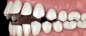
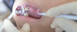
Such disocclusion can occur in any segment of the dentition. But most often it occurs in the frontal (front) area, that is, in the area of the incisors. Therefore, an even more complete name for this problem is vertical incisal disocclusion. It is characterized by the fact that only the lateral teeth close together, while the front teeth “hang” (do not close) and do not close vertically.
Vertical incisal disocclusion. Front view.
Vertical incisal disocclusion. Side view.
This is the reason for the “insidiousness” of this anomaly and the dislike of orthodontists for it. The fact is that open bite causes a huge number of relapses. That is, the treatment of vertical disocclusions in a huge number of cases is very unstable and all for one reason: there is no method, there is no apparatus that consolidates the success of treatment in the vertical plane. It’s the custom with orthodontists... If you’ve done it (cured it), fix it (just in case...). And it is right. Therefore, after any orthodontic treatment, retention devices and structures are installed. For anomalies in the sagittal plane, be it distal occlusion or mesial occlusion, such retention devices are available. There are also problems in the frontal plane (narrowing of the dentition, crossbite). But not in the vertical plane.
And therefore, in achieving a successful and stable result in the treatment of an open bite in an adult, it is very important to initially understand the reason for the occurrence of non-occlusion of the teeth vertically, in each specific case. After all, only by identifying and eliminating the cause can we “defeat” this most unfortunate and insidious open bite. No relapse. Once and for all.
Diagnostics is the first and key step in the treatment of open bite. Do you want to know why it’s impossible to do without diagnostics?
But, precisely, the reasons for vertical disocclusion are usually considered very simplistically and superficially. And is this why there are so many failures in correcting open bites?
Causes of open bite
One of the most common causes of open bite is the presence of bad habits in children. For example, such as: using a pacifier for a long time, placing the tongue between the teeth, sucking a finger or pencil.
A bad habit is putting your tongue between your teeth.
A bad habit is thumb sucking.
And it seems difficult to argue with this. Because mechanical impact on the dentition, of course, can lead to vertical disocclusion. But! This is a very superficial look at the cause of open bite. After all, thumb sucking and tongue thrusting are more likely a consequence than a cause.
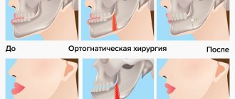
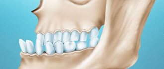
And the cause of open bite (vertical disocclusion) is always cranial problems (see “Cranial distortions”), that is, problems with the position of the cranial bones. And the resulting cranial dysfunctions are always reflected in the jaws (from which the teeth “grow,” as is known).
Treatment in installments
The Orto-Artel clinic offers installments for the entire process of treating any disease. Personal conditions are considered on an individual basis.
Find out more
or call 8 (495) 128-11-74
This is confirmed by thumb sucking in the womb. What bad habit are we talking about here? A bad habit is something acquired, something that appears throughout life. And thumb sucking while still in the mother’s womb is a subconscious act by which the child unconsciously tries to ensure the stability of the cranial bones. Thus ensuring the functionality of the cranio-sacral mechanism (for those who don’t know what it is, look at Wikipedia). After all, this mechanism is the most important factor ensuring the normal functioning of the central nervous system (CNS). That is, the main “computer” of the entire organism, regulating every single process taking place in it. And that is why every effort is being made to ensure the stability of this “computer”. Even in such a simple way as thumb sucking.
- Thumb sucking in utero during an ultrasound examination.
- Thumb sucking in early childhood, as a continuation of point 1.
- The figure clearly shows how the child unconsciously stabilizes the entire maxillary complex and adjacent cranial bones.
- Open bite as a consequence of cranial problems and “bad habits”.
As we see, tongue protrusion simply cannot but occur with such a vertical “gap” between the upper and lower teeth.
The tongue is too “curious” an organ to ignore such a tempting “hole”. And accordingly, tongue insertion, as well as insertion of other objects (including fingers and pencils), only aggravates the problem. That is, it is a “related product”.
Refusal of treatment
Malocclusion in the incisal region leads to various complications. First of all, conversational speech is disrupted, which entails the appearance of complexes in a child or an adult. The compensatory mechanism causes the facial muscles to be in constant overstrain, which causes myalgia or inflammation of the facial nerves.
Due to poor quality of biting and chewing food, various digestive disorders are possible. Dysfunction of the temporomandibular joint may develop, which causes blocking of the movements of the lower jaw. This phenomenon causes frequent or even constant headaches.
The periodontium in the frontal group is constantly under overload, so it begins to quickly wear out. Improper load distribution leads to injuries to the soft tissues of the upper palate.
To prevent the development of all these complications, treatment should be carried out at the initial stages of the development of the anomaly.
Deep incisal overjet, deep bite, deep traumatic bite
Treatment of deep bite
Treatment tactics depend on the form of deep bite, but the main tasks are as follows:
- promote dentoalveolar elongation in the lateral areas of the lower jaw, stimulate the eruption of chewing teeth by separating them,
- create a distal inclination of the lateral teeth,
- elimination (intrusion) of an existing dentoalveolar elongation in the anterior region or delay in the eruption of anterior teeth,
- when retruding the anterior teeth, create their vestibular inclination,
- in case of sagittal discrepancy of the dentition, correction of prognathia (more often) and progenia.
Treatment is carried out in various ways and methods, taking into account the pathogenesis of the clinical form of the anomaly, the age period (deciduous, mixed, permanent dentition). During the period of primary and early mixed dentition, it is necessary to normalize nasal breathing, eliminate existing bad habits, and perform myogymnastics. The following exercises may also be recommended:
- slowly push the lower jaw forward until the cutting edges of the lower incisors are positioned in front of the upper ones; hold in this position for 10 seconds, then slowly return to the starting position;
- A rubber tube is put on a wooden stick and placed between the front teeth, which are compressed and unclenched. Considering that a deep bite is often combined with a distal bite, you can add exercises from the corresponding complex.
In case of defects in milk teeth, their timely elimination by filling or prosthetics with inlays; in case of premature removal of milk or permanent teeth, prosthetics with appropriate structures or the use of devices that help preserve space for teeth that erupt later.
To eliminate a deep bite with a neutral relationship of the dentition (I class according to E. Engle), plates with a bite platform are used. The authors consider it necessary to emphasize that not with an inclined plane, but with a bite platform, since this does not require moving the lower jaw forward, but only a vertical adjustment of the bite. To prevent the lower jaw from moving forward or to the side, it is better to make the bite pad not smooth, but with imprints of the antagonist teeth. The most optimal way to fix a removable device is an Adams or A. Schwarz clasp, which are made of wire with a diameter of 0.8 mm.
The effect of these devices is manifested when biting with the lower front teeth in increasing the load on them, as a result of which the bone tissue of the jaws is rebuilt and dentoalveolar shortening occurs in this area. At the same time, the lateral teeth become separated and their dentoalveolar elongation (extrusion) occurs due to the fact that stratification (apposition) of bone tissue occurs over the entire periodontal surface of the sockets. Therefore, the ratio of the inside and extra-alveolar parts of the lateral teeth does not change significantly, since the teeth do not move forward, but grow together with the alveolar process towards each other on both jaws. However, more often only the lower teeth need to be moved. The bite block must be used constantly, and this must be continued to avoid relapse, even when the normal vertical dimensions of the lower third of the face are restored.
Vertical movement of teeth is possible due to intermaxillary traction, lever-like action of arches, the potential force of the NiTi arch itself, and compensating bends. In a mixed bite, a utility arch, rectangular, with a diameter of 0.40 x 0.40 mm, in brackets with a groove of 0.45 mm (0.18 inches) can be used to move teeth. But when using it, great caution and careful monitoring should be observed.
G.B. writes about risk factors and the need to control the amount of force. Ospanova, discussing the problem of root resorption during vertical movement of teeth, in particular with the help of an intrusive utility arch. Since only the first molar is used as a support, it is necessary to ensure that there is no significant extrusion of it. Due to the significant length of the arc, the load is dissipated and the initial force of the intrusion is weak, but its activation when placed in braces increases the force on the incisors, in particular in the direction of their vestibular inclination. This can be avoided by additionally creating oral torque on the incisors, but this will increase the extrusion of the molars. If you bend the utilitarian arch over the distal end of the tube on the molars, then their mesial displacement is possible, which is also undesirable. Therefore, it is better to wait for the formation of a permanent bite and then use intrusive arches. For complete optimal control of the anterior and lateral segments, the use of purstone segmental arches can be recommended.
If there is retrusion or crowding of the anterior teeth, it is necessary to first eliminate these disorders, and then proceed directly to deep bite therapy. Previously, and even now, removable devices with protracting springs are used for this purpose. Currently, if it is planned to use functional devices in such patients, and it is impossible to immediately move the jaw into a constructive bite position, the dentition should first be corrected using braces. The bite block does not interfere with the fixation of braces on the lower incisors.
You can first use a round wire arch to change the torque of the front teeth or heat-activated nickel-titanium arches, and then rectangular steel arches with a diameter of 0.45 x 0.45 mm. By the end of the second month, the dentition is usually aligned. In conjunction with these arches, if the deep bite is combined with prognathia or progenia, an appropriate intermaxillary rubber band can be applied while the growth process continues. As the sagittal anomaly is corrected, the incisal overlap may also decrease due to tooth extrusion. Before determining the constructive bite, the teeth should be open to better control the amount of movement. It is better to move the lower jaw forward no more than 3-7 mm, that is, until the front teeth come into direct contact.
The degree of separation of the dentition when eliminating supraocclusion of the lower and infraocclusion of the upper anterior teeth is determined by the size of the free interocclusal space. Typically, the bite pad is modeled in such a way as to separate the dentition by 2-6 mm, but this is purely individual, depending on the size of the incisal overlap and the interocclusal space at rest. With this disconnection, the chewing muscles will be in a moderate isometric contraction, and the entire load falls on the teeth in contact with the bite pad.
An Andresen activator is used, slightly modified in such a way (recommendation by W.poffit) that the linings on the upper lateral teeth and lower anterior teeth prevent their displacement in the direction of the occlusal plane, and the lateral areas of the lower jaw should not be limited in their movement. The R. Fränkel function regulator (FR1) can be effective in the treatment of deep bites with neutral occlusion of the first permanent molars.
A very important circumstance in the treatment of deep bite is the change in the inclination of the anterior teeth, since the creation of incisal-tubercular contact is considered a prerequisite for preventing the recurrence of the anomaly. Orthodontic treatment for the dentoalveolar form of deep bite, with a vertical position of the anterior teeth, is aimed at their vestibular deviation, which changes the depth of overlap. However, this is advisable in cases where the initial (before treatment) overlap was no more than 5 mm. With a larger incisal overlap, before making their vestibular deviation, it is necessary to reduce the supraocclusion of these teeth. It should be noted that with an incisal overlap of more than 8 mm, it is impossible to achieve incisal cuspal contact and depth of overlap, as with an orthognathic bite.
Orthodontic treatment of patients with deep skeletal bite is aimed at:
- change in the shape of the dentition and position of the front teeth,
- increase in interalveolar height,
- change in the depth of the incisal overlap.
Since almost all patients with this form of anomaly have a steep (vertical) position of the anterior teeth and a narrowing of the dentition, by expanding them, the orthodontist simultaneously achieves vestibular deviation of the anterior teeth, increasing the interalveolar height and reducing the depth of the incisal overlap by ~ 2-3 mm. Then, using removable and non-removable devices, grinding, you can further rebuild the vertical relationships.
With premature loss of primary molars and lack of timely prosthetics, resulting in mesial displacement of the first permanent molars, treatment is necessary as early as possible. Otherwise, the outcome of such a situation, in the absence of early treatment, is often the subsequent removal of premolars.
With an overlapping deep bite, characterized by a predominance of the size of the upper jaw, infraocclusion of the upper teeth and supraocclusion of the lower teeth of the same name, with retraction of the anterior portion of the lower jaw, a sharp inclination of the upper central incisors is often encountered orally, and the lateral ones vestibularly. The latter are often also rotated around the longitudinal axis. Patients come to the clinic not so much because of a deep bite, but more because of the incorrect position of the upper incisors. This form of anomaly in children with mixed dentition is difficult to treat, to speed it up Z.F. Vasilevskaya recommends a combined technique (surgical + orthopedic).
Schematic diagram of supervision: the vestibular part of the interdental gingival papillae on both sides of the lateral incisors is peeled off, the alveolar process is intersected with a thin fissure bur No. 3 in the vestibulo-oral direction to a height of ¾ of the length of the roots of the teeth being moved; it is necessary to ensure that the cutting line of the alveolar process is located at the same distance from the cortical plates of adjacent teeth; 5-7 days after surgery and postoperative phenomena have subsided, an arch can be applied. After moving the lateral incisors, the arch is replaced with a removable device with an inclined plane.
When treating a deep skeletal bite that is not accompanied by a change in the height of the lower third of the face, treatment is aimed at correcting the shape of the dental arches, the position of individual teeth, and eliminating supraocclusion of the lower anterior teeth. It is necessary to carry out treatment in such patients without disconnecting the dentition, preferably with the help of braces or other fixed devices.
Prosthetic treatment of patients with deep bite should be aimed at leveling the occlusal surface of the dentition by grinding and prosthetics with various structures. Indications for such treatment include:
- skeletal forms of anomaly that are not subject to orthodontic treatment and the patient’s refusal of surgical treatment;
- in the absence of a large number of teeth,
- ineffectiveness of orthodontic treatment or impossibility of its implementation for various reasons (severe general condition, remote place of residence, etc.) or refusal of the patient.
Prosthetic treatment is most effective for skeletal deep bites, when there is a large interocclusal space and it is possible to significantly change the interalveolar height. Preference should be given to fixed one-piece dentures and removable dentures with a metal base. A prosthesis with a plastic base requires more space; it is not applicable for multiple small defects or narrowing of the jaw. The same type of treatment should also have priority in cases of deep bite against the background of systemic periodontal diseases, since a prosthesis with a cast metal base allows better distribution of chewing pressure and prevents contact of the mucous membrane of the gingival margin with the prosthesis base.
In the process of orthodontic supervision of a patient with a deep or deep traumatic bite due to periodontal diseases, a partnership with a periodontist is necessary. Appliances and dentures must be modified taking into account the degree of pathological mobility of teeth and the amount of bone tissue resorption, the depth of the periodontal pocket, and the width of the attached gum. In other words, the preservation of bone tissue and the crown-to-root ratio have a significant impact on the prognosis when choosing abutment teeth for appliances and prostheses. The latter can be changed by preparing the tooth, using endodontoendossal implantation, or a specific design of removable dentures. In particular, for greater unloading of the remaining teeth with varying degrees of mobility, especially with terminal defects, it is preferable to use removable dentures with Roach clasps.
With favorable crown-to-root ratios, it is possible to regulate the load on the supporting teeth, even with a certain degree of pathological mobility, the correct distribution of the supporting and retention parts of the prosthesis, as well as the way they are connected to the base. In case of bone tissue atrophy, preference should be given to teeth with a crown-to-root ratio of 1:2.
A deep bite is often combined with other types of anomalies (most often with upper prognathism), which must be corrected before its immediate treatment. To treat a deep bite in combination with a distal bite, you can use the A.Ya. medical bite block. Katz, Andresen Goipl activators, R. Fränkel type I and II regulators, various bionators. In particular, the palters bionator, which also resembles an activator, but has practically no palatal base. In young patients with a deep bite, combined with a distal bite and a tendency to narrow the dentition, it is still better to use functional appliances than a facebow, since this sometimes stimulates growth and achieves an anterior position of the lower jaw. In addition, monoblock devices, especially the addition of additional elements (screw, spring), allow you to simultaneously expand the jaw.
Applying force to the first molars through a facebow with extraoral head or neck traction speeds up the treatment process. The choice of the type of extraoral traction depends on the nature of jaw growth and the direction of movement planned by the orthodontist during the treatment process. It should be kept in mind that when using a facebow with a cervical traction, the force applied to the maxilla will be downward and distal, and when connected to a head cap, it will be distal and upward. With their combined action and equality of forces, the direction will be purely distal, since the vertical components will be mutually destroyed, as if they are multidirectional.
Companies produce sets of ready-made face bows with intraoral components of various sizes, which should be pre-fitted according to plaster models, making the necessary adjustments. This treatment method is one of the most important components of the Vari Simplex Discipline, which uses the term “retractor” to refer to the face bow, which in turn consists of an internal and external part. The internal arch is inserted into special ring sleeves on the first molars of the upper jaw. The bushings can be placed closer to the gum or occlusal surface. In a gingival position, the advantage is that the force is applied closer to the center of rotation of the tooth, thus reducing its inclination. This position is preferable if only “distalization” of the molar is planned, without affecting the remaining teeth. RG Alexander recommends the occlusal placement of the sleeve, emphasizing its advantages: easier insertion into the tube for the patient and greater hygiene, as it is easier to clean.
The outer bow, attached to the head cap, neck strap or a combination of both, should be a few millimeters from the cheeks (5-10 mm) and end with hooks. The latter are necessary for fixing elastic or better spring rods. If the arc presses on the cheeks, the patient experiences discomfort, and if the distance is too large, it is difficult for the patient to sleep. The exception is situations when there is a need for different force effects of the arc on each side. For example, the dentitions close on one side along the first class. Angle, and on the other, according to class II, then with the latter more pressure is required and the outer arc on the same side bends almost at a right angle to the inner one. The length of the inner archwire should be slightly longer to increase the force applied to the upper first molar on that side.
The face bow (retractor) should be positioned so that the junction of the outer and inner bows is slightly anterior to the point of lip closure. It must be borne in mind that the fitting of the internal (dental) arch requires the closest attention. Sometimes it is necessary to adjust its loops, which need to be compressed or expanded to achieve this. The neck strap is adjusted in such a way that the initial force transmitted through the device is no more than 200-230 grams, and after adaptation it can be increased to 400-450 grams. After some time, the belt is pulled out and requires activation or replacement.
The patient must be taught how to use such devices and should not participate in mass games, ride a bicycle, etc., in order to avoid injury or damage to parts of the device. When using an arch, it is necessary to remember that all its sections are located in the same plane, the bends of the intraoral and extraoral parts must be symmetrical. A plastic coating is usually applied to the extraoral parts of the face bow, the color of which gives information about its size: yellow 83, pink 90, red 97, green 104, blue 111.
It should be borne in mind that in adult patients (in fact, after puberty and even somewhat earlier, in 12-14 year olds), with the action of extraoral apparatus, one can only count on the movement of teeth and partially alveolar processes; rotation of the lower jaw may also occur as a result of reciprocal action on the chin. It is useless to act on skeletal disorders in this way.
In the final period of the replacement and initial period (9-12 years) of the permanent dentition, they strive to use physiological processes in the formation of occlusion. The same orthodontic devices are used, but emphasis can also be placed on fixed devices: A. Katz guide crowns, E. Engle arches, brace systems.
In permanent dentition, it is advisable to carry out sequential disocclusion. The essence of this treatment is that a group of teeth is “excluded” from occlusion, initially 23 teeth, most often molars on the side that is not involved in chewing. This can be done by applying a mouthguard, and with the modern level of development of dentistry, it is much easier to apply bonding materials, preferably colored ones, so that they are more noticeable when they are ground off during the treatment process, as the deep bite is “opened”. Teeth excluded from occlusion “lengthen” and come into contact with antagonists, and teeth “released” from the aligner or after grinding off the composite, the teeth become separated and also “lengthen.”
Once a sufficient number of teeth are in contact and established at the new level, active treatment can be discontinued. This method is especially indicated for the elimination of anomalies associated with periodontal diseases, because it does not create additional load on the front teeth, as, for example, when using a bite block.
In permanent dentition, non-removable vestibular arch devices, including those with intermaxillary traction, are used to eliminate pronounced forms of anomaly. The system bracket can be used in combination with different types of bite blocks for the upper jaw. To accelerate the reorganization of bone tissue in the hypertrophied area of the alveolar process in the anterior region in adults, a complex method can be used: surgical (compactosteotomy) + orthodontic equipment.
When treating deep bites in adults, attention should be paid to issues of orthopedic treatment, that is, various types of prosthetics and combined, restructuring of the myostatic reflex (stretch reflex). The latter can be briefly explained as follows. In patients with a deep bite with central occlusion, the fibers of the masticatory muscles have a certain initial value, characteristic of excessive incisal overlap. When the bite is separated, for example, by a bite block, the fibers of the muscles that lift the mandible (mm. masseters, tstrongporales, pterygoidei medialis) are stretched, which increases their tone and causes the appearance of impulses in muscle receptors.
A reflex contraction of the levator muscles occurs, which through the periodontium creates tension in the area of the anterior teeth in contact with the bite pad. This constant tension increases during chewing and accelerates the restructuring of bone tissue. For retention after correcting a deep bite, you can use the same bite block as a retainer, but in an inactive state, that is, without separating the lateral teeth.
The goals and objectives of treatment of patients with secondary deep bite are determined by the reasons that caused this deformation (lack of teeth, systemic diseases of periodontitis, pathological abrasion). Treatment can be prosthetic or combined, and its choice is determined by the pathogenesis of secondary deep bite.