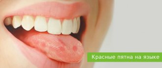
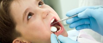
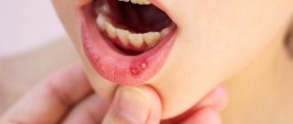
Sores or small ulcers that appear in the mouth are a rather unpleasant and painful sensation. This affects one's well-being, makes it difficult to eat, and can even affect a person's ability to speak. Sores in the oral cavity can occur in both adults and children. Let's consider the root causes, symptoms, treatment methods (including folk remedies), as well as methods of prevention.
Causes of sores in the mouth
Among the main reasons that can cause the appearance of ulcers on the oral mucosa are the following:
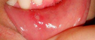
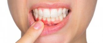
- Stomatitis (aphthous or herpetic). Both diseases are chronic. They differ only in the location of the wounds. With aphthous stomatitis, the ulcers are located on the cheeks, palate and tongue, with herpetic stomatitis - in the lower part of the oral cavity. The rashes are located in groups and may vary in size.
- Necrotizing periadenitis - first, compactions appear, which later develop into ulcers. The treatment is long-term.
- Poor oral hygiene. Most often, this factor causes unpleasant and painful rashes in the mouth of babies (dirty hands, pacifier and bottle not washed well).
- Mechanical damage - scratches as a result of eating hard food, hard teeth of a toothbrush, dentures or crowns with pointed ends. It is also possible that the oral cavity is damaged by instruments during a dental procedure or a thermal burn (from eating spicy or hot food).
- Other reasons are hereditary factors, frequent stress, lack of vitamins, thrush, and taking certain medications without medical supervision.
Methods for treating mouth wounds
Before starting treatment for wounds in the mucosa, you should understand the causes of the condition. If ulcerative defects are caused by another disease, it is necessary to fight the underlying pathology to reduce the size of the ulcers.
Wounds that appear from mechanical irritation with a brush or teeth are treated with antiseptic agents. They reduce the risk of bacterial flora attaching.
Therapy for the pathological condition is supplemented by diet, prescription of B vitamins, prevention of exacerbation of chronic pathologies, and elimination of stressful situations.
A patient with recurrent ulcers in the oral mucosa in the absence of treatment effect must undergo a set of tests to exclude a systemic disease of viral etiology (HIV, hepatitis).
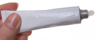
Medication
- Chlorhexidine is an antiseptic solution that helps quickly treat wound defects in the mouth. The drug has antimicrobial and disinfectant abilities. Allergic reactions to the composition of the drug are rare. It is suitable for children. Wounds treated with Chlorhexidine can heal in 5-7 days. Rinsing should be done 3-4 times a day.
- Xikain gel has three main effects: reduces pain, destroys pathogenic flora, and relieves inflammation. The medicine is not used in children.
- Asept is a popular spray based on Lidocaine. The drug simultaneously anesthetizes and disinfects the inflammatory focus. The spray is used 2-3 times a day.
- Stomatidin destroys pathogenic microorganisms and disinfects the oral mucosa. The drug is widely used to combat the symptoms of stomatitis. It has a healing effect if you simply bite your lip.
Folk
- Representatives of traditional medicine recommend treating a wound in the mouth after a bite with decoctions of oak bark, chamomile, and calendula. They can be mixed or cooked separately. Take 1 tablespoon of the plant per glass of boiling water. The oral cavity should be rinsed with warm infusion 3-4 times a day.
- Furacilin, soda, and salt will help get rid of the problem. To prepare the solution you will need 5 tablets of the drug, 1 tsp of kitchen ingredients. The listed components of the solution are mixed in a glass of water. You will need 4-5 rinses per day to achieve rapid healing of ulcerative defects.
- A recipe with propolis helps with painful scratches. Take 1 teaspoon of alcohol solution per glass of water and rinse. Propolis relieves discomfort and promotes rapid healing. The soreness goes away within a week.
- Rinses with carrot, horseradish, and cabbage juice are used against stomatitis.
- Kombucha treats wounds in the mouth that form after injuries to the mucous membrane. Improvement occurs after the first rinse.
How to treat wounds in the mouth?
Therapy should begin immediately as soon as at least one wound is discovered in the mouth. The treatment is complex using both medications and folk remedies in the form of rinsing.
Local therapy
Mouth wounds are treated topically using various ointments, gels and sprays. The most commonly used are such well-known disinfectants, disinfectants and analgesics that act locally as: Solcoseryl, Metrogyl Denta, Kamistat, Cholisal and Asepta. In addition to local medications, doctors prescribe rinsing. This procedure can be done using a solution of Chlorhexidine, Miramistin or Furacilin.
Review of effective ointments
- Bonafton is used in patients with herpetic lesions of the skin and mucous membranes. The drug has a negative effect on the herpes simplex virus. Suppresses the growth and reproduction of pathogenic microorganisms. The ointment is applied three to four times a day for 2 weeks.
- Solcoseryl is a medicine that is intended to improve metabolism and accelerate regenerative processes. The drug is prescribed for the treatment of inflammation of the cornea and blood vessels of the lower extremities. It fights the manifestations of stomatitis and gingivitis. The drug should be applied to the inflammation site 2 times a day. A positive effect is achieved 2-5 days after the start of treatment.
- Pimafucin has an antifungal effect. The active components of the cream suppress the vital processes of the microorganism. Pimafucin is intended for external use.
- Metrogyl Denta is an ointment for the treatment of wounds in the mouth. Consists of metronidazole and chlorhexidine. The products have disinfectant and antimicrobial properties. The gel is applied 2 times a day for 2-3 weeks.
What to rinse with?
- decoction of calendula flowers (1 tbsp per 250 g of water, boil the decoction for 15 minutes)
- saline or soda solution (1 tsp salt/soda per half glass of water)
- hydrogen peroxide (3%) – mix with water in a 1:1 ratio and wipe the area where the ulcers are located
- sea buckthorn oil and natural honey are also suitable as a rub, especially after meals
General therapy
Along with traditional medicine and local medications, a set of measures is also prescribed to increase the protective functions of the body as a whole. These may be immunomodulatory drugs, courses of vitamins and antiviral agents. For aphthous stomatitis, antihistamines and antibiotics may be prescribed. The appointment is made only by a doctor after a comprehensive examination and identification of the causes.
How and what to treat wounds on the oral mucosa
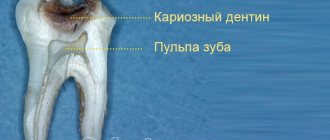
Defects in the mucous membrane are manifested by typical symptoms: pain that intensifies during eating, unpleasant odor, metallic taste. Pathology disrupts the normal rhythm of human life. He gets tired quickly and cannot eat due to severe pain.
Cheap antiseptics (iodine, brilliant green, peroxide), which are available in every home, will help remove unpleasant symptoms.
Gargling with hydrogen peroxide treats symptoms of acute tonsillitis. An antiseptic disinfects the oral wound from microorganisms that cause disease. The instructions for the drug indicate only external use. Representatives of traditional medicine advise diluting the medicine with water to avoid unpleasant consequences. Hydrogen peroxide is diluted: take 1 tsp per 120-150 ml of water. antiseptic. The solution should be warm to reduce irritation of the mucous membrane.
Before using the medicine, you should consult a medical specialist. Thus, specialists from the Ibn Sina dental clinic https://stomatologis.ru/ will help identify the true cause of the disease and determine the tactics for treating a wound in the mouth. Hydrogen peroxide is effective if the ulcers have a traumatic, bacterial etiology.
The medicine will not cope with fungi.
The medication can be used 4-5 times a day for 7-14 days. It's important not to overdo it. Incorrect dilution and frequent rinsing can lead to chemical burns of the mucous membrane. Following medical recommendations will help you avoid this pathological condition.
Prevention measures
For chronic stomatitis, it is important to undergo treatment courses in a timely and systematic manner. But even in this case, it is impossible to exclude a possible recurrence of the unpleasant disease.
Doctors recommend adhering to preventive measures so that unpleasant rashes in the mouth do not reappear:
- carefully monitor oral hygiene (brushing teeth and rinsing with antiseptics or decoctions of medicinal herbs)
- nutrition should be varied and balanced so that a person receives all the vitamins and microelements in the right quantities, they in turn help strengthen the body’s protective functions
- vitamin preparations – a person does not always get all the minerals and microelements he needs through food, so twice a year it is worth taking a course of vitamins prescribed by a doctor
When ulcers first appear on the oral mucosa or with chronic stomatitis, you should not self-medicate. Therapy, although complex, is selected individually, only after a full examination and diagnosis.
At LeaderStom dentistry, doctors of the highest category diagnose all diseases of the oral cavity using modern and high-tech equipment, and also prescribe an individual course of treatment.
Rules for processing and treatment in children
Ulcerative defects on the mucous membrane in children appear when personal hygiene rules are not followed. Children can injure the inner surface of the oral cavity with foreign objects. Before starting treatment, it is better to consult a pediatrician. Pharmaceutical drugs, when used incorrectly, cause allergic reactions in children's bodies. You can treat a wound in the mouth using special ointments, gels, and powders.
Children are prescribed Solcoseryl, Chlorophyllipt, Stopangin, Furacilin.
At home, you can use an infusion of calendula or chamomile to wash ulcers.
Pain syndrome in children older than six months is relieved with Ibuprofen and Paracetamol.
When treating the oral mucosa, you must adhere to the following rules:
- You cannot independently increase children’s doses of medications or the frequency of administration.
- If the child does not know how to rinse his mouth, then it is necessary to lubricate the wounds with antiseptics. Do not allow your baby to swallow the medicinal solution.
- If allergic rashes appear, stop treatment and consult a doctor.
- When treating a wound, it is necessary to follow the rules of using aseptic techniques. Before treatment, wash your hands and lubricate the inflammatory areas with a clean cotton swab with the applied medication.
In dentistry and maxillofacial surgery, much attention is paid to the study of the healing of wounds of the oral mucosa caused by various surgical instruments, mainly a scalpel, and more recently a laser and a radioknife [1, 3, 4, 6, 7]. The importance of studying this issue is due to the need to optimize the reparative process during periodontal surgery, removal of cysts and other formations, and reconstructive operations [5]. The choice of instrument for surgical intervention will create optimal conditions for the regeneration of tissue located in the surgical area. Surgical laser systems are being actively introduced into clinical practice, which, thanks to precise control of power and other parameters, create a minimal zone of thermal damage [2]. The bactericidal effect of the laser is well known, as well as the sterility of the electrode and the sterilizing effect of radio waves on the edges of the wound. The mechanism of action of the radio knife is to convert electric current into radio waves of certain ranges (AM-FM) with an input frequency of 3.8 MHz. A comparative analysis of reparative processes when using a scalpel, laser and radioknife on an identical experimental model was not carried out, which served as the basis for this study.
Material and methods
The experiments were carried out on 18 chinchilla rabbits weighing 3.5-4 kg. The animals were divided into three equal groups according to the instrument used to inflict the wound on the oral mucosa. In the 1st group, a laser was used, in the 2nd group, a radioknife, and in the 3rd group, a scalpel. The operation was performed under Zoleti anesthesia (Virbac Sante Animale, France). The drug was administered intramuscularly at the rate of 7.5 mg per 1 kg of rabbit body weight.
In the 1st group of the experiment, a dental laser unit Waterlaser Millennium (USA) was used, which operates on an Er, Cr: YSGG crystal (erbium, chromium, yttrium, scandium, gallium and garnet), abbreviated as erbium laser. The system emits photons at a wavelength of 2780 nm and a clock frequency of 20 Hz. The laser power can vary from 0 to 6 W, it has an adjustable air and water supply system. On the mucous membrane of the cheek of rabbits, on one side, two incisions were made with an erbium laser with a power of 1.5 W, 10-12 mm long, with a distance from the laser to the tissue of 0.5-0.8 cm. The wound was sutured. On the other side, two cuts were also made with a 6 W laser. The rabbits were removed from the experiment at 3, 6 and 11 days after surgery. According to the nature of the impact and the timing of the experiment, the wounds were divided into the following subgroups:
Ia - cut with a 6 W laser, 3 days.
Ib - cut with a laser power of 1.5 W, 3 days.
IIa - cut with a 6 W laser, 6 days.
IIb - cut with a laser power of 1.5 W, 6 days.
IIIa - cut with a 6 W laser, 11 days.
IIIb - cut with a laser power of 1.5 W, 11 days.
In the 2nd group, a radio knife was used, which was used to make an incision on the mucous membrane of the cheek, as in the 1st group. The radio wave surgical device Surgitron Dento-Surg (USA) is based on the use of the cutting properties of radio waves of various shapes. Surgitron Dento-Surg RPVC has 4 operating modes: fully rectified filtered wave for cutting; fully rectified unfiltered wave for cutting and coagulation; partially rectified unfiltered wave for coagulation; fulguration current (from Latin fulgur - lightning, a method of treating limited benign growths of the epithelium, hemocoagulation by cauterization with a spark without direct contact of the active electrode with the tissue). The current power in the device varies over a wide range. By setting the desired waveform and power, you can perform incision, coagulation, and fulguration.
In the 3rd, control group, a 10-12 mm long incision on the buccal mucosa was made with a scalpel, and the wounds were sutured. Material in groups 2 and 3 was also collected on days 3, 6 and 11 after damage. For this purpose, the animals were taken out of the experiment with an excessive dose of hexenal, the damaged area was cut out, the material was fixed in 10% neutral formalin, after the standard treatment it was embedded in paraffin, sections 4-5 μm thick were stained with hematoxylin and eosin, picrofuchsin according to van Gieson. The preparations were examined on an Olympus BX51 microscope, and computer microphotography was carried out using the Diamorph program.
Results and discussion
1st group (laser cut)
Subgroup Ia
— laser power 6 W, duration 3 days.
Laser cuts are represented by wound defects in which there is no epithelium (Fig. 1, a).
Figure 1. Wound caused by a laser with a power of 6 W (a) and 1.5 W (b), period 3 days. Microphotography. a — a large area of the wound surface, devoid of epithelium, is visible (the epithelium is visible at the edges of the defect); b - a relatively small defect is filled with a “plug” of exudate. There is granulation tissue at the edges and bottom of the wound. Regeneration of the epithelium is noted. Hematoxylin and eosin staining. ×100.
The bottom of the wound consists of partially necrotic mucous membrane and muscle tissue, as well as exudate of fibrin, neutrophilic leukocytes, macrophages and lymphocytes, with a predominance of neutrophils.
The epithelium located at the edges of the wound is thinned, its cells are dystrophically changed, and in places it is covered with a scab of necrotic tissue. In the deep layers of the mucous membrane, cellular infiltration is reduced compared to the superficial layers. The infiltrate consists of neutrophils, macrophages and lymphocytes, with a predominance of macrophages. The vessels are dilated, full of blood, and in some places diapedetic hemorrhages around the vessels are visible. Blood clots were often found in small vessels.
Subgroup Ib
— laser power 1.5 W, duration 3 days.
The tissue defect in the incision area is 3-4 times smaller than in the previous group. The defect itself is closed by a “plug” of fibrin, tissue detritus and neutrophilic leukocytes (Fig. 1, b).
Its bottom is to a small extent filled with necrotic muscles and fibrin elements, but mainly granulation tissue is formed there, consisting of newly formed capillaries and proliferating fibroblasts. Inflammatory infiltration in this tissue is weakly expressed. Unlike the previous group, epithelial regeneration is already determined here: from the edges of the cut, the epithelium creeps onto the wound defect.
The regenerating epithelial layer is characterized by poor cell differentiation and less thickness. In the deeper layers of the mucous membrane near the wound defect, small foci of muscle necrosis are observed, partially replaced by granulation tissue. Inflammatory tissue infiltration is significantly less than in the previous group.
Subgroup IIa
— laser power 6 W, duration 6 days.
At the site of the incision, a non-epithelialized defect remains, covered with exudate consisting of fibrin, leukocytes and necrotic tissue (Fig. 2, a).
Figure 2. Wound caused by a laser with a power of 6 W (a) and 1.5 W (b), period 6 days. Microphotography. a - the tissue defect is not completely covered by the epithelium, c from exudate. At the bottom of the wound there is granulation tissue with an inflammatory infiltrate; b - thickened differentiated epithelium covers the surface of the former wound defect. Underneath is fibrous scar tissue with a decreasing number of vessels. Hematoxylin and eosin staining. a — ×100; b - ×200.
The size of the defect is significantly reduced compared to the 3rd day. Marginal epithelialization is also noted: the growth of an epithelial layer on the surface of the defect under a “plug” of exudate. At the growth edges the epithelium is not differentiated, but closer to the intact epithelium it is already divided into layers. The defect is surrounded by granulation tissue containing numerous vessels, fibroblasts and collagen fibers. However, it should be noted that there is an increased neutrophil infiltration of this tissue.
In the deep layers of the mucous membrane, defects in muscle tissue are replaced by connective tissue. Giant multinucleated cells appear near the necrotic muscle fibers, carrying out their resorption.
Subgroup IIb
— laser power 1.5 W, duration 6 days.
By this time, the wound defect is already completely epithelialized (Fig. 2, b).
The epithelium is differentiated into layers, but thickened compared to intact epithelium. Under the epithelium is scar fibrous tissue consisting of collagen fibers and fibroblasts. The number of vessels and cells in it decreases significantly compared to the previous period, as does inflammatory infiltration. In the deep layers of the mucosa, small foci of muscle tissue necrosis still remain, which by this time are almost completely replaced by connective tissue, and the necrotic fibers are resorbed.
Subgroup IIIa
— laser power 6 W, duration 11 days.
The former wound defect at this time is already covered with epithelium, which is characterized by increased thickness and pronounced acanthosis, i.e. the formation of long outgrowths of the epithelial layer into the tissue (Fig. 3, a).
Figure 3. Wound caused by a laser with a power of 6 W (a) and 1.5 W (b), period 11 days. Microphotography. a - the former defect is covered with thickened epithelium with signs of acanthosis. Under the epithelium there is scar tissue with an increased number of vessels and fibroblasts; b — at the site of the defect, scar tissue is in the process of loosening and reverse development. It is covered with differentiated epithelium without signs of acanthosis. Hematoxylin and eosin staining. ×200.
Under the epithelium there is fibrous scar tissue, but it is distinguished by an increased number of fibroblasts and full-blooded vessels against the background of persistent inflammatory infiltration. In the deep layers of the mucous membrane, areas of muscle necrosis are mainly replaced by connective tissue.
Subgroup IIIb
— laser power 1.5 W, duration 11 days.
The former wound defect is covered by epithelium of normal thickness, and there is no acanthosis. The epithelium is well differentiated into layers and no longer differs from the intact epithelium (Fig. 3, b).
Scar tissue remains under the epithelium, but during this period it loosens compared to the 6-day period, which indicates the process of reverse development of connective tissue occurring in it. This reduces the density of the scar, bringing it closer to the structure of the mucous membrane. The number of vessels and cellular elements is close to normal, inflammatory infiltration practically disappears.
2nd group. Application of radio knife
In this group, on the 3rd day of the experiment, some animals had complete closure of the wound defect with differentiated epithelium, under which granulation tissue was formed, consisting of numerous vessels, proliferating fibroblasts and collagen fibers (Fig. 4, a).
Figure 4. Wound caused by a radio knife. Microphotography. a - period 3 days. There is differentiated epithelium over the former wound defect. The defect itself is filled with fibrosing granulation tissue rich in blood vessels; b - period 6 days. At the top there is a differentiated hyperplastic epithelium, underneath there is fibrous scar tissue with an increased number of cells and vessels; c — period 11 days. A narrow, loose scar, dense tissue is visible in the subepithelial space. The epithelium is slightly hyperplastic, growing into the former wound gap. Hematoxylin and eosin staining. ×200.
At the same time, the number of collagen fibers and the degree of their maturation were quite high, which indicated the relative maturity and beginning of fibrosis of granulation tissue. In other animals, a small wound defect remained with regenerating epithelium creeping onto its edges. In this group, apparently, as a result of the influence of the radioknife, an area of tissue necrosis appears, but in the case of its rapid rejection, as happened in the majority of animals in this group, epithelization of the defect and healing of the wound occurs. This process is more intense than in the control group.
On the 6th day, the wound defect is completely covered by the epithelium, which in this place is significantly hyperplastic. The epithelium is well differentiated. Under the epithelium there is granulation tissue, transformed into fibrous tissue (Fig. 4, b), which is characterized by an increased content of fibroblasts and macrophages, but the number of vessels there decreases, and the number of collagen fibers increases. Necrotic tissue is no longer detectable at this time, and inflammatory infiltration is almost completely absent.
By the 11th day of the experiment, the wound defect was epithelialized. The epithelium is hyperplastic to a lesser extent compared to the previous period, its structure is normalized, but partial ingrowth of the epithelium into the former wound gap is noted (Fig. 4, c).
The scar is thin, relatively loose, dense connective tissue is found only in the upper part of the scar. In the deep part of the former wound gap, moderate fibrosis of muscle tissue is determined. Thus, in the experimental group, by the 11th day, normalization of the tissue around the scar was determined.
3rd, control group. Using a scalpel
In this group, where wounds were inflicted with a scalpel and then sutured, the degree of wound healing varied among animals. In some animals, by the 3rd day only a small wound defect remained, onto which regenerating epithelium was creeping on both sides (Fig. 5, a).
Figure 5. Wound caused by a scalpel (control group). Microphotography. a - period 3 days. A small wound gap into which epithelium creeps on both sides. The wound defect is filled with proliferating fibroblasts. On the right, the remains of necrotic tissue with neutrophilic infiltration are visible; b - period 3 days. Fragment of the previous preparation. On the lower left there is proliferating epithelium, in the center there is fibrin, on top there is necrotic tissue with neutrophilic, macrophage and lymphocytic infiltration; c — period 11 days. A relatively thick, rough scar consisting of fibrous tissue. Hematoxylin and eosin staining. a — ×400; b — ×400; c - ×200.
It was distinguished by unclear differentiation of layers and dystrophic changes in cells at the edge of epithelialization. The bottom of the wound defect is filled with partially necrotic mucosal tissue during mechanical damage, but mostly the wound gap is filled with granulation tissue consisting of proliferating fibroblasts and thin collagen fibers. The tissue contains multiple vessels. Neutrophils and macrophages are detected, resorbing necrotic tissue. Focal infiltration of a few neutrophils, macrophages and lymphocytes is also visible in the granulation tissue.
The wound gap is partially closed with fibrin, filled with necrotic mucosal tissue and partially with granulation tissue (Fig. 5, b).
In this case, in the deep part of the wound defect, necrotic changes in the muscle fibers of the gums and foci of granulation tissue located between them are noted. However, in most animals the wound defect is still quite wide at this time; epithelial regeneration is detected only at the edges of the defect.
The outer layer of the wound defect is covered with a thick layer of fibrinous-leukocyte exudate, consisting of fibrin and numerous neutrophilic leukocytes, mostly destroyed, as well as elements of necrotic tissue. Under this layer are necrotic muscle fibers interspersed with foci of immature granulation tissue. This tissue, like the areas of necrosis, is infiltrated by macrophages, neutrophils and a small number of lymphocytes, with neutrophilic leukocytes predominating.
6 days after surgery in this group, in most animals, the wound defect is already covered by epithelium, and the newly formed epithelium is characterized by significant hyperplasia compared to intact epithelium and partially grows into the wound gap. The wound gap itself is filled with fibrosing granulation tissue, which in some places is still immature in nature: there are many fibroblasts, macrophages, lymphocytes and a few neutrophils.
In the deep parts of the wound gap, resorption of necrotic muscle fibers and their replacement with granulation or fibrous tissue is noted. In one of the animals, the wound defect is not yet completely covered by the epithelium; a narrow non-epithelialized gap remains filled with granulation tissue.
On the 11th day after the incision in group 3, the wound defect was completely epithelialized, but the epithelium in this area was still hyperplastic. At the site of the wound channel, a wedge-shaped scar is formed from dense fibrous connective tissue. This tissue consists of spindle-shaped fibroblasts and collagen fibers, oriented predominantly vertically. Some of the surrounding muscle fibers are atrophied and replaced by connective tissue. The scar in most animals of this group is relatively wide and rough (Fig. 5, c).
Conclusion
It has been shown that wound healing of the mucous membrane largely depends on the method of injury. 3 days after making the incisions at a laser power of 6 W, extensive wound defects formed on the mucous membrane, and epithelial regeneration did not occur. The bottom of the wound consisted of fibrinous-leukocyte exudate with extensive necrotic tissue debris. Large foci of muscle necrosis and pronounced inflammatory infiltration were visible in the mucous membrane. With a laser power of 1.5 W, at the same time, the wound defect was several times smaller, and marginal regeneration of the epithelium began, which, creeping onto the wound surface, partially flanked the “plug” of exudate and tissue detritus. At the bottom of the wound, granulation tissue formed with a large number of vessels and fibroblasts and moderate inflammatory infiltration. Necrotic changes in the mucous membrane and muscle tissue, as well as inflammatory infiltration, were significantly less than with a laser power of 6 W.
After 6 days of experiment with a laser power of 6 W, the tissue defect at the incision site decreased compared to a 3-day period, but was still greater than with a power of 1.5 W for this period. Granulation tissue formed at the bottom of the wound and was significantly infiltrated with inflammatory cells. In the deep layers of the mucous membrane, areas of necrosis remained with elements of giant cell resorption of necrotic muscle fibers. At the same time, with a laser power of 1.5 W, the epithelium completely covered the former wound defect and was already sufficiently differentiated, although it was thicker than the intact epithelium. Scar tissue formed at the site of the wound defect. After 11 days of experiment, the differences between the results of exposure to laser irradiation of different powers decrease. At a power of 6 W, the wound defect is already completely epithelialized, although the epithelium is distinguished by pronounced acanthosis. The connective tissue scar under the epithelium is still rich in blood vessels and cellular elements, and small foci of necrosis and inflammatory infiltration remain in the deep layers. With a laser power of 1.5 W, the epithelium covering the former wound defect was practically no different from the intact one, and the scar tissue underwent reverse development: it loosened, approaching the structure of the buccal mucosa.
Thus, a wound defect in the buccal mucosa caused by an erbium laser at an irradiation power of 1.5 W causes significantly less necrotic tissue changes and promotes faster wound healing than with an irradiation power of 6 W.
In the group of animals that received a wound to the buccal mucosa with a radioknife, already on the 3rd day the wound defect was almost completely epithelialized, and the newly formed epithelium was differentiated into layers, although it was distinguished by hyperplasia. In these animals, the granulation tissue of the wound canal had a mature character. In a minority of animals, at this time there was still a small wound defect with epithelium creeping onto the edges. The wound canal in a number of animals is filled with partially necrotic tissue, as a result of exposure to a radioknife, and partially maturing granulation tissue. In other animals, the necrotic tissue was almost completely resorbed by macrophages and rejected; their wound channel was filled with scarring granulation tissue. On the 6th day, the wound defect in all animals of this group was already completely covered by well-differentiated epithelium. Granulation tissue filling the wound channel is transformed into fibrous-scar tissue, necrotic tissue is completely resorbed, and there is no inflammatory infiltration. After 11 days, the scar becomes thin and loose, the tissue around it is completely normalized, the epithelium does not differ from the intact one.
When using a scalpel (control group), by the 3rd day, in most animals, epithelization of the wound defect was present only at the edges of the wound. Most of the defect is covered with fibrinous-leukocyte exudate. The wound canal is filled with connective and muscular necrotic tissue, as well as foci of immature granulation tissue. Inflammatory tissue infiltration was also noted. Necrotic muscle tissue remained deep in the wound channel and around it. By the 6th day, in most animals in this group, the wound defect is already completely epithelialized. The newly formed epithelium is characterized by hyperplasia and grows into the wound canal. The granulation tissue filling the wound channel matures, but does not turn into scar connective tissue, and inflammatory infiltration remains. On the 11th day, a wedge-shaped, relatively wide and rough scar made of dense connective tissue was formed at the site of the wound channel.
Thus, a wound defect in the oral mucosa, caused by an erbium laser at a power of 1.5 W or a radio knife, heals in a shorter time than when making an incision with a conventional scalpel. Necrotic tissue is quickly rejected and resorbed by macrophages. Granulation tissue in the wound channel matures and undergoes fibrous-scar transformation in a shorter time. Regeneration of the epithelium, its differentiation and complete epithelization of the defect occurs much earlier, and the scar that forms after healing remains thinner and more plastic.
What happens in the mouth after tooth extraction?
A wound formed in the mouth creates problems:
- it can bleed for quite a long time;
- food can get into it and cause repeated injury;
- it can fester, infecting surrounding tissues.
How long does it take for gums to heal after tooth extraction under normal conditions? After the surgeon removes the tooth, the healing process begins, and the ligament surrounding the removed tooth begins to contract. A blood clot forms in the wound itself, which will protect it from infection. After a couple of days, granulation tissue begins to form, which over time is replaced by osteoid tissue. As a result, after 2–2.5 weeks the edges of the wound come closer together, and then young bone is formed, over which the gums take shape.