Sometimes, after a short period of time, a second lesion forms in a filled, treated tooth—secondary deep caries. What could be the reason, what measures will the doctor take if he discovers caries under the filling? Read about this in our article.
According to experts, 98% of the world's population has fillings in their teeth. This means that at least once the carious lesion was treated. Many people believe that caries cannot occur twice in a treated tooth, but in reality this does not happen so rarely. This pathology is called recurrent or secondary caries.
In this article
- Why does caries form under a filling?
- What causes the filling to wear out?
- What are the symptoms of secondary caries?
- Treatment of secondary dental caries
- Prevention of caries under fillings
Causes of caries under fillings
As a rule, secondary caries is the result of a whole complex of different factors. Only a dentist can name the exact cause, based on medical history and examination of the unfilled tooth - after he sees what caries looks like under the filling and determines what factors could cause it.
Of particular importance in the formation of secondary pathology is the individual susceptibility of tooth tissue to caries, the composition of the filling material, as well as compliance with oral hygiene.
The main causes of caries under fillings:
- Application of high shrinkage fillings. Over time, such fillings reduce their volume in the carious cavity, which provokes detachment of the filling material from the edges of the hard tissues of the tooth. Bacteria penetrate into the spaces unfilled with a filling, provoking a new stage of caries development. Sometimes, caries damage can be seen with the naked eye along the edges of the filling, especially on the front teeth;
- Poor-quality cleaning of a carious cavity is a direct mistake by the doctor when filling: the affected tissue remains inside, in which bacteria are concentrated, continuing tooth destruction;
- Poor processing of the filling - rough grinding and polishing contribute to increased adhesion, which leads to accelerated plaque formation on the surface of hard tooth tissues, as well as the formation of chips of the filling material and microcracks.
Caries is a systemic disease that can occur not only due to the above reasons, but also due to general factors. For example, in regions of Russia with low fluoride content in the soil, more than 97% of the population (all age groups) suffer from primary and secondary caries. Also, the development of caries under a filling can be triggered by the patient’s refusal of immunomodulatory therapy during the treatment of concomitant diseases.
What determines the result of filling?
A tooth after filling can be destroyed by secondary caries for several reasons.
- When consuming cold and hot food and drinks at the same time (for example, cold ice cream and hot coffee or hot kebab and cold beer). In such cases, under the influence of a significant temperature difference, microcracks form in the tooth enamel and in the places of contact of dental tissues with the filling material.
- The seal may shift slightly when chewing on too hard, tough foods. This will be enough for a microcrack to appear.
- Malocclusion and pathology such as bruxism are accompanied by excessive friction of teeth against each other and cause accelerated abrasion of the filling composition and dental tissues.
- An insufficient level of hygiene leads to the settling of food particles and the formation of a significant amount of soft plaque in the fissures (tubercles on the chewing surface of the teeth) and in the interdental spaces. This accelerates the development of a bacterial infection.
Key differences between secondary caries and recurrent caries
Unlike secondary caries, recurrent caries occurs only at the site of treatment already performed. If lesions appear on the same tooth, but nearby, this is repeated caries. It is almost impossible to find out the cause of the development of recurrent or repeated caries. Only a doctor, based on examination data, can suggest why caries appeared under the fillings.
When diagnosing secondary and recurrent caries, the doctor must remember that both types can develop in combination due to a combination of various factors.
Excessive load
Mechanical trauma, of course, can cause not only the filling to fall out, but also the entire tooth. But less obvious, but more insidious situations should also be noted. For example, several years ago a filling was made from good material, in full compliance with the technology, strictly according to the indications, and over these years changes have occurred in the oral cavity - other teeth have been removed. If prosthetics did not follow, then the same filling began to work not only for itself, but also for absent neighbors. This increases the load on it and the risk of falling out.
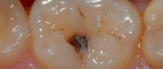
Symptoms of caries under a filling
In the vast majority of cases, secondary caries is asymptomatic. The first signs, with rapid development, may appear only 3-6 months after the filling is installed. If symptoms appear 2-4 weeks after filling, this is recurrent caries. It occurs due to improper treatment and can pose a serious threat to the tooth. To eliminate it, it is necessary to replace the old filling with repeated cleaning and disinfection of the carious cavity.
Classic symptoms of caries under a filling include:
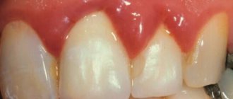
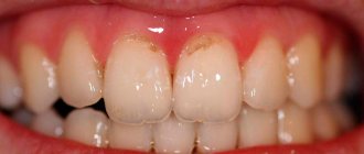
Darkening of the enamel near the edge of the installed filling - pigmentation can be located either at one edge or along the entire rim of the filling;- The causative tooth acquires a gray tint, through the enamel you can see what caries looks like under the filling - it will have a dark tint;
- Chips of the filling and tooth enamel appear, microcracks form (not always);
- The mobility of the filling increases. With prolonged development of caries, it may fall out;
- There is an unpleasant odor from the mouth, caused by the proliferation of bacteria in the formed microcracks;
- Increased sensitivity of teeth to cold, hot, sour and sweet;
- Aching pain appears, which intensifies during the process of biting;
- Acute tooth damage by secondary caries develops, accompanied by pain, swelling and bleeding.
Symptoms and signs
Recognizing signs of tooth decay by caries is not so easy; a filling and the inconvenience of self-examination do not allow one to detect darkening of the enamel or the beginning destruction of bone tissue. However, there are symptoms by which you can recognize the presence of the disease:
- pain on individual teeth and the lower part of the jaw;
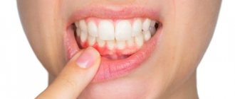
- inflammation of the gums in the area of the filled tooth, swelling;
- an unpleasant odor emanates from the mouth (if the tooth is rotting under a filling);
- the appearance of blood on the gums when brushing the mouth or as a result of a minor collision with blunt objects.
Bleeding from the gums of the affected tooth and aching in the jaw area are often observed. Carious lesions of bone tissue can also be detected visually. It is worth paying attention to the seal. Often it changes color or a dark brown rim appears along its edges. The formation of cracks and chips of tooth enamel is also observed.
Consequences of secondary caries
The lack of timely diagnosis and proper treatment leads to the development of complications in the form of acute or chronic tooth pulpitis with aching pain and all the ensuing consequences. At this stage, replacing the old filling with a new one, while simultaneously cleaning the carious cavity, will not help - preliminary treatment of pulpitis is necessary.
Delaying the treatment of pulpitis under a filling leads to partial or complete destruction of the hard tissues of the tooth. It is accompanied by the development of infection affecting the periodontal tissues with subsequent progression to periodontitis. In particularly severe cases, periodontitis affects the jaw bone - this can result in osteomyelitis of the jaw bone.
Advantages and disadvantages
- Plastic.
- Adheres tightly to dental tissues and prevents the penetration of bacteria.
- Unlimited processing time.
- Photopolymers accurately restore the structure of the dental unit and allow the doctor to carry out complex artistic restorations of the smile area.
- Versatility.
- Suitable for restoring anterior and chewing teeth.
- Light transmission and a large selection of shades.
- The light filling on the tooth is practically invisible.
- Durability.
- Modern photopolymer nanocomposites are characterized by increased strength, do not wear out, and last up to 5-7 years.
- They shrink.
- When using cheap photo composites.
- Possible incomplete polymerization.
- If the doctor does not comply with the treatment protocol, too large layers of material are applied.
In general, there are practically no disadvantages to light fillings. There are limitations that a competent dentist takes into account when installing.
Methods for diagnosing and treating caries under fillings
It is quite difficult to determine the presence of secondary caries in the initial stages, due to the absence of symptomatic and visual manifestations. The disease is diagnosed during a follow-up appointment using diagnostic equipment (hardware diagnostics).
Diagnostics includes two stages:
- Anamnesis and initial examination of the causative tooth in order to detect visible structural changes in hard tissues;
- Hardware diagnostics (physiography) is aimed at detecting hidden changes in the structure of dental tissues.
If a pathology is detected, treatment is prescribed taking into account the individual characteristics of the patient.
Continued action of the etiological factor that caused tooth decay
In case of caries, a filling not only restores lost tissue, but also prevents its further progression (if placed correctly). With a wedge-shaped defect, this does not always happen. Nevertheless, often a filling is the optimal way to solve the problem for some time with this disease. This is exactly the case when the dentist may not be at fault, and such a nuisance should be taken calmly. For more details, see Treatment of wedge-shaped defect.
Features of treatment of secondary caries
The use of one or another treatment regimen for secondary caries directly depends on the location of the lesion, its depth and the condition of the causative tooth. Treatment may include two options for developing the situation:
- Re-filling or replacing part of the filling in the affected area is performed only after treating the tooth and eliminating the carious lesion. In some cases, it may be necessary to remove the nerve and fill the canals.
- Surgical tooth extraction is used in extreme cases when the damage is too large and the doctor cannot save the affected tooth.
Expanding indications for tooth filling
Many patients are surprised, and some are even outraged, by the dentist’s statements about the need to restore the tooth with a more reliable structure - a crown or inlay (onlay) instead of a filling. Misunderstanding leads to suspicion of the doctor of incompetence or of a “scam” for an expensive restoration. “Why can’t you build up a tooth with a filling?” — Those who previously had a filling in the same place for several years are sincerely indignant, especially. It comes to the point that the patient runs away to a neighboring clinic, where the “kind” doctor saves his tooth from having a crown (sometimes charging an amount for “building up” or “artistic restoration” that is not much different from laboratory restoration), and then, two years later, a filling falls out or breaks off again.
There are medical indications and contraindications. They do not depend on the wishes of the doctor or patient. For fillings, the indication for installation is minor tooth decay. As a first approximation, we can say: insignificant - this is less than half the volume of the crown part of the tooth (although in reality everything is somewhat more complicated - the area of the lost chewing surface is taken into account, whether the cusps or cutting edge are affected, whether caries extends under the gum, how difficult it is to restore contact points, etc. .). If the tooth is more damaged, a filling is contraindicated. Of course, you may be lucky and an extensive filling will remain on the tooth for many years, but retention in the tooth is not the only requirement for a filling. It must also restore chewing function, have a tight contact point (so that food does not get stuck between the teeth), etc.
The loss of the “seal on the pin” is a direct consequence of violations of the indications. At a minimum, the “filling on the pin” should be covered and protected by an artificial crown, and at maximum, the tooth may have been so destroyed that it was contraindicated to preserve it even then, before pinning.
Why do some dentists follow the patient’s lead and make his dream of growing a tooth without a crown come true, while others are stubborn and refuse to do so? No, not because the former are good, and the latter are evil and greedy. It’s just that the “rescuers of hopeless teeth” agree to the “tuft of wool”, only the patient stayed and did not run away to another doctor. In addition, fillings and crowns are often done by different specialists: a general dentist and an orthopedic dentist, respectively. Moreover, the treatment of caries is the domain of the therapist; he does not always want to send the patient to another specialist, depriving himself of his daily bread. This is where the fillings are made. Even for the entire tooth, any volume of filling can be sculpted.
In case of small chips of enamel on the cutting edge, the fillings do not hold well. In this case, filling is also contraindicated, but instead, you can simply grind the enamel without restoring anything.
Methods for preventing caries under a filling
High-quality prevention of secondary caries involves the elimination of complications at the stage of initial installation of a filling - the doctor must ensure the correct method of caries treatment, antiseptic treatment of the hole for the filling and installation of the filling.
On the patient’s side, the most effective prevention of secondary caries under a filling is maintaining proper oral hygiene, as well as systematic examinations in a dental clinic: a month after the filling is installed and at least 2 times a year thereafter.
Preventive measures
Regular visits to the doctor will greatly help in assessing whether a filled tooth may have caries. It is important to have your teeth professionally cleaned twice a year and to ensure proper hygiene yourself - floss, brush your teeth twice a day, and use mouth rinses after meals. An early visit to the dentist will allow you to promptly detect the onset of the disease and take action - the doctor will promptly replace the old filling, preserving the maximum amount of living tooth tissue.
Category Caries Published by Mister stomatologist
Types of ceramic inlays
- inlay tab - restores the “cavities” of the tooth crown
- onlay tab - restores most of the chewing surface of the tooth crown
- overlay tab - restores the chewing surface and side walls of the tooth crown
The service life of the inlays is more than 10 years, while the filling will require replacement after an average of 5 years.
Now, when a dentist suggests restoring a tooth with an inlay, you know what benefits this will provide in the future.
What to do if your teeth are rotting
Rot inside the tooth is dangerous for the entire body. Therefore, at the first signs of decay of dental tissues, you should go to the dentist.
Before providing first aid, it is recommended:
- Rinse your mouth with a herbal decoction that has anti-inflammatory and antimicrobial effects. It's better to take chamomile and sage.
- Rinse your mouth with a pharmaceutical antiseptic: Chlorhexidine, Miramistin.
- Thoroughly clean the surface of the dentition to prevent the accumulation of soft plaque and its transformation into hard tartar: bacteria multiply under its surface and spoil the enamel even faster.
- You should not try to remove pus yourself by picking at the enamel with a sharp object. This action will provoke even greater destruction of damaged tissue.
- On the side of the jaw where the rotten tooth is located, it is better not to chew anything so that food does not penetrate into the carious holes.
- After eating, you need to rinse the remaining pieces of food with a weak saline solution or boiled water.
What is the danger of this disease?
Caries is dangerous because there are no symptoms for a number of years. It is detected more often in an advanced stage, when tooth extraction becomes the solution.
Visual diagnosis is difficult because all destructive processes occur inside the tooth. Pronounced plaque or tartar hides any stains on the enamel.
The root is hidden under the gum and external irritants do not affect it until a certain time. On the other hand, the root walls are thin, so they are destroyed quickly and with complications.
Dental restoration
Nowadays, it is no longer enough to simply cover a tooth defect with a filling; patients place increased demands on the appearance of their teeth.
Dental restoration - restoration of aesthetics, anatomy and functionality of the tooth.
With the help of artistic restorations the following problems can be solved:
- caries and its complications;
- dental injuries;
- color changes;
- restoration of crooked teeth;
- closing gaps between teeth.
Direct and indirect composite dental restoration
Based on the manufacturing method, a distinction is made between direct and indirect restorations.
- Direct composite dental restorations involve the restoration of a tooth directly in the mouth by a dentist using composite materials. Our clinics use layer-by-layer tooth restoration technology. Pre-treatment of the tooth is carried out very carefully and carefully with high-quality small diamond burs and instruments, this allows minimal trauma to the living tissues of the tooth. The material is applied layer by layer to the tooth. In this case, many of its shades are used to recreate the natural appearance of the tooth. This is facilitated by the highly artistic abilities of our dental therapists. Each layer hardens under the influence of a light beam. Loss of material is excluded, since it connects with tooth tissue at the micron level. As a result, the restored tooth gains strength and does not differ from the rest.
For fillings and restorations, we use materials from the Swiss company Dentsply, which has been supplying the global dental community with high-quality materials for more than a century. We also widely use materials from the manufacturer Kerr, which has proven clinical effectiveness when used in leading dental clinics in the world for more than 100 years.
- With the indirect method, the doctor processes the tooth for restoration, takes an impression and sends it to the laboratory, where models of the dentition are cast from the impression and the restoration is made directly on the model. Then it is given to the doctor in finished form, and he fixes it on the tooth. For indirect restorations, along with composite materials, ceramics are used, which are superior to composites in aesthetics and reliability.