Gingivitis is inflammation of the gums. The gums are inflamed in the vast majority of the world's population. Therefore, it is a common joke among dentists that it is possible to diagnose any patient, even if he did not have time to enter the office. This diagnosis, as you may have guessed, is “gingivitis.” This article will discuss the classification, pathogenesis and causes of gingivitis.
Gingivitis is the earliest stage of gum inflammation. Over time, the inflammation becomes more severe, the connection between the gum and tooth is destroyed (epithelial attachment - number 3 in the figure), and “gingivitis” changes to “periodontitis”.
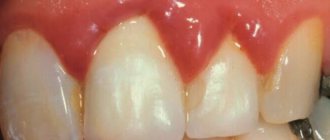
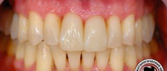
To easily identify pathology, you need to remember the norm. Let's remember what a normal, non-inflamed gum looks like.
Classification
There is no generally accepted classification. Morphologically, catarrhal, ulcerative-necrotic, hypertrophic and atrophic forms of Gingivitis are distinguished; according to the nature of the course - acute and chronic gingivitis. According to the prevalence of the process, a localized form is distinguished (the so-called papillitis - inflammation of individual gingival papillae) and a generalized form (the so-called marginal Gingivitis - inflammation of the entire gingival margin).
According to the clinical course, they are distinguished: mild form - the inflammatory process is localized in the cervical part of the gums (there is no pathological dental-gingival pocket); moderate-severe - inflammation of the gingival papilla and the mucous membrane of the alveolar part of the gums and the presence of a pathological dental-gingival pocket up to 4 mm deep, as well as a severe form, when the entire mucous membrane of the gums is inflamed up to the transitional fold and the depth of the dental-gingival pocket is more than 5 mm. Hypertrophic G., in addition, is divided into the first, second and third degrees of hypertrophy of the gingival papillae.
Based on the etiological basis, they distinguish between G. traumatic, infectious, allergic, G. due to drug intoxication, systemic diseases (hypo- and avitaminosis, endocrine, gastrointestinal tract, blood system, etc.)” and also G. due to periodontal disease.
To express the prevalence of the inflammatory process of the gums in numbers or percentages, the so-called. papillary-marginal-alveolar index (PMA), which is calculated by adding up the assessment of the gingival condition of each tooth; at the same time, the number of teeth at the age of 6 to 11 years is conventionally considered equal to 24, from 12 to 14 years - 28 and from 15 years - 30; inflammation of the papilla is assessed as 1, cervical part of the gum - 2, alveolar part of the gum - 3: RMA (in%) = (sum of indicators × 100)/(3 × number of teeth) Etiology. The most common local causes of G. are unsatisfactory hygienic condition of the oral cavity and hron, mechanical trauma: tartar, decayed tooth, overhanging filling, poorly fitting crowns. G. can be caused by physical. damage to the mucous membrane (acids, alkalis, thermal burns, ionizing radiation, etc.), as well as microorganisms contained in dental plaque. Ulcerative-necrotic G. occurs with the participation of spindle-shaped rods (Bac. fusiformis) and spirochetes (Borrelia vincentii). Common causes include bacterial and viral infections, diseases of the gastrointestinal tract. tract, endocrine changes during puberty, pregnancy, allergic reactions, intoxication with heavy metal salts, vitamin deficiencies, blood diseases. A predisposing factor is a decrease in the body's resistance.
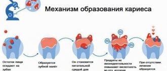
Causes of gingivitis
A huge number of bacteria, including pathogenic ones, are constantly present in the oral cavity. With a decrease in immunity (general or local), pathogenic flora begins to actively multiply. This is usually due to poor hygiene and plaque deposits. Malocclusion and crooked teeth are the reasons why food particles remain in the mouth for a long time and serve as food for pathogenic bacteria. It is they who cause the development of the disease. In rare cases, gingivitis is viral or fungal in nature.
The likelihood of developing the disease increases against the background of a weakened immune system, a chronic source of infection, hormonal or endocrine disorders, improper dental treatment, poor nutrition, and lack of vitamins and microelements.
Pathogenesis
The pathogenesis of gingivitis is determined by the cause of the inflammatory process. G., arising as a result of locally acting causes, is manifested by a local inflammatory reaction of the epithelium and connective tissue of the gums (with a predominance of alteration, exudation or proliferation). G. in general diseases reflects the patterns of pathogenesis of the main patol. process. With mechanical irritants or exposure to weak solutions of acids and alkalis, catarrhal G. (acute or chronic) usually occurs; at significant concentrations of chemicals. irritants - ulcerative-necrotic lesions of the gums. Allergic reactions, intoxication with bismuth, mercury, lead, and lack of vitamin C, as a rule, lead to acute or chronic catarrhal inflammation, which then takes on an ulcerative-necrotic character. Blood diseases, diabetes mellitus, gastrointestinal diseases. tract is often accompanied by acute or chronic, catarrhal G., usually recurrent; in case of dysfunction of the gonads - catarrhal or hypertrophic G., etc.
Nutrition rules
The diet of a person suffering from gingivitis should include as many fresh fruits and vegetables as possible.
- Apples and pears contain pectin and microelements that accelerate the regeneration process.
- A large amount of vitamin C, which helps strengthen blood vessels and reduce bleeding, is found in citrus fruits.
- Vegetables are rich in fiber and antioxidants. By including cabbage, carrots and zucchini in the menu, you can speed up metabolic processes in the body and reduce the time of gum regeneration (healing).
- Berries (blackberries, currants, raspberries) enrich the body with vitamins and minerals, increasing its overall immunity.
You should not eat food containing fast carbohydrates (flour products, potatoes, sweets) in large quantities, as they contribute to the formation of soft plaque.
Pathohistology
Catarrhal Gingivitis is characterized by thickening of the epithelium of the gingival papilla with the phenomena of parakeratosis (see) and acanthosis (see). The cytoplasm of the cells of the spinous layer is granular, the clarity of the boundaries between cells is disrupted. In the nuclei of cells, the phenomena of vacuolar degeneration are often detected (see). In the connective tissue of the stroma there are limited or diffuse lymphoid-macrophage infiltrates with an admixture of plasma cells, in the acute period - with an admixture of segmented granulocytes. In acute cases - swelling of the connective tissue of the gums, expansion of capillaries, venules, swelling of the endothelium, parietal hyaline thrombi. Signs of connective tissue disorganization with mucoid swelling are determined.
In ulcerative-necrotic G., a picture of an acute inflammatory process is observed with the presence of surface defects of the epithelium, pronounced acanthosis, parakeratosis, and infiltration of segmented nuclear granulocytes. Lymphoid-macrophage infiltrates and necrobiosis phenomena are detected in all layers of the gums (see Necrosis). In the stroma there are foci of moderately pronounced plasmorrhagia, the number of capillaries is increased, their lumen is unevenly expanded.
Hypertrophic G. is characterized by hyperplastic growths of the epithelium with pronounced vacuolization of the cytoplasm of the cells of the spinous layer. In the connective tissue - edema, plasmorrhagia of the walls of blood vessels, perivascular infiltrates consisting mainly of lymphoid and plasma cells, an increased number of mast cells with degranulation phenomena, fibrosis in the deep parts of the stroma.
Atrophic G.: pronounced thinning of the epithelial layer, the phenomenon of vacuolar degeneration of the cytoplasm of the cells of the spinous layer; in the connective tissue - thickening of collagen fibers with moderate plasmorrhagia, less often with fibrinoid deposits, moderate lymphoid-macrophage infiltration with the presence of plasma cells, single leukocytes and mast cells.
Folk remedies
To speed up treatment, you can use various rinses and baths from a decoction of medicinal herbs. Recipes for making infusion:
- Celandine and oak bark have astringent properties. They relieve swelling and bleeding from gum inflammation. Crushed oak bark and dried celandine herb are used. To prepare the infusion, they are mixed: two tablespoons of each are poured with two glasses of boiling water. After the infusion has cooled at room temperature, you should rinse your mouth four times a day while the symptoms persist.
- Pour 200 ml of boiling water over a tablespoon of sage and leave for 20 minutes. Wait for the broth to cool and rinse your mouth after each meal. You can also prepare an infusion of chamomile. It has bactericidal properties and accelerates the gum healing process.
If you use traditional medicine, then discuss your actions with a periodontist. Advice from unenlightened people should be ignored.
Clinical picture and diagnosis
Rice.
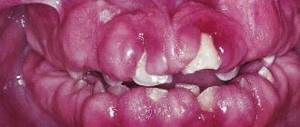
1. Normal gums. Rice. 2. Catarrhal gingivitis: diffuse hyperemia and swelling of the gum mucosa, especially the gingival papillae. Rice. 3. Ulcerative gingivitis: dirty gray plaque and areas of ulceration of the cervical part of the gums. Rice. 4. Hypertrophic gingivitis: gingival papillae bulge, covering part of the tooth crown. Rice. 5. Atrophic gingivitis: the cervical part of the gum is thickened, the necks of some teeth are exposed. Rice. 6. Scorbutic gingivitis: the mucous membrane of the gums is swollen, loose, bluish in color; In the cervical part, the gums lag behind the necks of the teeth. For catarrhal
Gingivitis (tsvetn. fig. 1 and 2) gingival papillae and the mucous membrane of the gingival margin are hyperemic, swollen, and bleeding; with hron, during the course, patol, tooth-gingival pockets are formed. Patients complain of sore gums and bad breath.
With ulcerative-necrotic
G. (tsvetn. fig. 3) the mucous membrane of the gums in the area of the interdental papillae or the entire gingival margin is sharply hyperemic, areas of necrosis have the appearance of a dirty-gray, easily rejected mass. The gum has an uneven, scalloped edge; it bleeds and is sharply painful. The lesion can be limited (area of 1-4 teeth) or involve the entire gum. Patients complain of general malaise, bad breath and severe pain in the gums when eating; body temperature can rise to 39°.
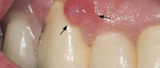
For hypertrophic
G. (tsvetn. fig. 4) are characterized by enlarged gingival papillae, which can cover part of the tooth crown in the form of a roller. The gingival margin, especially the gingival papillae, is loose (edematous stage) or dense. At the first stage there are no complaints; significant hypertrophy (grades 2 and 3) is accompanied by bleeding and some pain when chewing.
Atrophic
Gingivitis (tsvetn. fig. 5) is considered as a manifestation of the dystrophic process in the periodontium; it is characterized by exposure of the roots, usually of individual teeth. Patients complain of fleeting pain from temperature and chemicals. irritants. Some authors consider atrophic G. to be a symptom of periodontal disease (see).
The acute course of G. is characterized from the very beginning by pronounced phenomena of alteration and exudation, which after 10-12 days decrease and disappear, or G. acquires a chronic course. Sometimes inflammation of the gums occurs as a primary chronic. with mildly expressed phenomena of alteration and exudation, periodically exacerbating. Catarrhal and ulcerative-necrotic G. should be considered as different stages of the same process: having begun as an acute catarrhal form with a weakening of the body's defenses, G. can turn into an ulcerative-necrotic form; in turn, the outcome of the ulcerative-necrotic form of G. can be the catarrhal form.
With Gingivitis, which is a symptom of general diseases or intoxications of the body, the picture of gum damage is specific. When the body is intoxicated with mercury, a characteristic black border appears along the edge of the gum, the gingival papillae are swollen, bleed, and ulcerative-necrotic lesions occur that easily spread to other areas of the oral mucosa; patients complain of a metallic taste in the mouth, increased salivation, pain when eating and bad breath. Poisoning with lead and bismuth causes catarrhal or ulcerative-necrotic G. with a characteristic gray border along the edge of the gums; Often erythematous or bullous rashes also appear on the skin.
Manifestations of drug allergies are more often observed when taking antibiotics, sulfonamides, iodine preparations, salicylates in the form of catarrhal and ulcerative-necrotic G. In catarrhal G., the mucous membrane of the gums is bright red and swollen; the appearance of erosions and necrotic areas may be preceded by blistering rashes. Changes in the oral cavity are in most cases accompanied by skin rashes. Patients complain of pain when eating, increased salivation, decreased appetite, body temperature rises to 37.5-38.5°.
Gingivitis with hypo- or vitamin deficiency C begins with swelling of the gingival papillae (tsvetn. Fig. 6), hyperemia and sharp bleeding of the gums, then turning into ulcerative G. with the spread of the process to other areas of the oral mucosa, hemorrhages in the oral mucosa and subcutaneous tissue various parts of the body (see Scurvy). First, tooth mobility occurs, then they fall out.
In diseases of the hematopoietic system, changes in the oral cavity often appear much earlier than the signs of the underlying disease. Acute leukemia is characterized by pale oral mucosa with hemorrhages; The gingival margin is loosened and bleeds when touched lightly. In severe cases, necrotic G. occurs, the gingival papillae are covered with a gray coating; increased salivation is observed, which is replaced by its decrease. G. with hron, leukemia is characterized by gradual atrophy of the gums and changes in the bone tissue of the alveolar process, slight mobility of the teeth; gingival papillae are swollen and hyperemic, bleeding. There is no suppuration in leukemia. X-rays show osteoporosis, a violation of the structure of the bone tissue of the interalveolar septa.
In the initial stage of periodontal disease, a slight hyperemia of the gum edge is detected in certain groups of teeth, then the swelling of the gingival papillae intensifies, and patol is formed. dental-gingival pockets, subgingival tartar is deposited, teeth become mobile, suppuration and bad breath appear; characterized by uneven severity of patol, the process in different parts of the gums and hron, course.
During examination, first of all, the presence of traumatic factors, the form and stage of G. are determined.
It is necessary to differentiate between Gingivitis, which occurs as an independent disease, and Gingivitis, which accompanies other diseases of the body (for example, ulcerative-necrotic Gingivitis with gum damage, characteristic of blood diseases, vitamin C deficiency, mercury, bismuth intoxication), based on anamnesis and tests blood, urine, cytol. examination of the affected area of the oral mucosa, etc. In diabetes (patients complain of dry mouth, paresthesia), it is important to determine blood sugar levels. G. allergic etiology, including drug intoxication, is diagnosed with Ch. arr. based on medical history and results of allergic diagnostic tests (see).
Toothpastes
No matter how painful and unpleasant it may be, a sick person needs to brush their teeth twice a day. It is forbidden to spare or injure, so a toothbrush should be of medium hardness. Toothpaste should be chosen with an anti-inflammatory and healing effect; it should contain sage, chamomile, calendula, and yarrow. They reduce swelling and bleeding, strengthen the gums.
Whitening toothpastes are not used for gingivitis because they contain abrasives that can cause injury when brushing and should be put aside until the gums are healthy again.
Treatment
Treatment should be comprehensive, taking into account the etiol, factor and underlying disease, against the background of which G. developed.
Locally: 3% hydrogen peroxide solution, 0.25% chloramine solution, 1% iodinol solution, enzyme preparations (trypsin, chymotrypsin, chymopsin, ribonuclease, deoxyribonuclease), vitamin preparations that normalize epithelization) (oil solution p vitamins A, E, haloscorbine); in case of an allergic nature of the disease - ointments containing corticosteroids, etc.
A prerequisite for successful treatment for any form of G. is the elimination of tartar, denture defects, and fillings; removal of decayed teeth is carried out after the acute phase of G is cured. In the hypertrophic form, after elimination of acute inflammatory phenomena, surgical excision of overgrown areas of the gum mucosa is indicated.
Physiotherapy is widely used: in acute and aggravated G., microwave therapy is used to relieve pain (see). The emitter is applied to the skin of the cheek according to the lesion for 5 minutes. (course of treatment 5-6 procedures). Electrophoresis is used to administer vitamins C, P, B1, calcium chloride and other drugs (see Electrophoresis, medicinal). In order to destroy granulation tissue in patol, dental-gingival pockets, diathermocoagulation is used (see). Physiotherapy is contraindicated for neoplasms and decompensated diseases of the cardiovascular system.
General treatment is carried out by dentists together with doctors of other specialties. For G. of allergic origin, in addition to stopping contact with drugs that cause allergies, desensitizing and antihistamine therapy is prescribed. In case of intoxication with salts of heavy metals, it is necessary to isolate the patient from occupational hazards; antidotes are prescribed internally. In case of dysfunction of the gonads, diabetes, blood diseases, scurvy and other diseases, treatment is carried out by doctors of relevant specialties.
Prevention
Personal oral hygiene, elimination of factors that provoke gingivitis; systematic prof. examinations by a dentist and dispensary records of patients with chronic diseases in which G. phenomena may occur, as well as persons employed in enterprises with occupational hazards; sanitation of the oral cavity (see).
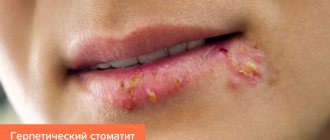
See also Stomatitis.
Bibliography:
Novik I. O. Diseases of the teeth and oral mucosa in children, M., 1971, bibliogr.; Popova Yu. P. Clinical manifestations of leukemia on the oral mucosa and their therapy, Dentistry, No. 3, p. 25, 1969; Guide to therapeutic dentistry, ed. A. I. Evdokimova, p. 372, M., 1967, bibliogr.; Severova E. Ya. and Bokanova Zh. V. Manifestations of drug allergies in the oral cavity, Dentistry, No. 6, p. 28, 1968; Chechel A.P. About hypertrophic gingivitis in the treatment of patients with epilepsy, ibid., No. 4, p. 93, 1970; Pindborg JJ Atlas of diseases of the oral mucosa, Copenhagen, 1973, Bibliogr.
E. V. Borovsky.
Causes of gingivitis
Gingivitis occurs for a number of reasons; they are usually divided into two groups: internal and external.
Internal ones include:
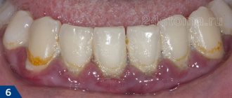
- Lack of hygiene. Bacterial plaque accumulates on the teeth, which forms a dense film. If it is not removed, within 2-3 days it crystallizes and turns into tartar. This is a constant source of infection in the oral cavity. In addition, the stone injures the gums, and an infection develops as a result of bacteria entering the wound.
- Lack of vitamins leads to decreased immunity and insufficient nutrition of gum tissue.
- Tooth growth is a natural process during which the integrity of the gums is compromised. If an infection occurs at this time, inflammation develops and gingivitis occurs.
- Malocclusion, in which a tooth or several teeth constantly injure the gums.
- Diseases of the gastrointestinal tract, which lead to a lack of vitamins and microelements, increased acidity of saliva.
External causes of gingivitis:
- An infection that came from outside. Often develops against the background of acute respiratory viral infections and other respiratory diseases, when eating poorly washed vegetables and fruits.
- Mechanical damage of any nature, including during infection of a wound resulting from inaccurate dental treatment or removal of tartar.
- When exposed to radiation or while taking certain medications.
- Smoking.
- The presence of multiple foci of caries.
Most often, gingivitis occurs in children: their immune system is still developing, so it cannot always recognize the infection in time and cope with it. Children often put dirty hands and objects into their mouths, which cause tissue damage and infection.