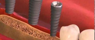
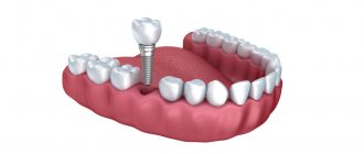
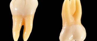
Bone grafting is the augmentation of bone tissue, which is often very necessary for dental implantation. This is due to the fact that immediately after the removal or loss of a tooth, the bone tissue gradually begins to decrease due to lack of load. As a result, its size may simply not be enough to install an implant, and this deficiency will need to be filled. So if you have lost your teeth a long time ago and have only now decided to undergo implantation, then you will most likely need bone grafting.
TYPES OF BONE PLASTY
There are several main types of bone grafting, which are used depending on the area of future implantation:
- On the sides of the lower jaw, the width and height of the alveolar process usually decreases, and the distance from the upper part of the bone tissue to the mandibular canal decreases significantly. If there is not enough bone tissue in this area, the jaw can move forward. The plastic surgery here is simple - you just need to build up the bone tissue;
- In front of the upper and lower jaw, a sharp and thin alveolar ridge is often formed. Plastic surgery in this area is a little more difficult, since the aesthetics of the final result is very important;
- On the sides of the upper jaw, plastic surgery is necessary because if there is a lack of bone tissue, the implant will penetrate into the maxillary sinuses or sinuses. Here plastic surgery is carried out using a special technology - sinus lifting. Sometimes it is required even immediately after tooth loss, if the sinuses are physiologically too large.
Symptoms
A fracture of the alveolar process of the upper jaw has several characteristic features. The most common symptoms:
- sharp pain
- inability to completely close your mouth,
- occurrence of malocclusion,
- dysfunction of chewing and swallowing,
- hematomas,
- dislocations and mobility of one or more teeth,
- severe swelling of soft tissues,
- bleeding.
If you suspect an alveolar bone fracture, you should seek medical help as soon as possible.
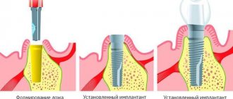
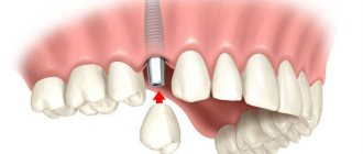
How is bone grafting performed?
As we noted above, there are different methods of bone grafting, but in general it follows the same scheme:
- Anesthesia;
- Providing access to bone tissue - incision or puncture of mucous membranes;
- Laying the selected bone material;
- Suturing the wound until healing.
If it was necessary to restore a small amount of bone tissue, then it is possible to carry out implantation simultaneously with bone grafting. If the bone atrophy was severe, then you will need to wait six months until the bone blocks completely take root.
Periostitis of the jaw - symptoms and treatment
Treatment of periostitis is always surgical; if you deviate from this rule, there is a serious danger of a limited focus of inflammation flowing into a more dangerous - diffuse form, which will require different treatment tactics and often hospitalization of the patient.
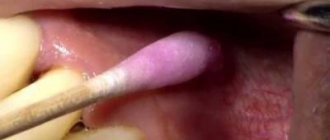
Treatment of periostitis is standard and consists of eliminating the source of primary inflammation and evacuation of pus, i.e., removing the causative tooth (in cases of unfavorable treatment prognosis) and surgically opening the site to its entire width, followed by drainage.
The procedure is usually performed under local anesthesia; in some cases, drug sedation is indicated; anesthesia is also possible. You need to understand that the introduction of an anesthetic into tissues strained by exudate (fluid accumulation) is an extremely painful procedure, therefore, the method of pain relief before periostotomy (opening an abscess) has its own specifics: conduction anesthesia is preferable (a type of anesthesia when the nerve is blocked to the operation site) followed by local anesthesia performed superficially with a thin needle, without immersing the needle into the abscess.
The diseased tooth must be removed in the case of an unfavorable therapeutic prognosis: the presence of large foci of bone destruction, incorrect previously carried out treatment of the canals, resulting in their perforation or blockage by a fragment of an instrument, etc. In cases of a favorable prognosis, endodontic treatment is carried out, which involves the removal of pulp decay from the canals, appropriate mechanical and medicinal treatment of the canals with a special instrument and powerful antiseptics to eliminate infection in them, followed by temporary filling with calcium preparations and temporary filling of the tooth. Subsequent X-ray monitoring is very important in order to confirm the effect of the canal therapy: the focus of destruction in the bone should decrease and subsequently disappear.
To evacuate pus, periostotomy is performed (dissection of the mucosa and periosteum) along the entire length of the infiltrate along the transitional fold.
As a rule, the surgeon detects a characteristic symptom - detachment of the periosteum from the bone, whereas in a healthy state the periosteum is firmly attached to the cortex. At this stage, the abscess is emptied, or the absence of pus is detected along with the periosteal reaction. The surgeon uses a blunt instrument to probe the entire abscess cavity to identify isolated lesions. In the case of the presence of pus, irrigation (washing) of the subperiosteal space with antiseptics is usually carried out, followed by the introduction of drainage (usually a strip of glove rubber) into the wound to prevent its edges from sticking together. The wound is not sutured; the patient is given a dressing after a day or two to remove the drainage [8].
In the absence of contraindications, antibacterial therapy (in most cases, semisynthetic penicillins) and NSAIDs (nonsteroidal anti-inflammatory drugs) are prescribed. In case of severe edema, desensitizing drugs are prescribed. It is also necessary to prescribe analgesics or their direct intramuscular administration after surgery, since in the first hours after treatment, when the effect of the anesthetic wears off, severe pain symptoms appear. It also makes sense to locally cool the infiltrated area with ice for several hours to reduce bleeding and swelling. The cooling time must be assigned based on the patient’s subjective feelings, and not on specific time periods.
Almost always, this treatment leads to a positive result: within a few hours the patient experiences relief, decreased pain and swelling. Although the infiltrate in the form of a moderately painful compaction will persist for several days.
A follow-up visit the next day is necessary to confirm the results of the treatment and possible removal of the drain if there is no discharge from the wound. Subsequently, the patient is observed by a doctor for 3-5 days; it is quite legal to issue a certificate of incapacity for work for this period. After the pronounced symptoms of inflammation disappear, they begin to continue treatment of the causative tooth.
In rare cases, for example, when the incision is of insufficient length or when the drainage falls out and the edges of the wound stick together, treatment may be complicated due to a delay in the evacuation of residual pus from the lesion. In these cases, the incision should be widened and drainage should be ensured by introducing drainage.
Materials for bone grafting
Bone material is required for tissue augmentation during bone grafting. There are four main options for bone grafting materials:
- Autogenous transplant. This graft is taken from the patient's own bone. Typically, a bone block is taken from the jaw in the area of the wisdom teeth, from the chin, or from another area. The survival rate of such implants is almost one hundred percent, however, the collection of donor material is required, that is, an additional, rather difficult operation, so such transplants are rarely used;
- Allograft. Such grafts are taken from the bone material of another person who has donated the organs for medical purposes. Bone blocks are carefully selected, sterilized and processed so that the body eventually perceives them as its own bone material;
- Xenograft. For its production, the blood of cattle, most often cows or bulls, is used. The bones are carefully sterilized and prepared in a special way so that the human body perceives them as its own bone. Xenografts survive quite well;
- Alloplastic grafts made entirely of artificial materials. They are different. For example, absorbable grafts do not replace, but rather stimulate the person’s own bone tissue, which ultimately grows faster, and the implant itself then dissolves. There are also non-absorbable grafts that serve as a lattice on which the bone grows.
Diagnostics
At the initial stage, the doctor will listen to the patient’s complaints and find out the mechanism of injury. Next, the specialist proceeds to an examination, which allows him to obtain a lot of valuable information. In severe displaced fractures, the diagnosis can be made already at this stage.
The mobility of fragments in a complete fracture can be determined by palpation. Based on these data, the location and direction of the fracture can be assumed.
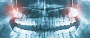
Accurate diagnosis is carried out on the basis of radiographic examination. The image clearly visualizes the fracture line and the direction of displacement of the fragments. Computed tomography can provide more detailed information. This technique makes it possible to take pictures of layer-by-layer sections of the damaged area, as well as determine the condition of the soft tissues and the position of the fragments relative to other anatomical structures.
When a fracture occurs in the area of the alveolus, the teeth suffer. The dentist must determine which ones can be attempted to be preserved. For this purpose, the viability of the pulp is determined using EDI. In doubtful cases, the study is repeated after 1-2 weeks; the pulp may undergo necrosis or restore viability. Based on the results obtained, further tactics are determined.
POSSIBLE COMPLICATIONS AFTER BONE PLASTY
Bone grafting is a fairly complex operation, so complications after it are not that uncommon. Any mistake by the surgeon can cause:
- Inflammation of the wound;
- Development of suppuration;
- Complete rejection of the bone block.
But soft tissue swelling after bone grafting is not a complication, but a completely common occurrence. As a rule, after three days the swelling completely subsides. Hematomas can also occur in the plastic surgery area, but they are also safe for human health.
Treatment methods for fractures
Treatment of fractures of the alveolar process of the upper jaw includes several stages:
- pain relief,
- antiseptic treatment,
- repositioning of all fragments,
- immobilization with special structures.
Displaced fractures require complex treatment. In case of serious injuries, the wound is inspected, the sharp edges of bone fragments are smoothed and the mucous membrane is sutured. The bone fragments are returned to the correct position and securely fixed with staples or splints. To prevent complications in the first days after injury, antibacterial therapy and rinsing the mouth with herbal decoctions are prescribed.
With timely provision of qualified assistance, bone callus is formed within 8 weeks. The speed of recovery depends on the individual characteristics of the body.
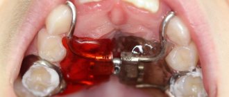
Elimination of a defect in the alveolar process of the upper jaw due to perforation of the maxillary sinus
Perforation of the bottom of the maxillary sinus is one of the most common complications in dental practice that occurs directly during the operation of removing teeth of the upper jaw. The resulting communication between the oral cavity and the maxillary sinus requires the doctor to take urgent measures to close the defect, since the oroantral anastomosis is in the future the gateway for the penetration of odontogenic infection from the oral cavity into the sinus cavity [2, 3]. A significant number of works are devoted to the issues of diagnosis, prevention and treatment of perforations of the maxillary sinus [2, 5, 7].
The relevance of research
With traditional methods of treatment, the bone tissue defect is not restored, only the mucous membrane is sutured, which leads to disruption of the shape of the alveolar process, and in 9-30% of cases - to divergence of the wound edges and the formation of persistent fistulas [7].
The issues of bone grafting of oroantral perforations aimed at compensating for bone loss for the purpose of further rational prosthetics are not sufficiently covered.
Recently, directed regeneration of bone structures has been widely used. The scientific literature covers a number of clinical methods of treating this category of patients using various osteoplastic materials [1, 3, 4, 6]. Unfortunately, many of the drugs used have certain disadvantages, which dictates the need to search for new, more advanced materials [1, 5, 6].
Currently, given the large number of operations to close the oroantral communication, the search for a method to prevent inflammatory complications is also relevant. There are only a few studies devoted to studying the effectiveness of using polymer-based drugs in the treatment of patients with perforations of the floor of the maxillary sinus.
In the literature, which describes a large number of methods for plastic closure of oroantral perforations, there are no most convenient and acceptable methods of operations using osteoplastic agents intended for both outpatient and inpatient doctors.
Purpose of the study
Increasing the effectiveness of treatment of patients with perforations of the maxillary sinus through the use of osteoplastic materials in eliminating the oroantral communication.
Material and methods
A total of 84 patients of both sexes aged from 19 to 65 years with perforation of the bottom of the maxillary sinus without pronounced clinical and radiological signs of sinusitis, who were undergoing outpatient treatment in the regional dental clinic of Stavropol and the department of maxillofacial surgery of the 4th clinical clinic, were under observation. hospitals in Stavropol from 2008 to 2012
Most often, perforation of the maxillary sinus occurred in the age group from 19 to 40 years (59.5% of cases), that is, the most socially active, with a predominance of males. In 13% of patients, perforation of the bottom of the maxillary sinus was accompanied by the penetration of a foreign body into the sinus (tooth root or filling material); in 87% of patients there were no foreign bodies in the sinus. Oroantral perforation occurred more often during the removal of the first molars (69.1%), less often during the removal of the second molars (16.6%) (Table No. 1).
Table No. 1. Variants of localization of perforation of the maxillary sinus (n = 84).
Localization of perforation | Number of patients | % |
| 1st premolar | 4 | 4,8 |
| 2nd premolar | 6 | 7,1 |
| 1st molar | 58 | 69,1 |
| 2nd molar | 14 | 16,6 |
| 3rd molar | 2 | 2,4 |
Depending on the surgical treatment tactics, patients with perforations of the maxillary sinus floor were divided into 5 groups. The characteristics and size of the study groups are presented in Table No. 2.
Table No. 2. Distribution of patients by treatment methods depending on the size of the defect (n = 84).
Treatment method | Defect size | |||
Group I< 5 mm | Group II5-7 mm | III group> 7 mm | ||
| 1 | "Collost" | 6 | 7 | 6 |
| 2 | "KollapAn-M" | 5 | 6 | 6 |
| 3 | "Osteoplast" | 5 | 8 | 4 |
| 4 | "Collost-gel" + "Osteoplast" | 4 | 7 | 5 |
| 5 | "Kollapan-gel" + "Osteoplast" | 3 | 8 | 4 |
In patients of all groups, filling of the bone defect in the perforation zone was carried out using osteoplastic materials separately or in combination. For clinical study, a domestically produced osteoplastic biocomposite material, KollapAn-M (Intermedapatit LLC), was taken, which is a combination of synthetic hydroxyapatite and collagen. Additionally, the preparations “Collost” in the form of blocks and crumbs, “Collost-gel” based on bone collagen of animal origin (Biopharmholding CJSC) and “Osteoplast” (Liko LLC) based on bone collagen and sulfated glycosaminoglycans were used.
The analysis showed that all identified oroantral perforations can be divided into three groups depending on the size of the defect:
- Group I - 23 patients with a defect size of up to 5 mm;
- Group II - 36 patients with a defect size from 5 to 7 mm;
- Group III - 25 patients with a defect size of more than 7 mm.
The timing of patients' admission to the clinic from the moment of oroantral anastomosis occurred varied. The largest number of patients - 41.6% - were admitted on the first day after tooth extraction. According to patient medical histories, left-sided perforations were the most common - 56%, right-sided ones were recorded in 44% of patients.
When examining patients, clinical and anamnestic data were taken into account, including complaints, how long ago the oroantral communication was formed, its location, size, as well as the results of additional research methods.
X-ray examination, in addition to standard methods, included computed tomography (CT), the results of which assessed the condition of the bone tissue of the operated area at 3, 6, 12 months.
Endoscopic examination was performed using a rigid endoscope from Rami (Italy) with viewing angles of 0, 30, 70° and a working tube diameter of 4 mm. The specified characteristics of the device made it possible not only to insert the working part of the tube through the oroantral communication (if the size is more than 5 mm) into the sinus, but also to examine its walls. The endoscopic picture was recorded on video. The choice of method for plastic closure of the oroantral defect depended on its size (Table No. 4).
Table No. 4. Methods of plastic closure of oroantral perforations depending on their size (n = 84).
Defect plastic method | Group IBone defect < 5 mm | Group IIBone defect 5-7 mm | III groupBone defect > 7 mm |
| Suturing soft tissue under perforation | 23 | 31 | 0 |
| Trapezoidal flap from the vestibule of the oral cavity | 0 | 5 | 25 |
The size of the bone defect, its location and shape were determined using computed tomography data in various modes (Fig. 1 a, b).
Rice. 1a. Computed tomography in 3D mode, perforation of the maxillary sinus, oroantral anastomosis. Rice. 1b. Computed tomography in 3D mode, perforation of the maxillary sinus, oroantral anastomosis.
In 64.3% of patients with small and medium-sized perforations up to 7 mm, the mucous membrane under the perforation area was sutured. To do this, the edges of the wound around the oroantral defect from the side of the oral cavity were refreshed, the mucous membrane around was separated subperiosteally, and two releasing parallel incisions were made, going from the vestibule of the mouth to the palate. If necessary, an additional incision was made on the palatal side at a distance of 5 mm medially from the oroantral defect.
Plastic closure in patients with a defect size of more than 7 mm in 35.7% of cases was performed according to the method of A. G. Mamonov, B. V. Kononov (1973) with a trapezoidal mucoperiosteal flap cut from the vestibule of the oral cavity. After de-epithelialization of the trapezoidal flap, the mucoperiosteal flap was peeled off along the edge of the socket on the palatal side and the edge of the trapezoidal flap was inserted into the resulting pocket (Fig. 2 a, b).
Rice. 2a. Filling the cavity with osteoplastic material. Rice. 2b. Mobilization and suturing of the mucoperiosteal flap.
The study of mucociliary transport of the nasal mucosa was performed on days 3, 7, and 14 after surgery using a saccharin test. For this purpose, grains of food saccharin from Hergestellt (GMBH, Germany) were used. One grain of saccharin weighing 0.3 g was placed on the surface of the inferior turbinate, 1 cm from its anterior end. The patient was asked to perform one swallowing movement per minute and the time was measured. When the taste sensation of sweets appeared in the oral cavity, the time of mucociliary transport was noted.
When statistically processing the research results, nonparametric methods were used: Mann and Whitney, Kruskal-Wallis and the X² criterion (Gubler E. G., Genkin A. R., 1973). Differences with p <0.05 were considered statistically significant.
Results and discussion
Analysis of the timing of epithelization of the wound surface showed that in patients of groups 1-3, wound epithelization averaged 7 days, in patients of groups 4-5 - 5 days. In groups 4-5, there was earlier hemostasis, fewer relapses, swelling, pain syndromes, and accelerated healing of soft tissues of the wound surface compared to groups 1-3. The data obtained were confirmed during repeated CT studies performed in the postoperative period 3, 6 and 12 months after plastic surgery of the oroantral anastomosis (Table No. 3).
Table No. 3. Dimensions of the bone defect before and at various times after surgery according to computed tomography data (in mm).
Study groups | Groups | Before treatment | In 3 months | In 6 months | After 12 months |
| 1 | 2 | 3 | 4 | ||
| "Collost" | <� 55—7 > 7 | 3,8±0,25,2±0,3 7,1±0,4 | 2,3±0,33,2±0,2*1 5,4±0,3*1 | –*1,22,0±0,2*1 3,1±0,3*1 | –*1,2–*1—3 1,9±0,1 |
| "KollapAn-M" | <� 55—7 > 7 | 3,7±0,35,4±0,4 7,0±0,5 | 2,1±0,1*13,0±0,2*1 3,5±0,5*1 | –*1,21,4±0,1*1,2 2,1±0,3*1 | –*1,2–*1—3 –*1—3 |
| "Osteoplast" | <� 55—7 > 7 | 3,6±0,25,1±0,3 6,9±0,3 | 2,0±0,1*12,9±0,2*1 3,6±0,3*1 | 1,2±0,08*1,22,0±0,2*1,2 2,8±0,3*1 | –*1—3–*1—3 –*1—3 |
| "Collost-gel" + "Osteoplast" | <� 55—7 > 7 | 3,9±0,15,4±0,2 7,2±0,5 | –*11,1±0,08*1 2,2±0,1*1 | –*1,2–*1,2 1,3±0,3*1,2 | –*1,2–*1,2 –*1—3 |
| "Kollapan-gel" + "Osteoplast" | <� 55—7 > 7 | 3,5±0,35,1±0,4 7,7±0,3 | –*12,2±0,1*1 3,3±0,2*1 | –*1,21,2±0,08*1,2 1,9±0,2*1,2 | –*1,2–*1—3 –*1—3 |
* 1, 2, 3: ratio of the result to the original value.
Data from X-ray studies in patients of groups 4-5 confirmed that bone tissue regeneration in the area of the defect was completed by 3-4 months. Complete restoration of bone tissue occurred by 6 months of observation. In patients of groups 1–3, bone tissue restoration in the early stages was much slower. The results of the clinical course of the postoperative period indicate that the introduction of the drugs “Collost-gel” + “Osteoplast” and “Kollapan-gel” + “Osteoplast” into the bone cavity helps to reduce the intensity of the main clinical signs (pain, swelling, temperature reaction) compared with 1-3 groups.
An integrated approach using modern osteoplastic biomaterials in the form of blocks, crumbs, gels and optimal methods of bone grafting allows for the recovery of patients even on an outpatient basis. Inflammatory phenomena of the sinus mucosa significantly inhibited the transport function of the ciliated epithelium until it was completely blocked. The dynamics of restoration of the time of mucociliary transport of the sinus mucosa into the nasal cavity depended on the duration of the perforation - the earlier the patient underwent surgical intervention to eliminate the oroantral defect, the faster the functions of the sinus mucosa were restored. In cases of elimination of “fresh” perforation (up to 3 days), the mucous membrane of the nose and maxillary sinus was practically not subject to reactive postoperative phenomena and local inflammatory processes caused by the pathological process were quickly stopped.
Thus, an integrated approach using modern osteoplastic biomaterials in the form of blocks, crumbs, gel and optimal methods of bone grafting makes it possible to achieve stable recovery of patients even in an outpatient setting.
The developed treatment and diagnostic algorithm makes it possible to reduce the treatment time for patients by an average of 2-3 days, prevent postoperative inflammatory complications, thereby preventing the occurrence of maxillary sinusitis, and reduce the risk of relapse of postoperative oroantral communication. The developed technique for eliminating the oroantral defect allows not only to prepare the alveolar process of the upper jaw for further prosthetics, but also contributes to the rapid restoration of the transport function of the nasal mucosa and maxillary sinus.
conclusions
The choice of method for plastic closure of oroantral perforations depends on the size of the bone tissue defect, the dimensions of which should be determined using computed tomography. The use of osteoplastic materials to fill the oroantral bone defect is an effective and reliable method for restoring bone and soft tissue in the area adjacent to the maxillary sinus.
LITERATURE
1. Belozerov M. N. Evaluation of osteoplastic properties of various biocomposite materials for filling jaw defects / M. N. Belozerov // Author's abstract. dis. Ph.D. honey. Sci. - M., 2004. - 23 p. 2. Bocharova I. A. Restoration of bone tissue of the alveolar process during perforation of the maxillary sinus in conditions of directed tissue regeneration / I. A. Bocharova // Author's abstract. dis. Ph.D. honey. Sci. - Voronezh, 2008. - 24 p. 3. Losev F. F. Bone grafting using membranes: indications for use, possible errors and violations of the principle of action of directed tissue regeneration / F. F. Losev, A. V. Zharkov // Dentistry. - 2010, No. 6. - P. 27-32. 4. Panin A. M. New generation of osteoplastic materials (development, laboratory and clinical substantiation, clinical implementation). Author's abstract. dis. doc. honey. Sci. - M., 2004. - 52 p. 5. Ivanov S. Yu. Clinical results of using various osteoplastic materials for sinus lifting / S. Yu. Ivanov, A. F. Bizyaev, M. V. Lomakin, A. M. Panin // New in dentistry. - 2009, No. 5. - P. 51-53. 6. Ivanov S. Yu. Development of biomaterials for osteoplasty based on bone tissue collagen / S. Yu. Ivanov, A. M. Panin, D. N. Volodina // Clinical dentistry. - 2005. - No. 4. - P. 21-23. 7. Rasmusson L. A. Closure of local defects of the alveolar process // New in dentistry. - 2011, No. 5. - P. 40-43.