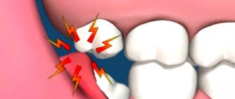
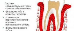
Symptoms of this disease will be:
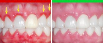
- hyperemia, swelling, burning and hemorrhages from the gums;
- hypersensitivity to cold, hot, sour foods;
- aesthetic defect of the gums.
Often, hypertrophied gums act as a mechanical obstacle when chewing food, which contributes to injury, penetration of pathogenic microorganisms into damaged areas and aggravation of the inflammatory process. Diagnosis of hypertrophic gingivitis is carried out through examination, palpation examination of the patient’s gums, determination of dental indices, as well as based on the results of additional studies (radiography and CT of the dental system). Therapeutic tactics include the use of local anti-inflammatory drugs, physiotherapeutic procedures, sclerotherapy, diathermacoagulation of the gum papillae and surgical excision of hypertrophied tissue (gingivectomy).
According to statistical data, the children's category of the population suffers from this disease most often, especially children and adolescents in the pre-pubertal and puberty period (from the beginning to full puberty), since the phenomenon of hypertrophic gingivitis is often associated with hormonal development.
Hypertrophic gingivitis is one of the forms of gingivitis (inflammation of gum tissue) and is less common in comparison with other forms (catarrhal, atrophic, ulcerative necrotic).
Causes of hypertrophic gingivitis
The causes of the development of hypertrophic gingivitis can be both local and general factors, but most often a combination of them occurs. Local factors include:
- malocclusion (deep and open bite);
- crowding of teeth in the frontal (front) group;
- dental deposits (dental plaque and tartar);
- low frenulum attachment;
- mechanical trauma to the gums due to improper installation of fillings;
- irrationally selected dentures;
- poor oral hygiene when wearing orthodontic appliances;
- taking medications to prevent unwanted pregnancy;
- changes in hormonal status.
In addition to these factors contributing to the development of hypertrophic gingivitis, the disease often develops during puberty, pregnancy and menopause. Subsequent causes of hypertrophic gingivitis include:
- endocrine diseases (diabetes mellitus, thyroid pathology);
- tuberculosis;
- nephropathy;
- hypovitaminosis;
- leukemia
Internal causes of gingivitis are:
- physiological growth of teeth (in some cases, a growing tooth can injure the mucous membrane of the gums);
- an incorrectly installed filling or the edge of a decaying temporary tooth;
- poor nutrition and lack of vitamins in the child’s diet;
- decreased immunity.
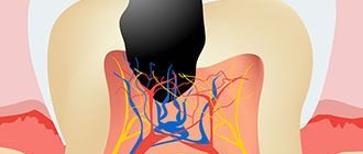
Internal causes of gingivitis also include various infections, namely when there is a carious lesion in the oral cavity.
Gingivitis and periodontitis: cause and effect relationships
Hypertrophic gingivitis can manifest itself as an independent disease or occur during an exacerbation of periodontitis. Despite similar symptoms (pain, bleeding gums), the medical history has a completely different scenario: the mucous membrane becomes inflamed, a hypertrophy mechanism is triggered in its cells, and the volume of periodontal tissue increases. But! The integrity of the dental epithelial junction is not compromised and no pathological changes occur in the bone tissue. Inflammation does not penetrate deeper than the upper layer of the gum epithelium.
While periodontitis is characterized by a much more serious clinical picture: the inflammatory process first covers the mucous membrane, and then goes deeper - it affects the lower layers of periodontal tissue and reaches the bone.
The danger of hypertrophic gingivitis (like any other) is that it can develop into periodontitis, which is more difficult to treat and leads to serious consequences, including tooth loss. Why is this happening? The fact is that when periodontal tissue swells and false periodontal pockets form, the problem grows like a snowball. The gums become very sensitive and react with acute pain and burning to the slightest irritation. In this case, complete teeth cleaning is simply not possible and the number of bacteria in the oral cavity increases rapidly, worsening the disease every day. This is why it is so important to seek dental care in time to clear plaque from your gums and teeth. This is the only way to minimize the bacterial factor and reduce the likelihood of periodontitis.
Classification of hypertrophic gingivitis
Based on the prevalence of pathological changes, localized and widespread hypertrophic gingivitis are distinguished. The localized process of gingivitis occurs on a small section of the gum. With widespread gingivitis, the gums become inflamed on both jaws. Depending on the type of hyperplastic processes, hypertrophic gingivitis can occur in two forms:
- edematous (inflammatory) form, which is accompanied by swelling of the connective tissue fibers of the gingival papillae, vasodilation, lymphoplasmacytic infiltration of the gum tissue;
- fibrous (granulating) form, in which proliferation of connective tissue fibers of the gingival papillae, thickening of collagen fibers, parakeratosis phenomena are detected with minimal severity of edema and inflammatory infiltration.
In both forms, professional hygienic treatment and anti-inflammatory therapy in combination with high-quality individual oral hygiene are especially important.
It should also be noted that in dentistry there are three degrees of hypertrophic gingivitis according to the nature of the growth of gum tissue, namely:
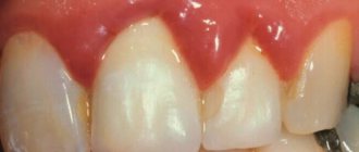
- slight hypertrophy of the gingival papillae at the base, when the overgrown edge of the gum covers the crown of the tooth by 1/3;
- medium degree, in which there is a progressive increase and dome-shaped change in the shape of the gingival papillae, when the overgrown gum half covers the dental crowns;
- severe degree, in which hyperplasia of the gingival papillae and gum margins is pronounced; they cover the dental crown by more than 1/2 of the height.
Treatment of ulcerative gingivitis
The treatment regimen is as follows:
- Treatment of ulcers: first, anesthesia is performed, then with the help of enzymes and instruments, the gums are cleared of ulcers;
- Fighting dental plaque. This stage includes mandatory antiseptic treatment of the entire oral cavity and superficial anesthesia using a gel;
- Taking antibiotics (metronidazole orally);
- Local antibacterial therapy with metronidazole;
- After healing begins, local wound-healing preparations are used (oil solutions of vitamins A, E, sea buckthorn);
- Anti-inflammatory therapy;
- Antiallergic drugs;
- After recovery – treatment of diseased teeth;
- General restorative therapy: vitamins, possible prescription of immunomodulators.
In case of ulcerative gingivitis, they are required to issue a certificate of incapacity for work (sick leave) for several days.
Symptoms of hypertrophic gingivitis
In the edematous form of hypertrophic gingivitis, the patient experiences symptoms such as:
- enlargement and swelling of the gingival papillae;
- burning;
- painful sensations;
- bleeding gums when brushing teeth and eating;
- hypertrophy of interdental papillae;
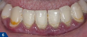
- bright red gum color with a glossy sheen;
- presence of dental plaque;
- the formation of false periodontal pockets containing detritus.
In addition to the expansion and proliferation of capillaries, in the edematous form of hypertrophic gingivitis, abundant and varied cellular infiltration is observed (leukocytes, plasma and mast cells, lymphocytes).
The fibrous form of hypertrophic gingivitis manifests itself as follows, namely:
- keratinization of the epithelium according to the type of parakeratosis;
- thickening of the epithelium and proliferation in depth of connective tissue;
- thickening of the walls of blood vessels;
- inflammatory infiltration.
In this case, the gums have an uneven, bumpy surface and acquire a pale pink color. With this form of gingivitis, the gums are absolutely painless and do not bleed on contact. Upon examination, soft and hard subgingival deposits are revealed. In addition, the epithelial attachment is not impaired. At first, this form of gingivitis usually does not bother patients. As the process develops (moderate and severe), patients note pronounced bleeding of the gums, a specific odor emanating from the oral cavity, gum defects visible to the naked eye, and a large amount of plaque on the surface of the teeth.
What can trigger chronic gum hypertrophy?
It is important to understand that there is no difference between the concepts of “hypertrophic gingivitis” and “chronic hypertrophic gingivitis”. The growth of periodontal tissues does not happen overnight; it is a long-term process, the consequences of which may become noticeable after several months, or may not show themselves for several years. It is precisely because of the long course characteristic of this disease that it always has a chronic form.
One of the causes of hypertrophy is considered to be advanced gingivitis. It, in turn, is provoked by pathogenic bacteria that actively multiply in dental plaque. This means that regularly ignoring the rules of oral hygiene can sooner or later have such serious consequences that the problem cannot be solved without surgical intervention.
However, no matter what threats long-standing plaque deposits pose, most often chronic gingival hypertrophy occurs not due to bacterial, but due to mechanical effects. Situations in which there is a risk of seriously injuring the gums are not that uncommon. The problem can be caused by:
- incorrectly selected and, as a result, uncomfortable dentures;
- error during installation of braces;
- inaccurate filling and installation of a filling of the wrong size, which comes into contact with the mucous membrane and irritates it;
- the presence of large deposits of tartar;
- malocclusion.
But the main danger is that even those patients who deal only with experienced dentists and carefully follow their instructions are not immune from hypertrophy. Other factors that, at first glance, have nothing to do with dentistry, can also serve as a trigger and start the irreversible process of division of the basal cells of periodontal tissue.
Rare causes
- diabetes mellitus and various types of disorders in the endocrine system;
- the presence of malignant tumors in the body;
- infectious diseases of the respiratory tract;
- diseases of the gastrointestinal tract;
- problems with the cardiovascular and nervous systems;
- chronic vitamin deficiency.
Rarely, in dental practice there have also been cases where mucosal hypertrophy occurred as a result of pregnancy. The fact is that during this period women experience a sharp change in hormonal levels. If, in addition to the whole body, you also have to experience an acute shortage of useful microelements, then this may not have the best effect on the condition of the oral cavity in general and on the gums in particular.
Diagnosis of hypertrophic gingivitis
During a visual examination, the dentist diagnoses that the patient’s interdental papillae and gum margin are increased in volume. The subsequent stage of examination of a patient with hypertrophic gingivitis includes:
- determination of the hygiene index;
- determination of periodontal index;
- analysis of papillary-marginal-alveolar index (PMA);
- biopsy;
- examination of gum tissue;
- conducting the Schiller-Pisarev test;
- hygiene assessment test according to Fedorov-Volodina.
If these tests show positive results, this indicates the presence of an inflammatory process. The patient also undergoes radiography, which includes intraoral, panoramic and orthopantomography. In addition, patients with hypertrophic gingivitis and concomitant diseases are examined by other doctors of the relevant profile.
Classification
External manifestations of gingivitis are manifested by its appearance. According to the form and differences in the clinical picture, the disease is divided into four groups:
| ulcerative-necrotic | characterized by a severe course, the standard manifestations of gum inflammation are accompanied by deterioration of the condition, hyperthermia, and ulcers form on the mucous membrane. |
| catarrhal | the simplest form and manifests itself in inflammation of the interdental papillae and marginal gums. At the same time, it becomes smoothed, relief is lost, bleeding occurs during probing and a large amount of both soft and mineralized plaque is determined. |
| atrophic | a pathological process opposite to the previous one is observed - a loss of soft tissue occurs, which leads to the formation of recessions. The disease is common among children. |
| hypertrophic | There is an overgrowth of soft tissues, due to which the crowns of the teeth are partially or completely hidden by the gum, which hurts and is very itchy, and also bleeds. |
Inflammation of the gums is not only an independent disease - some of its forms are concomitant and indicate the presence of disturbances in the functioning of the body.
Treatment of hypertrophic gingivitis
The main thing in the treatment of hypertrophic gingivitis is to determine the cause of the onset and development of the disease. In patients with hypertrophic gingivitis, treatment includes eliminating traumatic factors, namely:
- replacement of fillings;
- dental restoration;
- elimination of prosthetic defects;
- orthodontic treatment;
- plastic surgery of the frenulum of the lips and tongue.
After removing plaque and mandatory polishing of the tooth surface, a treatment method for hypertrophic gingivitis is selected depending on its type.
For the edematous form of hypertrophic gingivitis, treatment methods such as:
- removal of dental plaque;
- treatment of the oral mucosa with antiseptics;
- periodontal applications;
- oral baths;
- rinsing with herbal decoctions;
If these anti-inflammatory measures do not give the desired effect, then the patient undergoes sclerotherapy. It involves the injection of solutions of calcium chloride or gluconate, glucose, and ethyl alcohol into the gingival papillae under local anesthesia. To reduce swelling and inflammation in hypertrophic gingivitis, hormonal ointments and injections of steroid hormones are rubbed into the gingival papillae. The subsequent method may be deep injection sclerotization of a solution of hydrogen peroxide and glucose into the apex of the gum papilla.
In the treatment of fibrotic hypertrophic gingivitis, conservative methods, as a rule, do not have the desired effect. In this regard, the patient is injected into the papillae between the teeth with a drug (lidase ampoule diluted in a solution of novocaine), and cryodestruction of the hypertrophied papillae is also performed. In severe cases, the specialist resorts to surgery and performs excision of an area of hypertrophied gum, followed by anti-inflammatory therapy with hydrocortisone or heparin ointment.
Along with these treatment methods, the patient is prescribed one of the following physiotherapeutic procedures:
- electrophoresis with heparin;
- pinpoint cauterization of gum papillae (diathermocoagulation);
- galvanization;
- darsonvalization;
- ultrasound;
- laser therapy;
- gum massage
The main criterion for effective treatment of hypertrophic gingivitis is the complete disappearance of external changes in the appearance of the gums and pain, normalization of dental indices and the absence of false periodontal pockets.
Vincent's ulcerative-necrotizing gingivitis -
This type of gingivitis is officially called “Vincent ulcerative-necrotizing gingivitis.” Sometimes the terms Vincent's gingivitis or ulcerative gingivitis are used. This is the most severe form of gingivitis, which is accompanied by symptoms of intoxication of the body. There are acute and chronic forms of this disease (Fig. 12-15).
Causes of occurrence: critically poor oral hygiene plays a significant role in the development, when there is a significant increase in the mass of microbial plaque on the teeth (especially fusobacteria and spirochetes). Under these conditions, the local immunity of the oral mucosa is no longer able to cope with large amounts of toxins released by pathogenic bacteria. As a result, foci of mucosal necrosis and ulceration occur.
The triggering factor that initiates the development of necrotizing ulcerative gingivitis against the background of poor oral hygiene can be a sharp decrease in immunity or an exacerbation of severe concomitant chronic diseases of the body. But these factors are only predisposing; the main reason is poor hygiene and the accumulation of microbial plaque and/or tartar.
Acute ulcerative-necrotizing gingivitis: photo
Chronic ulcerative-necrotizing gingivitis: photo
Necrotizing ulcerative gingivitis: symptoms and treatment in adults, upon visual examination, you can find that the gums are covered with a whitish or yellowish coating, there are areas of gum ulceration, and some of the gingival papillae are necrotic. In the acute course of the disease, patients complain of high fever, loss of appetite, headaches, putrid breath, bleeding and pain in the gums (Fig. 12-13). In the chronic course of Vincent's gingivitis, the symptoms are less pronounced (Fig. 14-15).
How to cure ulcerative necrotizing gingivitis - treatment is carried out exclusively by a dentist, and urgently. The basis of treatment is the removal of dental plaque, including mandatory scraping of necrotic plaque. Plaque along with dental deposits can be easily removed using a conventional ultrasonic tip (scaler), followed by removal of plaque residues with a curettage spoon. Next, antibiotics, antiseptic rinses, and anti-inflammatory drugs are prescribed.
- Antibiotic therapy - prescribed antibiotics must be effective against fusobacteria and spirochetes, therefore a combination drug of amoxicillin and clavulanic acid, Amoxiclav, is usually prescribed in a tablet.
(for adults - tablets of 500 mg amoxicillin + 125 mg clavulanic acid, which are used 3 times a day - during the first day of the disease, and 2 times a day for the next 6 days). In parallel with Amoxiclav, you need to take the antibiotic Trichopolum (Metronidazole) - 500 mg 3 times a day, for a total of 7 days. In parallel with this, you should use antiseptic rinses with a 0.2-0.25% chlorhexidine solution, as well as a gum gel, for example, Cholisal.
Important: the use of antibiotics and antiseptics for self-medication (without removing deposits and necrotic plaque) will lead to the transition of acute necrotizing gingivitis to a chronic form. As a result, you will get gradual increasing necrosis of the gums, exposure of the roots of the teeth, as well as constant intoxication of the body. Therefore, an urgent visit to the dentist and removal of dental plaque is mandatory! After inflammation subsides, agents are prescribed that accelerate the epithelization of the mucous membrane, for example, Solcoseryl Gel.
Forecast and prevention of hypertrophic gingivitis
For all types of gingivitis, the prognosis is favorable only if the patient consults a doctor in a timely manner before the process progresses into a more advanced form. It should be remembered that hypertrophic gingivitis is prone to relapse, so it is important during its treatment to eliminate all factors that could provoke its manifestation.
In turn, methods for preventing hypertrophic gingivitis include compliance with certain rules, namely:
- exclusion of mechanical injuries to the gums;
- regular oral hygiene;
- proper care of teeth and gums;
- therapy of endocrine diseases;
- visiting the dentist.
Therapy in pregnant women
Problems traditionally arise with the treatment of pregnant women, since most medications are not recommended in this situation. Therefore, treatment for gingivitis must first and foremost be safe.
Treatment regimen for gingivitis in pregnant women:
- They start by removing tartar using ultrasound; this is absolutely safe for the baby, although some are mistaken, considering ultrasonic waves harmful.
- Herbal preparations for treating the oral cavity. For both treatment and prevention, rinsing the mouth with a decoction of oak bark is recommended. The drug Maraslavin has proven itself to be the most effective. This is a collection of five herbal ingredients that have anti-inflammatory, antiseptic effects, reduce swelling and bleeding of the gums. The solution is diluted with water one to one, and the mouth is rinsed 3 times a day.
- Chlorhexidine and Furacilin mouth rinses are safe.
- In case of severe bleeding gums, you can use: hydrogen peroxide or applications of Heparin ointment and Aspirin.
- Physiotherapy in the gum area is also safe for the expectant mother and baby.
- It is important to maintain good oral hygiene. From the very beginning of pregnancy, it is good to use antibacterial toothpastes, which work better against plaque. And if this paste also contains herbal anti-inflammatory ingredients, then this is an ideal option.
Such pastes are available in the Lakalut, Parodontol, President and many other brands. To rinse the mouth after eating, it is recommended to use plant-based balms-rinses.
