What does a visiograph look like and when is it used?
Dental radiovisiographs are small in size. To take an image, a small sensor of the device is placed in the patient’s oral cavity and transmits the result of the image directly to the computer.
The visiograph consists of several components:
- The basis is the high-frequency apparatus in the photo above, which sets the desired direction of particle movement;
- Small wired or wireless sensor;
- Analogue-digital system providing image digitization;
- A PC or laptop that allows you to view, store, and send images via email.
The wired visiograph is equipped with a special sensor that is applied to the tooth. Every radiologist should know how to take targeted photographs of teeth using such equipment. Using a wired connection, the image is transferred to the computer screen. A wireless radiovisiograph has a scanner capable of reading an image and displaying it on the screen.
The radiation dose during the examination is small and amounts to 2-3 μSv. Compared to this device, fluorography creates an exposure of 500-800 μSv.
The resulting image can be stored in an archive and on a hard drive. A visiograph belongs to the category of diagnostic and treatment dental equipment and allows you to obtain a clear image of a diseased tooth.
How profitable is this?
And now the main question is why buy it. This is where elementary arithmetic comes to the rescue. The average cost of one package of film (1 package – 50 pictures) is 1250 rubles. The average daily throughput of the X-ray room is 15 patients. Taking into account the defect rate of 1/5, this is 20 shots per day. It turns out that one package of film is enough for 2.5 days. 144 packages are needed per year with a total cost of 180,000 rubles. And this does not take into account chemical reagents. With the average cost of a photo for a client being 120 rubles, the revenue will be 864,000 rubles. Thus, the net profit will be 684,000 rubles per year.
Let's now look at the visiograph. Its average price on the Russian market (with a resource of 400,000 images) is 130,000 rubles. Of course, there are obvious benefits. Add to this the simplest maintenance (you will have to clean it with soapy water or isopropyl alcohol), and a rather long service life.
You can purchase a visiograph by following the link.
What does a radiovisiographic image reveal?
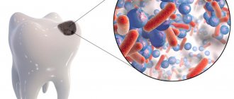
A digital radiovisiograph allows you to identify any foci of infection, the location of which is difficult to determine visually. A visiogram is done in case of possible occurrence of the following diseases:
- Caries;
- Pulpitis, which causes inflammation of the tissues surrounding the tooth;
- Small neoplasms in the oral cavity;
- Periodontitis.
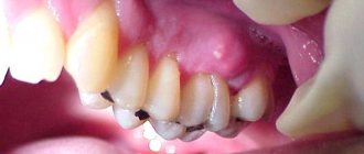
The image allows you to detect hidden foreign bodies that have entered the canal. Any foci of inflammation, granulomas and cysts are painted black, which stands out sharply against the gray background of bone and connective tissue. With the help of detailed visual images, the dentist will be able to make an accurate diagnosis and prescribe appropriate treatment.
Radiovisiography allows you to determine the depth of tissue damage and control the quality of filling.
Indications and contraindications for the procedure
List of main indications for use of the equipment:
- Prevention of pulpitis, caries, periodontitis;
- Viewing the condition of a filled tooth, decayed gums, damaged bone tissue, or installed implant.
Radiovisiographic examination allows complete control of the treatment process. It will help avoid many complications. This method is safer than x-ray examination. It does not pose a health hazard.
Radiovisiography can be performed during pregnancy and lactation.
The list of relative contraindications includes:
- Mental illnesses;
- Neurological disorders.
Dental clinic No. 2
What is a visiograph and how does it differ from an x-ray?
This one of the frequently asked questions is akin to the difference between a car and a traffic light... It seems that both concepts have some kind of connection, but it is somehow difficult to compare them. It's the same here. A radiovisiograph is a system that receives x-ray radiation, transforms it into digital form and displays the image on a computer screen. Roentgen (who is Wilhelm Conrad) is a long-dead German physicist who gained worldwide fame for his discovery of short-wavelength rays with enormous penetrating power. The physicist himself called these rays X-rays (in English today they are called exactly that - X-ray), but now we often call them X-rays, and in everyday life simply “X-rays”. The unit of radiation power was also called the x-ray. Now it is clear that a visiograph and an x-ray are completely different things. If we compare the visiograph with anything, it is with x-ray film, which it is universally replacing from all areas of medicine.
Is it true that a visiograph is safer than a regular film photograph?
When asked about such a comparison, they mean the radiation exposure that the patient receives when using different techniques. In this sense, indeed, a visiograph is preferable, since its sensor is much more sensitive than the best film. Therefore, to obtain a high-quality image using a visiograph, much shorter shutter speeds are needed. To take a picture on film, the shutter speed is 0.5-1.2 seconds. To obtain the same image using a visiograph sensor – 0.05-0.3 sec. Those. 10 times shorter. As a result, the radiation exposure received by the patient when using a visiograph is reduced to an insignificant minimum.
How many pictures can you take at one time? And in general, isn’t it harmful when treating a large number of teeth that you have to take a lot of X-rays?
This is the most pressing question asked about x-rays. Either as an echo of Chernobyl, or because of life safety lessons that come to mind, but in our society there is a very strong phobia for everything that is even remotely connected in our heads with radiation. Any extra photo often raises questions about radiation sickness, or “will I glow in the dark?” Therefore, I will try to explain in more detail here. First from the point of view of naked science.
To measure the amount of radiant energy applied to living tissue, various units are used - joule per kilogram, gray, rem, sievert, etc. In medicine, X-ray procedures typically measure the dose received by the entire body during one procedure—the effective equivalent dose, measured in sieverts. According to SanPiN 2.6.1.1192-03, when carrying out preventive medical x-ray procedures and scientific research, this dose should not exceed 1000 μSv (microsievert) per year. Moreover, here we are talking specifically about preventive studies, and not about therapeutic ones, where this bar is much higher. What is 1000 µSv? Is it a lot or a little? Remembering the famous cartoon, the answer is simple - depending on what you measure it in. 1000 μSv is approximately:
500 targeted images (2-3 μSv), obtained using a radiovisiograph 100 of the same images, but using good X-ray film (10-15 μSv) 80 digital orthopantomograms* (13-17 μSv) 40 film orthopantomograms (25-30 μSv) 20 computed tomograms* (45-60 µSv)
So, as you can see, even if we take 1 picture on a visiograph every day throughout the year, in addition to a couple of 3D computed tomograms per year, and the same number of orthopantomograms, then even in this case we will not go beyond the safe limits doses There is only one conclusion - there is no need to be afraid of receiving a significant dose during dental interventions. No matter how hard you try, it is unlikely that you will be able to go beyond the permissible values. To make it clear, below are the doses required to produce any serious health consequences: 750,000 µSv - short-term minor change in blood composition 1,000,000 µSv - mild radiation sickness 4,500,000 µSv - severe radiation sickness (50% die irradiated) a dose of about 7,000,000 μSv is considered absolutely lethal
All these figures are incomparable in their significance with the doses we receive in everyday life. So, even if, for some reason, several pictures are taken in a row at once, and the day before you were already “exposed” by doing an orthopantomogram, you don’t need to panic and run to the store to buy a Geiger counter or type “the first symptoms of radiation sickness” into an Internet search engine. . To calm yourself down, it’s better to “detoxify” with a glass of red wine. There will be no point in this, but the mood will immediately improve.
Is it possible to do x-rays for pregnant women?
I will not expand on the topic that it would be better to prepare for pregnancy in advance, including “preparing” your own teeth at the dentist in advance. Yes, so as not to run away later with acute pain and be killed by doubts whether this or that manipulation will harm the developing baby... Therefore, let’s leave the lyrics and look at the bare facts and common sense. Without phobias, prejudices, speculations and myths. So, is it possible to do x-rays for pregnant women? Here's what they write to us about this in the documents (SanPiN 2.6.1.1192-03):
7.16. Pregnant women are prescribed for X-ray examination only according to clinical indications. Studies should, if possible, be carried out in the second half of pregnancy, with the exception of cases when the issue of termination of pregnancy or the need for emergency or emergency care must be decided. If pregnancy is suspected, the question of the admissibility and necessity of an x-ray examination is decided based on the assumption that there is a pregnancy...
7.18. X-ray examinations of pregnant women are carried out using all possible means and methods of protection so that the dose received by the fetus does not exceed 1 millisievert for two months of undetected pregnancy. If the fetus receives a dose exceeding 100 mSv, the doctor is obliged to warn the patient about the possible consequences and recommend terminating the pregnancy.”
In general, the conclusion from these two main points is simple and clear. In the first half of pregnancy, it’s definitely not worth taking pictures, but in the second half - 1 mSv for a visiograph - this is practically unlimited.
I would also like to add here that I have often encountered the militant obstinacy of this opinion: an x-ray at the dentist during pregnancy is an absolute evil. It’s better, they say, to screw up a tooth, to cure crooked canals... there are a lot of teeth, pregnancy is more important. Moreover, such sermons are given not only by lay patients who have little understanding of the essence of things, but also often by dentists themselves, who have forgotten their school physics course. To resolve this doubt, we must understand that sources of ionizing radiation are not only found in medical offices. And you don’t have to live next to Chernobyl (and now Fukushima) to receive some doses from the environment around us every day. After all, every second we are affected by both natural sources (sun, water, earth) and man-made ones. And the doses received from them are much greater than those received from an x-ray of a tooth. For clarity, we can give one simple example. As you know from a school physics course, the sun emits electromagnetic energy in a wide range, not only in infrared (heat), visible (light), ultraviolet (tan), but also in x-rays and gamma radiation. Moreover, the higher you are from the surface of the earth, the more rarefied the atmosphere is and, therefore, the weaker the protection from sufficiently strong radiation from the sun. And after all, while “fighting” radiation at the dentist, the same people often calmly fly south to bask in the sun and eat fresh fruit. Moreover, during a 2-3 hour flight “for a healthy” climate, a person receives 20-30 μSv, i.e. the equivalent of approximately 10-15 images on a visiograph. In addition, 1.5-2 hours in front of a cathode ray monitor or TV gives the same dose as 1 picture... How many pregnant women, sitting at home, watching TV series, hanging out on the Internet, think about how many pictures they “took” while watched another program, and then discussed it with friends on the forum and social networks? Almost no one, because the average person does not associate all this with ionizing radiation, unlike an image in a doctor’s office.
And yet, dear expectant mothers, prepare for pregnancy in advance. For many people, visiting the dentist still remains stressful. And it’s not so much that anesthesia or x-rays can be harmful during this period, but what is important is your peace of mind and the absence of unnecessary worries (of which many already have more than enough during this period).
What is the best protection to use if you need to take a picture of a pregnant woman? Is it better if the doctor puts 2 protective aprons on me?
The number of aprons does not matter! See above. In contact radiography, the apron essentially protects not from direct radiation, but from secondary, that is, reflected. To X-rays, the human body is an optical medium, just as a glass cube is to a flashlight beam. Point the beam of a flashlight at one of the faces of a large glass cube, and, regardless of the thickness and direction of the beam, the entire cube will be illuminated. It’s the same with a person - you can swaddle him completely in lead and shine only on his head - at least a little, but it will reach every heel. So, under two aprons with a good lead equivalent, it will simply be harder for a pregnant woman to breathe.
Is it possible to do x-rays for nursing mothers? And if possible, then what about feeding the child after the procedure?
Can. X-rays are not the same as radioactive waste. By itself, it does not accumulate in the biological environment. If you give a loaf of bread a lethal dose, it will not mutate, get radiation sickness, or begin to “foul.” X-rays differ from light rays only in wavelength and have a direct damaging effect only under certain conditions. If you shine a flashlight into a bucket of water and turn off the flashlight, the light won't stay in the bucket, right? The same is true in a protein-fat solution, which are many biological fluids (including breast milk) - the radiation passes through, weakening in denser tissues. So, with such a load, which is necessary to work with a visiograph, the milk itself is unlikely to do anything. As a last resort, to reassure yourself, you can skip one regular feeding. Another thing is that the breast tissue itself during lactation is, of course, more susceptible to the harmful effects of radiation. But, again, we are talking about doses more powerful than is necessary for digital radiography (of course, subject to all protective measures and without “shooting” 20 times anywhere).
Difference from X-ray and computed tomography
Computed tomography allows you to obtain a three-dimensional image of the jaw to examine it in all planes. A visiographic device helps to examine each problematic tooth individually. A similar examination is carried out before the therapeutic procedure, and the method is also used to monitor the treatment process.
X-ray can only create flat images. Even interproximal radiography is not always able to determine the presence of tumors in the oral cavity. Using a radiovisiograph, you can see a tumor in the initial stage of its formation.
Radiovisiography is safer than radiography; the level of dangerous examination in the first case is relatively low. The signal from the visiograph passes to a special CCD sensor or microcircuit that is more sensitive than x-ray film.
Example of resulting images
Advantages and disadvantages of a visiograph
The main advantages of radiovisiography:
- compactness of the device;
- image accuracy;
- the ability to instantly view a diseased tooth on a computer screen with multiple magnification of the resulting image, storing the image on a PC and transmitting it via a local network and e-mail;
- safety of the procedure;
- speed of manipulation;
- editing a digital visiogram, using various filters to improve the visual perception of the picture.
Radiovisiography of the tooth is carried out in a fairly low spatial resolution. This is the main disadvantage of the procedure. The device's sensor does not clearly show the difference between different structures with unequal densities. But thanks to the ability to adjust the contrast and brightness of the image, details become more visible.
Despite the fact that doctors do not prohibit performing the procedure during pregnancy, the expectant mother should warn the attending physician about her situation.
What does it have to do with x-rays?
Digitized image.
Many people mistakenly believe that a visiograph is the same as an x-ray. It has nothing in common with x-rays. A visiograph is a digital equipment that does not require chemical reagents or X-ray film to obtain an image. This directly affects the exposure time, because the digital output method provides instantaneous image acquisition on a computer monitor. Therefore, a special room is not required to place the visiograph. It can be placed next to the dental unit, in close proximity. The patient does not need to leave the dental chair to take the image. Thus, the patient's appointment time is significantly reduced.