Where are the alveoli of the teeth, what do they look like in the photo, why are they needed in the mouth?
Most people associate the term “alveolus” with lung tissue, but the same term is used to refer to the sockets for teeth in the human oral cavity. How many of these holes are there in the mouth? What functions do they perform, what do they look like and where are they located? Are the alveolar sockets and processes in the jaws susceptible to pathological processes? Let's figure it out together.
What are alveoli, why are they needed in the mouth?
The roots of all 32 human dental units are located in special socket-shaped depressions called alveoli.
Their main purpose is to ensure reliable attachment of a canine, incisor, premolar or molar in the jaw.
If a pathological process develops in the alveolar socket, the tooth unit will begin to loosen and may even fall out. In addition to an aesthetic defect, such processes lead to a deterioration in diction (the patient begins to lisp), as well as problems with chewing food.
Where are the dental alveoli located and what do they look like?
Where the alveolar sockets and processes are located and what they look like can be seen in the photo. In general, a single socket looks like a hole in the jawbone. There are such depressions in both the upper and lower jaws of every person. Their formation begins during the period of intrauterine development. It occurs in parallel with the formation of the rudiments of milk and molars.
The alveolar sockets of the upper and lower jaw differ slightly from each other in structure. Each of them includes blood vessels, a nerve and a special spongy tissue (it provides the “porosity” of the structure of the human jaw element in question), as well as cells responsible for controlling the balance between the growth of bone tissue and its destruction.
The alveoli, in which multi-rooted teeth are located, differ from the sockets intended to accommodate canines and incisors. If a molar or premolar has several roots, then partitions are formed in the alveolar “socket” that separate them from each other. At the top, the alveolar septum (interdental or interradicular) is always thinner than at the bottom. The sockets of canines and premolars are deeper than those of incisors and molars.
Alveolitis and other pathological processes in the alveolar sockets
Pathological processes can develop in the alveolar sockets, the most common of which is alveolitis. This is an inflammation of the walls of the alveolar socket, which can be encountered by a person who has undergone tooth extraction.
Failure by the patient to comply with the doctor’s recommendations after extraction, along with mistakes made by the dentist during extraction, are common causes of alveolitis. Additional infection factors:
- abundant plaque on tooth enamel, which becomes a breeding ground for actively reproducing pathogenic microorganisms,
- periodontitis,
- carious lesion,
- chronic inflammatory process in the gums.
A person can suspect the development of alveolitis by the appearance of a number of characteristic symptoms. In general, they are similar to the signs of any inflammatory process - this is an increase in temperature (local or general), general weakness due to intoxication of the body. Alveolitis is also accompanied by the following symptoms:
- there is an unpleasant odor from the hole,
- lymph nodes located close to the site of localization of the inflammatory process swell,
- swelling appears, which eventually spreads to the patient’s face,
- the hole is covered with a grayish coating,
- after an operation to remove a molar or premolar (less commonly, an incisor or canine) a blood clot does not form in the wound,
- the patient feels pain at the site of inflammation (sometimes it radiates to the temple or ear).
Inflammation is not the only pathological process that can develop in the alveolar socket or process. The transformation of the alveolar processes under the influence of changing loads on the jaws continues throughout a person’s life, and can sometimes lead to their fracture. Such damage can only be repaired through surgery.
Alveolar process atrophy is a pathology that cannot be called rare, and modern dentists are able to diagnose and treat it.
There can be many reasons for atrophic changes - from osteoporosis and mechanical damage (most often due to trauma) to the long-term absence of one of the incisors, canines, molars or premolars (for example, if after extraction of a unit the patient refuses prosthetics for a long time).
Loading…
Symptoms of the disease
As a rule, symptoms of the disease appear within a few days after surgical tooth extraction. The signs of alveolitis are exceptional and cannot be confused with anything else . Blood thickens in the hole. After this, severe pain appears in the area of tooth extraction. It spreads to neighboring areas. The pain does not subside, but only intensifies.
This is followed by an increase in body temperature to 38-39 degrees, this is due to the spread of infection. A pronounced chill begins to be felt. Henna forms in the wound, which is accompanied by an unpleasant odor. The area around the hole becomes inflamed and red. In some cases, inflammation of the lymph nodes is observed.
It is worth noting that if a person has even a few of the above symptoms, then this is a good reason to consult a dentist. The earlier treatment begins, the more effective it is. If treated inappropriately, serious complications may occur.
Pathology
To consider the issue of alveoli in the oral cavity in more detail, we should also dwell on pathological processes. Among the conditions affecting the dental cells, it should be noted:
- Alveolitis.
- Atrophy of the alveolar process.
- Developmental anomalies.
- Fractures.
Inflammation of the socket, or alveolitis, most often occurs after tooth extraction, when periodontal tissue and the wall of the cavity itself are damaged. This is possible if there are difficulties with extraction, due to poor processing and infection, as well as as a result of a decrease in the patient’s general immunity. Pain appears at the site of the extracted tooth, the temperature rises, and bad breath occurs. On examination, the gums are swollen and red. Inflammation continues for 10–14 days.
Atrophy of the alveolar sockets is observed with tooth loss and untimely prosthetics, as a result of osteoporosis or osteomyelitis. The depth of the cells decreases, and their thin walls are destroyed. The process may have an abnormal shape or size due to congenital characteristics. And with injuries, a fracture of the alveoli can occur. Then there is severe pain in the jaw, bleeding from the oral cavity, the teeth have incorrect contact, and the gums and cheek on the affected side swell.
Having familiarized yourself with the basic information about the alveoli, now, probably, there is no one who, looking into his mouth, will not find the place where they are located. This is where the teeth grow from the jaw. You should always remember that for any questions of a dental nature, it is strongly recommended to consult a doctor. And in case of pathology, this is the only way out to solve the health problem.
The human lungs are a paired respiratory organ. The lungs are located in the chest cavity, adjacent to the heart on the right and left. They have the shape of a semi-cone, the base of which is located on the diaphragm, and the apex protrudes 1-3 cm above the collarbone. The right lung consists of 3, and the left lung of 2 lobes. The skeleton of the lung is formed by tree-like branching bronchi. Each lung is covered with a serous membrane - the pulmonary pleura - and lies in the pleural sac. The inner surface of the chest cavity is covered with parietal pleura. On the outside, each pleura has a layer of glandular cells that secrete pleural fluid into the pleural fissure (the space between the wall of the chest cavity and the lung). On the inner (heart) surface of the lungs there is a depression - the hilum of the lungs. They enter the bronchi, the pulmonary artery, and exit two pulmonary veins. The pulmonary artery branches parallel to the branching of the bronchi. Lung tissue consists of pyramidal lobules (25 mm long, 15 mm wide), the base of which faces the surface. The apex of the lobule includes a bronchus, which by successive division forms 18-20 terminal bronchioles. Each of the latter ends with a structural and functional element of the lungs - the acini. The acini consists of 20-50 alveolar bronchioles, divided into alveolar ducts; the walls of both are densely dotted with alveoli. Each alveolar duct passes into the terminal sections - 2 alveolar sacs. So, the lungs are paired organs located in the chest cavity. The right lung is larger in volume than the left. Each lung has an irregular cone shape, with the base pointing downward and the apex pointing upward. In the lung there are three surfaces (diaphragmatic, medial, costal) and three edges that separate these surfaces from each other - lower, anterior, internal. The hilum of the lung is located on the medial surface of the lung. Through which the bronchi, pulmonary artery and nerves enter the lung, and two pulmonary veins and lymphatic vessels exit. All these formations make up the root of the lung. Alveoli (diameter - 0.15 mm) are hemispherical protrusions and consist of connective tissue and elastic fibers, lined with thin transparent epithelium and intertwined with a network of blood capillaries. In the alveoli, gas exchange occurs between the blood and atmospheric air. In this case, oxygen and carbon dioxide pass through the process of diffusion from the red blood cell to the alveoli, overcoming the total diffusion barrier of the alveolar epithelium, basement membrane and blood capillary wall, with a total thickness of up to 0.5 μm, in 0.3 s
When you hear the word “alveolus,” the first thing that comes to mind is the structure of the lung tissue. But alveoli are not only found in the lungs. Dentists also use this term. Alveoli are the sockets in which the roots of the teeth are located. The structure of dental alveolar cells, their functions and possible pathologies will be discussed below.
Fracture treatment
The first thing to do is to put the broken section in the correct position. You absolutely cannot do this on your own. An exceptionally qualified doctor is able to perform this procedure and performs it under local anesthesia. After this, a smooth splint-brace or splint-kappa is applied. The first is used when healthy teeth are preserved next to the fracture. Fixation is recommended for a period of one to two months, depending on the severity of the fracture.
If the teeth fall into the fracture line, and the ligaments holding them in the alveolus are damaged, they are removed. In another case, the vitality of the pulp (the tissue that fills the tooth cavity) is checked. If it is dead, it undergoes endodontic therapy (“treatment inside the tooth”; usually the pulp is removed, and the vacant space is filled with filling material). If the tissues are relatively healthy, they are constantly monitored and checked for viability.
Wounds received along with a fracture of the alveolar process are treated and freed from small fragments. In some cases, stitches are required.
Particular attention is paid to children whose permanent teeth are located in follicles. First, their viability is checked: if they are dead, they are removed
Treatment can be carried out either inpatient or outpatient, depending on the severity of the injury. For about a month after damage to the upper or lower jaw, eating solid food is contraindicated. It is also necessary to carefully monitor oral hygiene.
Hygiene rules
To maintain the alveoli in good condition, it is necessary to follow some hygiene rules:
- properly balance daily nutrition;
- brush your teeth twice a day: in the morning after breakfast and before bed;
- systematically visit the dentist for preventive purposes;
- give up bad habits (alcohol, smoking, drugs);
- Wash your hands frequently and properly.
Oral hygiene will prevent the development of alveolar disease
- enriching the body with vitamins. In the summer - natural immunization in the form of vegetables and fruits; in autumn and winter - in the form of a complex of vitamins;
- any trauma to the oral cavity should be avoided;
- It is not recommended to pick your teeth with toothpicks and other devices;
- Use dental floss to remove tiny bits of food.
Oral hygiene is an integral part of human life. Hygiene measures must be observed no matter where the person is. Pay attention to your body's signals in a timely manner, and you will be healthy.
Dental alveolus and alveolar process
Dental alveolus and alveolar process. That part of the upper or lower jaw in which the teeth are strengthened is called the dental or alveolar process (processus alveolaris). It consists of two walls: the outer (buccal, or labial) and the inner (oral, or lingual), which stretch along the edge of the jaw in the form of arcs (Fig. 96).
On the upper jaw they converge behind the third large molar, and on the lower jaw they pass into the ramus of the jaw. The space between the walls of the alveolar process is divided in the transverse direction with the help of bone partitions into a number of dimples - dental sockets or alveoli, in which the roots of the teeth are located.
The bony partitions that separate the tooth sockets from each other are called interdental partitions (Fig. 97).
In addition, in the sockets of multi-rooted teeth there are also interroot septa, dividing them into a number of chambers in which the branches of the roots of these teeth are located (Fig. 98). Establishing a diagnosis
The interradicular septa are shorter than the interdental septa and extend from the bottom of the corresponding alveoli. The edges of the alveolar processes and interdental septa do not reach the neck of the tooth (cemento-enamel border) slightly. Therefore, the depth of the dental alveolus is somewhat less than the length of the root and the latter protrudes slightly from the jaw bones. Under normal conditions, this part of the tooth root is covered by the edge of the gums (Fig. 99).
Both walls of the alveolar process on the buccal and lingual sides consist of a compact bone substance that forms the cortical plate of the alveolar process. It consists of bone plates, which in some places form typical Haversian systems (Fig. 100).
The cortical plate of the alveolar process, covered by the periosteum, passes into the bone of the jaw body without a sharp boundary. The thickness of this plate is not the same in different parts of the alveolar process. It is thicker on the lingual side than on the buccal side. In the region of the edges of the alveolar process, the cortical plate continues into the wall of the dental alveolus. The thin wall of the alveoli consists of densely spaced bone plates and is penetrated by a large number of Sharpey's fibers. These fibers are a continuation of the collagen fibers of the pericementum. The wall of the dental alveolus is not continuous. It contains numerous small holes through which blood vessels and nerves penetrate into the periodontal fissure.
All spaces between the walls of the dental alveoli and the cortical plates of the alveolar process are filled with spongy bone. The interdental and interroot septa also consist of the same spongy bone. The degree of development of the spongy substance is not the same in different parts of the alveolar process. In both the upper and lower jaws there is more of it on the oral side of the alveolar process than on the vestibular side. In the area of the anterior teeth, the walls of the dental alveoli on the vestibular side are almost closely adjacent to the cortical plate of the alveolar process, and here there is very little or no cancellous bone. On the contrary, in the area of large molars, the dental alveoli are surrounded by wide layers of spongy bone.
The crossbars of the cancellous bone adjacent to the lateral walls of the alveoli are located mainly in the horizontal plane.
In the area of the bottom of the dental alveoli, they take on a more vertical arrangement, parallel to the long axis of the tooth. This arrangement of the cancellous bone struts in the circumference of the dental alveoli ensures that chewing pressure from the pericementum is transmitted not only to the wall of the dental alveolus, but also to the cortical plates of the alveolar process, or, in other words, to the entire periodontium.
The spaces between the crossbars of the spongy bone of the alveolar process and the adjacent areas of the jaws are occupied by bone marrow. In childhood and adolescence, it has the character of red bone marrow. In adults, it is gradually replaced by yellow, or fatty, marrow. Remnants of red bone marrow are retained longest in cancellous bone in the area of the 3rd molar. The conversion of red bone marrow to yellow occurs at different times in different people. Sometimes red bone marrow persists for a very long time. Thus, Meyer observed large remains of it in the alveolar process of a 70-year-old man.
How to treat
The alveoli in the mouth are located near the facial nerve, so most often the symptoms include acute pain, in most cases also involving trigger points. Pain syndrome with alveolitis is relieved with non-steroidal analgesics. Nimesulide helps best. If the pain is severe, you can inject Ketorol or Diclofenac.
Antibacterial drugs are used as etiotropic drugs. The choice should be based on whether any antibiotics have been taken in the last 2 months, as well as intolerance options.
In the initial stages, Lincomycin helps well. Dentists also prescribe Cifran OD. The standard dose is 500 mg once a day.
The course of treatment should not be less than 7 days. If these deadlines are not met, resistance to this group of antibacterial agents usually develops. For pathologies of the gastrointestinal tract, Flemoxin solutab is used.
Local treatment is performed by a dentist. At the same time, food debris and necrotic tissue, if any, are carefully removed. Then the mucosal area is treated with antibiotics and antiseptics.
If the facial nerve is damaged, physiotherapeutic treatment is prescribed. Usually this is ultrasound or laser. Warming up is not recommended.
Among folk remedies, only rinsing with chamomile infusion is allowed. An alternative is to irrigate the oral cavity using an alcohol solution of Eucalyptus tincture diluted with water.
The sockets (alveoli) in the mouth after tooth extraction should be treated as correctly as possible. After all, they are close to the ENT organs, to which inflammation spreads in case of infection. Therefore, you should not ignore your doctor’s advice on hygiene and prevention of complications.
This is important for long-term dental health.
Causes of alveolitis
Alveolar process
Fortunately, alveolitis appears quite rarely . Some factors are identified as the cause of the disease:
- recent surgical tooth extraction;
- open wound in the gum area;
- reduced immunity or weakened body;
- poor quality dental intervention;
- penetration of tartar into the wound area;
- poor oral hygiene.
Treatment is prescribed by a doctor. It is strictly forbidden to treat alveolitis on your own. Standard mouth rinse is not effective.
The disease provokes an infection that can only be overcome by antibiotics and anti-inflammatory drugs. Painful sensations before visiting the dentist can be temporarily overcome with painkillers:
"Ketanov" is a drug that has anti-inflammatory and mild antipyretic effects. The strength of the analgesic effect can be compared with morphine. Available in tablets and solution for injection. After oral administration or intramuscular infection, it begins to act within 40 minutes.
"Brustan" is a drug that has anti-inflammatory, analgesic and antipyretic effects. Available only in tablets. It differs in that it is quickly absorbed and excreted from the body.
"Nimesil" is an anti-inflammatory, analgesic and mild antipyretic drug. Available only in powder form with an orange or lemon scent. It is quickly absorbed, after 30 minutes a visible effect is already felt.
Nimesil
Pathological conditions
Among the pathologies leading to defects of the alveolar sockets
, the following can be distinguished:
- developmental defects;
- inflammatory process in the alveoli;
- injuries, fractures;
- atrophic processes in the tissues of the alveolar process.
The alveolar process may have an inferior structure from birth with deviations in shape and size, which interferes with the correct formation of dental cells. Defective sockets, in turn, interfere with the correct formation of the dentition.
Alveolitis (inflammation of the dental cell)
occurs as a result of tooth extraction, accompanied by trauma to the periodontium and the alveoli itself. The reason may be the patient’s reduced immunity, poor-quality treatment, or infection. The condition is accompanied by redness and swelling of the gums, pain, increased body temperature, and the appearance of an unpleasant odor from the mouth. Alveolitis can last 1.5-2 weeks.
Trauma to the alveoli occurs as a result of a strong blow when the wall of the tooth socket is fractured. Symptoms of this condition will be: bleeding, swelling of the gums and cheeks from the injury, severe pain, displacement of one or more teeth, and their possible loss.
Atrophy of tooth sockets
can be a consequence of osteomyelitis and osteoporosis, and can also be triggered by tooth loss and lack of timely prosthetics. The depth of the alveoli decreases, this process is accompanied by the destruction of their walls.
The alveoli in the mouth are depressions in the human jaw, which are located on the upper and lower jaw. They are located directly in the roots of the teeth. It is generally accepted that they are responsible for smiling and chewing food.
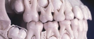
Where the alveolus is located in the mouth can be seen in the photo below. When expanded, the location of the alveoli is clearly visible. Outwardly, it looks like a kind of depression in the human jaw.
Normally, a person has 32 alveoli, 16 each in the upper and lower jaws, respectively. During the period of human life, the structure of the alveoli changes; this happens individually for each person. This usually depends on his lifestyle. The structure of the jaw plate begins in the womb.
The tooth germs are separated from the dental plate, and bone crossbars begin to build around them, which leads to the formation of the walls of the dental alveoli. The rudiments of permanent teeth are located together with milk teeth in the same alveolus.
Alveoli of teeth
The alveolar process appears only during the period of teething in infancy. Also at this time, a restructuring of the alveolar bone occurs, this is due to the fact that the eruption of teeth is accompanied by tissue changes. Next, the bone outgrowths seem to merge and form cells surrounding each tooth separately.
Alveolitis of the tooth socket
Alveolitis of the tooth socket is a dental disease, the appearance of which is caused by infection of the wound in the mouth during tooth extraction. When a tooth is surgically removed, the gums are often damaged and the tooth socket is injured. This leads to an inflammatory process. In most cases, the wound heals within one or two weeks.
However, if infection occurs, the process is delayed for a long time. To prevent the development of the disease, it is necessary to ensure that infection does not occur. To achieve this, it is highly recommended to pay more attention to oral hygiene.
What are dental alveoli and where are they located?
Alveoli in the mouth are the depressions in the jaw plates that are located on the alveolar teeth. Normally, their number corresponds to the number of teeth - 16 on each jaw. During human life, the structure and structure of the alveoli undergo individual changes associated with the natural processes of aging.
Alveola is translated as cell. The term is used in pulmonology, dentistry and other medical fields. The alveoli have a spongy structure penetrated by:
- a network of nerve endings that ensure their sensitivity;
- a network of blood vessels that supply the alveolar processes with nutrients.
The walls of the dental alveoli are divided into internal, located closer to the throat, external, located on the side of the lips, and interdental.
From the inside, the sockets are separated by thin bone partitions, the location of which depends on the structure of the roots of the teeth.
Their inside is covered with a spongy plate, the size of which exactly matches the size of the tooth located in the socket. The outer edge is covered by a cortical jaw plate.
alveoli in the mouth
Elastic fibers predominate in the composition of alveolar tissue. The main function of the remaining cells is the constant restoration and renewal of bone tissue. The balance between the processes of its destruction and growth depends on them.
Bone tissue contains both organic and inorganic particles. Its main components are:
- proteoglycans;
- osteoclasts;
- collagen;
- osteocytes;
- osteoblasts.
If a tooth falls out or is removed, the hole heals quite quickly. A few weeks after extraction, the gums heal, and after a few months, a new gum cover is completely formed.
Functions of alveolar cells
The alveoli are designed to securely attach the teeth to the jaw. Their structure ensures the stable position of the teeth and prevents them from moving and falling out.
Thanks to dental alveoli, people can chew food without the risk of incisors, canines and molars becoming loose or falling out, unable to withstand the load.
Between the sockets and teeth there are connective fibers of periodontal tissue.
Penetrating into the upper layers of the bone tissue of the tooth and the walls of the cells, periodontal tissue firmly binds them, which contributes to the correct position of the tooth in the socket.
Additionally, the periodontium acts as a shock absorber, reducing the load on the dentition and slowing down its destruction.
Formation of dental cells
alveolar ridge
The formation of alveoli begins in the perinatal period. When the tooth germs are separated from the jaw plate, bone crossbars are formed around them - the future walls of the alveoli. The cells are completely formed during the eruption of dental units.
The formation of the alveolar process occurs in infancy under the influence of the restructuring of the alveolar bone caused by tissue changes that accompany the eruption of primary incisors, canines and molars. Subsequently, the bony outgrowths fuse together and form sockets surrounding each tooth.
In adulthood, the structure of the alveoli of the teeth undergoes the opposite changes: atrophic processes are activated in the bone tissue and pulp, as a result of which the elasticity of the collagen fibers in the socket decreases.
The rate of development of atrophic processes depends on the state of the body and bone tissue, the quality of oral hygiene and nutrition.
The solution to the problem should be approached comprehensively: attention must be paid to all factors that may affect the condition of the cells. Dentists evaluate the condition of the alveolar sockets based on the clarity of diction and how firmly the teeth stand in the dentition
Dentists evaluate the condition of the alveolar sockets based on the clarity of diction and how firmly the teeth stand in the dentition.
What are the features of the upper alveoli of the teeth?
alveolitis
Regardless of which jaw the alveoli are located on, there are no significant differences in their structure. The only peculiarity of the upper alveoli of the teeth is that their structure affects diction and intelligibility of speech, which is due to the close location of the alveolar process and the palate.
The alveoli are susceptible to a number of dental diseases, the most dangerous of which is alveolitis. The disease can cause relaxation of the alveolar tissue, which can cause the tooth to shift, become loose, or even fall out. If you suspect that your teeth have begun to shift, you should immediately contact the dentist.
Alveolus in dentistry
The recess in the jaw in which the tooth root is located is what the alveolus is. Its wall is formed by a compact substance that looks like a plate. It contains osteocytes, as well as salts of calcium, phosphorus, zinc and fluorine, therefore it is quite hard and durable. The plate is attached to the bone beams of the jaw and has periodontal cords in the form. It is also abundantly supplied with blood and is intertwined with nerve endings. After tooth extraction, a strongly protruding wall of the outer part of the socket and bone septum remains. The alveoli of the teeth heal within 3-5 months by first forming granulation tissue, which is replaced by osteoid tissue, and then by mature bone tissue of the jaw.
The alveoli in the mouth are depressions in the human jaw, which are located on the upper and lower jaw. They are located directly in the roots of the teeth. It is generally accepted that they are responsible for smiling and chewing food.
Where the alveolus is located in the mouth can be seen in the photo below. When expanded, the location of the alveoli is clearly visible. Outwardly, it looks like a kind of depression in the human jaw.
Normally, a person has 32 alveoli, 16 each in the upper and lower jaws, respectively. During the period of human life, the structure of the alveoli changes; this happens individually for each person. This usually depends on his lifestyle. The structure of the jaw plate begins in the womb.
The tooth germs are separated from the dental plate, and bone crossbars begin to build around them, which leads to the formation of the walls of the dental alveoli. The rudiments of permanent teeth are located together with milk teeth in the same alveolus.
Alveoli of teeth
The alveolar process appears only during the period of teething in infancy. Also at this time, a restructuring of the alveolar bone occurs, this is due to the fact that the eruption of teeth is accompanied by tissue changes. Next, the bone outgrowths seem to merge and form cells surrounding each tooth separately.