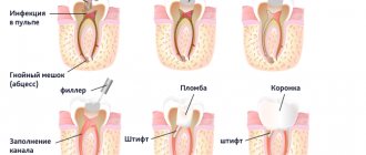
EDI for pulpitis
When the pulp is inflamed, the readings range from 20 to 100 microns.
- acute focal 20-25 microns (this means that the pathology has not yet affected the root part and develops in the coronal part)
- acute diffuse 20-50 µA
- chronic fibrous 30-40 microns
- chronic gangrenous 60-100 mA
In addition to caries and its complications such as pulpitis and periodontitis, this type of diagnosis is also used for other conditions. For example:
Equipment for EDI
Electroodontometry is a popular and informative method for obtaining information about the condition of the soft tissues of teeth. The doctor evaluates the current strength at which the tooth responds to the procedure. The research uses modern foreign and domestic devices that allow diagnostics to be made with high accuracy. Among the imported devices, Vitapulp, Gentle Plus, Pulptester are often used, but it is worth considering that on the models the scale is presented not in μA value, but in conventional units.
The following models of domestic devices are used: EOM-1 and 3, OD-2, IVN-01, Analytic. OD-2M is a modernized device that makes it possible to use both alternating and direct current. It is inconvenient for a doctor to work with EOM-3 independently, so the help of an assistant is required.
To obtain results, the patient first takes an X-ray, which helps the doctor determine which areas need to be examined. Diagnosis of EDI is not very informative in the following cases:
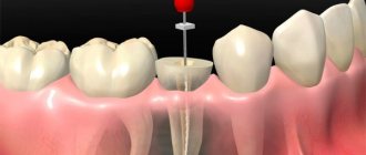
EDI technique
Before performing the EDI procedure, the dentist needs to prepare the device - turn it on and check the operation of the signal light. If at this stage the doctor does not encounter any difficulties, he begins to prepare the patient. He needs to be seated in a chair and a rubberized mat placed at his feet. Next, the dentist begins performing diagnostics.
Carrying out EDI includes the following stages:
- Drying the tooth with cotton wool or a cotton swab. It is strictly prohibited to use alcohol or ether for these purposes.
- Check for plaque or caries. If there is a problem, the dentist needs to get rid of it and thoroughly dry the mouth before the procedure.
- Placement of electrodes in the correct places. The active electrode is installed on the teeth, the passive electrode is installed on the back of the hand.
- Current supply. At this stage, the patient may feel a burning sensation or excess heat. In any case, he should warn his doctor every time about strange sensations. When discomfort occurs, the nurse should record the threshold electrical current.
During the examination, the doctor must ensure that the active electrode does not touch the gums and mucous membrane of the oral cavity, and also dry the enamel from time to time so that it does not become wet. The electrical excitability of one tooth is checked twice, and at the end the dentist makes a conclusion based on the average value.
- patients with artificial teeth;
- patients with pacemakers;
- patients with amalgam fillings;
- children under 5 years old;
- patients with intolerance to electric current.
Technique of the procedure
- The patient is seated and a rubber mat is placed under his and the doctor’s feet.
- The tooth being diagnosed is isolated from saliva and dried with cotton wool.
- The active electrode is placed on the teeth, the passive electrode is given to the patient’s hand or fixed on the back of the hand, depending on the model of the device.
- A current is supplied, during which the patient may feel warmth, a slight burning sensation, or a jolt. He immediately notifies the doctor about all his reactions by sound or gesture (raising his hand, for example).
It is very important that during EDI the active electrode does not touch the gums and mucous membrane of the oral cavity, and the enamel does not become wet; for this, it is periodically dried.
- EOM-1 and 3;
- OD-2;
- OD-2M is a modernized odontometer that allows the use of alternating and direct current from the city network.
Equipment for EDI
Electroodontometry is a popular and informative method for obtaining information about the condition of the soft tissues of teeth. The doctor evaluates the current strength at which the tooth responds to the procedure. The research uses modern foreign and domestic devices that allow diagnostics to be made with high accuracy.
The following models of domestic devices are used: EOM-1 and 3, OD-2, IVN-01, Analytic. OD-2M is a modernized device that makes it possible to use both alternating and direct current. It is inconvenient for a doctor to work with EOM-3 independently, so the help of an assistant is required.
Diagnostics in dentistry is carried out to identify pathological changes in tissues. It competes with radiography and checking the condition of teeth using a laser, but the first method does not always have the desired effect, and transillumination is applicable exclusively on the front teeth. Both methods help to detect the problem, and electroodontodiagnosis provides information about its nature.
To obtain results, the patient first takes an X-ray, which helps the doctor determine which areas need to be examined. Diagnosis of EDI is not very informative in the following cases:
- trigeminal neuralgia;
- osteomyelitis of the jaw;
- neuritis of the facial nerve;
- in case of a fracture of the lower jaw, if the fragments have shifted.
During one study, it is not advisable to check more than 3-4 teeth in a row affected by pulpitis and deep caries. The body adapts to the action of the current, and inhibitory processes develop in the medulla oblongata. Oral sensitivity returns to normal after about 60 minutes.
To avoid cross-infection, the mouthpiece and active electrode are sterilized and disinfected before each patient. Other surfaces require regular disinfection, but sterilization is not required. The device charges the battery or connects it to the network. The doctor selects the angle of attachment of the active electrode and inserts it into the desired socket on the control unit, then the device is turned on and configured. It is advisable not to twist the wires of the device.
Electroodontometry
Electroodontometry (EOM) is a diagnostic analysis method that allows you to assess the condition of the dental pulp through the conduction of electric current. The use of this method is safe for patients of different ages with contraindications to X-rays and CT. Depending on the functional and morphological state of the pulp, its nerve endings have varying degrees of excitability. When exposed to electric current, the patient may feel mild pain in the form of tingling or burning. A decrease in the sensitivity of nerve tissue may indicate that pathological changes are occurring. The purpose of the study is to determine the possibility of curing the tooth.
Electroodontodiagnostics in modern dentistry
Electroodontodiagnosis (EDD) is a method of dental research based on determining the threshold excitation of pain and tactile receptors in the dental pulp when an electric current passes through it. The process of studying the electrical excitability of teeth is called electroodontometry (EOM). The current generated by EDI devices and used for EOM is called diagnostic current. It should be emphasized that EDI gives an idea not so much about the state of the dental pulp itself, but rather characterizes the integrity and functionality of its sensitive nervous system. As is known, during various pathological processes in the hard tissues and pulp of the tooth, not only the histological structure and hemodynamic processes in the pulp change, but also degenerative processes occur in the nerve receptors, which is manifested by a change in their electrical excitability. At the same time, it must be remembered that changes in EOM indicators can occur under various pathological conditions of the periodontal tissues and sensory nerves of the maxillofacial region.
Normally, the dental pulp reacts to the electric current passing through it with minor pain, a tingling sensation, a feeling of a slight jolt, a weak electric shock, etc. The high sensitivity of the pulp to the action of irritants is explained by the large number of sensory nerve endings located in the subodontoblastic nerve plexus of Rashkov, the odontoblastic layer, and predentin.
Dental caries , as the process progresses and the carious cavity deepens, causes the development of changes in the pulp, leading to a decrease in the sensitivity of nerve receptors: deposition of replacement dentin, changes in the odontoblast layer, initial degenerative processes in the nerve elements. The listed phenomena can gradually lead to a slight decrease in EOM indicators.
Acute forms of pulpitis are accompanied by severe pain, however, EOM indicators, as a rule, decrease slightly, and sometimes remain at the level of the physiological norm. This is due to the fact that the sensitivity of nerve receptors is influenced, first of all, by the duration of the pathological process and the degree of dystrophic changes in the dental pulp, and not by the severity of inflammatory phenomena. As is known, in acute forms of pulpitis, significant degenerative processes in the nerve elements of the pulp do not occur due to the transience of the process. At the same time, a significant decrease in the electrical excitability of the pulp and the absence of positive dynamics in EOM indicators during therapy (for example, with a biological treatment method) indicate the irreversibility of the pathological process and the ineffectiveness of the ongoing therapeutic measures, which is an indication for the use of extirpative treatment methods.
Chronic forms of pulpitis occur with irreversible atrophy of the cellular elements of the pulp, its replacement with coarse fibrous connective tissue, progressive dystrophic changes in the nerve fibers, and a change in the excitability threshold of the nerve receptors of the pulp. This leads to a significant, 5-6 times, increase in EOM indicators compared to the physiological norm. The decrease in electrical excitability of the pulp is even more pronounced with the death of its coronal part - chronic gangrenous pulpitis. It should be emphasized that during exacerbations of chronic forms of pulpitis, EOM indicators do not change, remaining at a level corresponding to the degree of dystrophic changes in the nervous apparatus of the pulp. EOM also provides valuable diagnostic information for “residual” pulpitis.
Periodontitis is characterized by total necrosis of the dental pulp. In this case, the sensitive nerve endings of the periodontium react to irritation by electric current. The diagnostic current values increase significantly - usually more than 10 times compared to the physiological norm. Often, the subjective sensations of patients during EOM also change - tactile sensations predominate: blow, push, etc. In some cases, there is no reaction to the diagnostic current at all.
Non-carious lesions of hard dental tissues , if there are no secondary inflammatory-dystrophic processes in the pulp, are accompanied by only minor changes in electrical excitability. With pathological abrasion of hard dental tissues, even with significant loss of enamel and dentin (III degree according to Bracco), EOM indicators increase only 1.5–2 times, and in the initial stages of the disease remain within normal limits, which is evidence of the absence of serious pathological processes in pulp and is associated mainly with changes in the electrical conductivity of hard tissues. With wedge-shaped tooth defects, EOM indicators increase 2–3 times compared to the norm, which is quite understandable from the point of view of the dynamics and severity of dystrophic processes occurring in the dental pulp in this form of pathology. With hyperesthesia of the hard tissues of the teeth, the electrical excitability of the pulp is either within the physiological norm, or, in severe forms of this pathology, slightly increases.
Periodontal diseases may be accompanied by secondary degenerative processes in the dental pulp. At the same time, there was no unidirectional trend in changes in EOM indicators: the electrical excitability of the dental pulp in this category of patients can be either increased or decreased or be within the physiological norm. In this regard, in periodontal patients, EDI has a relatively low diagnostic value and is used only to identify possible complications, such as retrograde pulpitis.
Traumatic, inflammatory and oncological processes in the maxillofacial region quite often occur with damage to the nerves that provide sensitive innervation. In this case, the EOM indicators of one or several adjacent teeth may change. Such manifestations are most typical for traumatic injuries of teeth and jaws, diffuse inflammatory processes (sinusitis, osteomyelitis of the jaw), tumors, neuritis of the branches of the trigeminal nerve, side effects of radiation therapy for diseases of the maxillofacial area. At the same time, it was noted that in pain syndromes of central origin, for example, in trigeminal neuralgia, changes in the electrical excitability of dental pulp receptors do not occur, which is an important diagnostic criterion.
Orthodontic treatment is associated with fairly large non-physiological forces acting on the teeth for a long time. It is believed that a decrease in the electrical excitability of a tooth during the active stage of orthodontic treatment indicates excessive load and the danger of developing dystrophic changes in the dental pulp, up to its death.
Temporary (baby) teeth are not recommended to be examined using EOM. This is due, first of all, to the difficulty of adequate contact between a doctor and a child patient, the difficulty of obtaining reliable “feedback” when taking measurements and, as a consequence, the low degree of reliability of the results obtained.
Teeth that are in the stage of root formation , due to the structural features and development of the sensory apparatus of the pulp, also pose a significant problem for EOM research. During the initial period of tooth eruption, electrical excitability is absent or sharply reduced. As the tooth develops, electrical excitability increases and reaches normal only by the time the roots are fully formed. In this regard, in this category of patients, the indicators of electrical excitability of teeth that are at the same stage of root formation as that of the tooth under study are taken as the physiological norm. There is also evidence that the above-described pattern of changes in EOM indicators makes it possible to monitor the dynamics of tooth development from the moment of its eruption to the complete formation of roots.
One of the most modern, functional, ergonomic and informative devices for EDI, in our opinion, is the PulpEst device, developed and manufactured by Geosoft-Dent. This device generates a pulsed diagnostic current with the following characteristics: frequency – 3 pulses/s; amplitude – from 0 to 180 V.
EDI is one of the most reliable clinical research methods, allowing the dentist to obtain important diagnostic information about the condition of the dental pulp in the absence of pronounced clinical and radiological symptoms, to monitor the dynamics of the pathological process, and to evaluate the effectiveness of the treatment. There is also no doubt that the value of this method is often overestimated, which leads to annoying diagnostic errors.
This is especially evident when specific digital values of EOM are strictly associated with specific dental diagnoses. With a competent and scientifically based approach, combined with technologically correct implementation and adequate logistical support, EDI research can take its rightful place in dental practice, becoming an invaluable aid in difficult clinical situations.
When is an electroodontometric study necessary?
Electroodontometry in dentistry is prescribed in the following cases:
- differential diagnosis of caries depth;
- diagnosis of pulpitis;
- identification of tumors on the roots of the tooth;
- injury to the maxillofacial region;
- inflammatory process of the maxillary sinus;
- inflammatory destructive bone disease;
- chronic focal or hematogenous anaerobic infection;
- jaw tumors of various origins;
- inflammation and damage to peripheral nerves;
- radiation damage to tissues;
- treatment with orthodontic appliances.
Reasons for getting incorrect results
When EDI is used in dentistry, the readings may not always be correct. False positive reactions are possible if:
- There is contact between the electrode and a metal part, for example, a bridge or filling.
- If the patient is not explained in detail what to expect and how to proceed, he may raise his hand prematurely.
- Poorly treated pulp necrosis.
- There is no good isolation from saliva.
In some cases, it is possible to obtain false negative results:
- Before the procedure, the patient drank alcoholic beverages and took sedatives and painkillers.
- During preparation, the nurse made poor contact between the electrode and the tooth enamel.
- The patient recently suffered a dental injury.
- The device is not plugged in or the batteries are dead.
- The tooth has recently erupted and the apex has not fully formed.
- Incomplete pulp necrosis.
- The electrical circuit breaks because the doctor is wearing rubber gloves.
Electroondometric diagnostic procedure
Before the procedure, the doctor must explain to the patient the essence of the method and the purpose of its use, warn about possible discomfort and describe the technique. The patient must understand what actions he needs to perform when a pain signal occurs. An important condition for using the device is its disinfection and sterilization.
Electroondometry stages:
- Preliminary cleaning of teeth from plaque and stones to obtain a more accurate result.
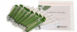
- Attaching a neutral electrode to the patient. It may take the form of a part of a diving apparatus hose. The electrode does not touch the area being analyzed.
- Drying the tooth with cotton swabs and separating it from saliva with napkins or gauze.
- Applying a conductor gel to the active electrode for better contact with tooth enamel. A cap can act as a conductor, which must first be moistened.
- Place the electrode on sensitive teeth and turn on the current supply. Monitoring by the doctor to ensure that the mucous membrane does not come into contact with the active electrode.
- If pain occurs, the patient gives a hand signal to the dentist. At this moment, the current supply stops, and the result obtained is recorded.
- Repeat the procedure to obtain the most accurate data.
Diagnosis of caries in children
Female Dentist, vector illustration
The dentist can prescribe any method for diagnosing dental caries for a child. But recently, private clinics have given preference to the special device “Diagnodent”.
The device consists of two small displays with a control panel. The essence of the device is that laser beams are reflected from healthy and diseased teeth differently. Any deviation from the norm is recorded by the device, and the doctor draws conclusions about the presence and degree of development of the disease.
The advantages of diagnostics with this device cannot be overestimated:
- Caries is detected at an early stage of development, and the device shows even minimal damage to the enamel.
- Caries is visible on the adjacent areas of the teeth, as well as at the root of the tooth and under fillings.
- Diagnostics using the device is absolutely harmless and painless. That is why it is recommended for children and pregnant women.
Prevention of caries
Regular visits to the dentist's office will help avoid many dental problems. But besides this, we should not forget about preventive measures. First of all, caries prevention involves strengthening tooth enamel with minerals.
To keep your tooth enamel strong and healthy, you need to watch your diet and take vitamins. You need to eat foods that benefit the body in general and teeth in particular. At the same time, you need to avoid harmful foods. In addition, monitor oral hygiene and use special toothpastes with a whole range of minerals and trace elements.
Pros and cons of using the apical patency technique (“Patency”)
Arguments in favor of creating apical patency (“Patency”)
If you do not push a significant amount of infected dentin beyond the apex, then, of course, you can get a positive effect, since this technique:
- Provides significantly more accurate root canal length measurements using apex locators.
- Probably, it makes it possible to carry out better disinfection in the periapical area (this statement remains in doubt).
The following question has not yet been finally resolved. Is it possible, by carefully “opening” the apical foramen and “leaving it open” for the duration of the medicated dressing in the root canal, to speed up the process of tissue healing, as well as improve the effect of the medicated dressing on the tissue of the periapical region (provided there is a sealed temporary filling in the coronal region) ? Then it is possible (!) that with further permanent filling of the root canal, the filling will fit more tightly in the apical area, since the likelihood of apical microleakage (“Leakage”) will decrease. All these assumptions are currently only theoretical. No serious research has been conducted in this area (yet).
Counterarguments
This procedure can be compared to walking on a steel cable over an abyss (most likely more (?) or perhaps less bacteria will penetrate into the periapical region). Prof. Hülsman believes that when treating teeth with viable pulp (vital extirpation), such an “apical patency” technique is more likely to cause harm. Teeth with viable pulp have a “natural apical patency” (“Patency”). A “living” tooth always (!) for physiological reasons has a largely open apical foramen. Prof. Hülsman emphasized that the technique of creating apical patency (“Patency”) makes sense only when treating teeth with non-viable (avital) pulp and if there are changes in the apical periodontal area on the initial radiograph.
The advantages of universal removal of tools beyond the root apex are currently more likely to resemble theoretical conclusions. At the end of his report, Prof. Hülsman asked the following question: “Is it advisable to use this technique?” At present it is not entirely clear, but this topic is the subject of very heated debate among German specialists in the field of endodontics. Discussions at the end of the conference of the German Society of Certified Dental Endodontists (VDZE) confirmed this.
Discussions
The first dentist to speak during the discussion noted that many teachers at German universities, as before, are very critical of the deliberate removal of instruments beyond the apical foramen. Prof. Hülsman agreed with him, and also noted that the purpose of such extension beyond the apex is often only to achieve “patency” of the root canal during its development and nothing more... Beyond the apical narrowing, as was previously accepted, nothing is prepared. This also applies to cases with extensive changes in the apical periodontal area...
Another dentist who spoke, on the contrary, proposed using irrigation solutions, or rather sodium hypochlorite solutions, to penetrate far into the periapical region (!). In his opinion, it is highly advisable to carefully “soak” this periapical area with sodium hypochlorite solution to kill bacteria.
The dentist from Bad Homburg, Dr. Oliver Pontius, then added that cleaning in the apical area is still more effective after using the apical patency technique (“Patency”). As a result of such manipulations, the branches of the root canal in the area of the “apical delta” with all the complexities of their anatomical structure become at least slightly more accessible. Oliver Pontius considers this another additional benefit of using Patency, in addition to obtaining more accurate electrometric results. In all likelihood, Dr. Pontius is one of the proponents of the new technique for creating apical patency.
Prof. Huelsman noted after Dr. Pontius' presentation that when measuring the working length of the root canal using electronic instruments, despite the use of an apical patency technique, deviations in the results (by approximately 0.5 mm) can still be observed. Argument regarding better cleaning of the root canal in the apical region by Prof. Hülsman questioned it. He believes that good manual skills and technique can prevent the apical region of the root canal from becoming blocked by too much dentin debris and pulp debris. This can be achieved by frequently changing or cleaning instruments, as well as repeated and copious rinsing of the root canal with irrigating solutions.
High-quality and thorough mechanical preparation of root canals, as well as irrigation only within the root canal (i.e. up to the apical constriction) as an (old) solution to the problem? To this, Dr. Pontius replied that he still believed that the technique of using extreme mechanical debridement of root canals (as in the past...) using very large diameter instruments (ISO 100 and more) had already become obsolete. It no longer makes sense to use it at this time.