The purpose and essence of the method
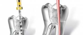
Retrograde filling is an alternative to the conventional method of root canal treatment. The purpose of retrograde filling is to prevent pathogenic microflora from entering through the dental root canals into the peri-apical area (periodontal, alveolar bone). This technique ensures high-quality closure of the apical zone.
The essence of the method is apiectomy, i.e. surgical resection of the tooth root in the apical part. The canal prepared in this way is processed and then closed with filling materials.
Retrograde canal filling - what is it?
The operation is performed to prevent pathogenic microflora from entering the periapical area.
Essentially, this is an alternative to traditional treatment, but through access to the root system in the gum. The technique is based on resection of the upper part of the root followed by filling. There may or may not be inflammation or destruction in this area. In the first case, patients complain of pain, swelling of the gums, and the presence of fistulas. In the second case, the problem is more often detected by chance using an x-ray, which is taken to examine adjacent dental units.
Indications for use
The retrograde filling technique is used if the patient has the following indications:
- Impossibility of secondary treatment due to poor quality of manipulations for filling canals with cement compounds, pastes or similar materials.
- The root canal is not obturated tightly enough , and there are foreign bodies in the canal, namely fragments of endodontic instruments, anchor pins, stump inlays.
- Irreversible processes of overgrowing of root canals of various etiologies - the presence of an inflammatory process up to oncological tumors, pathological age-related changes, the result of improper treatment.
- Root canals are abnormally long or have excessive curvature, making endodontic intervention ineffective or impossible.
- Channel roots have apical deltas or expand into a funnel shape.
- The teeth have protective crowns (metal-ceramics, porcelain, etc.) or permanent structures are installed, while the previously given indications are available in various combinations.
The presence of a fistula - a tooth canal - is one of the main factors in preparing the gums and performing filling procedures through the apex of the tooth.
The surgical procedure is used in the presence of destructive changes in the peri-apical tissues of the teeth.
In this case, patients exhibit the following symptoms:
- swelling of the gums and inflammation;
- canal fistulas are formed;
- the presence of pain of varying intensity.
Patients seek help from dentists when their symptoms worsen. In some cases, the periosteum is opened, followed by medication therapy . Inflammatory processes are stopped with the help of antibiotics.
In cases where the patient does not have pronounced symptoms of destructive changes, but is still concerned about the condition of the teeth under the crowns, it is necessary to take a panoramic radiograph to identify problems with canal filling.
Composition and properties of Filtek filling material, indications for use.
Read here about the rules for using glass ionomer cement for fillings.
At this address https://www.vash-dentist.ru/lechenie/zubyi/plombyi/idealnyiy-variant-spektrum.html you will find expert reviews about the Spectrum filling.
Materials used
The traditional retrograde filling material is amalgam. The advantages of amalgam include good shape retention and low shrinkage.
The use of amalgam is associated with a number of difficulties:
- the technology requires thorough drying of the cavity surface before filling with material;
- excess amalgam getting on the tissue leads to darkening of the gums, which is undesirable in the smile area;
- The technique is quite labor-intensive.
Regular development of innovative dental materials for filling allows manipulations to be performed with less time and good quality.
COE
Zinc oxide eugenol cements (ZOE) are made from eugenol and zinc oxide mixed in certain proportions. This composition hardens in 10-12 hours.
The positive properties of COE include the following characteristics:
- inertness to dental tissues, have good biological compatibility;
- work as additional antiseptics;
- thermal conductivity is low,
- do not affect the change in color of hard tooth tissues.
In terms of their manipulation and physico-chemical properties, COEs are inferior to varieties of other dental cements.
This type of cement is used for the following purposes:
- installation of temporary fillings;
- performing manipulations for intermediate dental restoration;
- fixation of therapeutic inserts required in the treatment of deep carious cavities;
- installation of crowns, both permanent and temporary;
- securing gaskets to provide thermal insulation;
- sealing the root canals of the anterior teeth;
- fixation of gingival dressings with medications.
The most popular COEs are represented by the following names: “Cariosan” (DentalSpofa), “Cavitec” (Kerr).
COEs containing eugenol inhibit the polymerization reaction, so combined use with composite materials is undesirable.
Intermediate restoration pastes
This material makes it possible to perform manipulations in periapical tissues. The results of using these pastes make it possible to fill a dental cavity more reliably in comparison with the traditional material - amalgam.
The material used in modern dentistry for filling root canals, the procedure technique.
In this publication we offer reviews from experts about cement fillings on teeth.
Follow the link https://www.vash-dentist.ru/lechenie/zubyi/kofferdam-v-stomatologii.html to become more familiar with the types of rubber dam used in dentistry.
Super-EBA
An improved version of COE is Super EBA cement. The components of the material are:
- liquid fraction : a) eugenol – 37.5%; b) ethoxybenzene acid – 62.5%;
- powdery fraction : a) zinc oxide – 60%; b) aluminum oxide – 30%; c) natural polymer – more than 6%.
The practical use of Super EVA cement gives good results (more than 96% of patients experienced healing after a year).
Super EVA cement occupies an intermediate position in quality between COE and glass ionomer materials.
The preparation of a working cement mixture is done as follows:
- A small amount of the liquid fraction is applied to the surface of the glass and generously sprinkled with a powdery composition. The result of mixing should be a homogeneous shiny mass with good adhesion to the glass surface.
- The resulting mass is carefully rolled into a cone approximately 1 mm thick and up to 3 mm long.
- Filling is performed in portions. To speed up the hardening of the cement mass, it is recommended to cover the finished filling with a hot, damp cotton swab.
- Excess cement is removed with a round plugger, a curette, then everything is polished with a diamond-tipped bur.
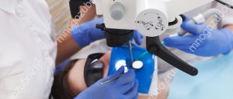
Intermediate and final results of the work are examined under microscopic magnification.
Glass ionomer cements
Modern filling materials are represented by glass ionomer cements, which receive excellent reviews from dentists.
Glass ionomer cements (GIC) have a heterogeneous structure, which is filled with aluminosilicate glass . Polyacrylic acid serves as a framework for the matrix.
In practice, GICs are used as materials for restoring and filling teeth, and also as insulating liners.
In addition, polyacrylic cement may contain polyitaconic, polymylenic acid. The following components can be used as a catalyst:
- hydroxyethyl methacrylate;
- wine acid;
- camphorquinone.
The powder fraction is composed of the following substances:
- the addition of quartz gives greater transparency , lengthens the setting time (the dentist has a reserve of time when working), and the strength characteristics of the cement are somewhat reduced;
- aluminum oxide reduces working time , gives greater strength, acid resistance and opacity;
- calcium fluoride increases strength , imparts color rendition and resistance to the effects of carious processes;
- Aluminum phosphates make it possible to select color shades , stabilize the structure, and increase strength;
- barium salts and inclusions of other metals allow radiography to be performed with greater contrast.
The advantages of glass ionomer cements are the following:
- practicality of use, the composition fills prepared cavities in 1-2 stages of application, while dentin and enamel do not require an etching procedure;
- performance of the material in humid environments;
- provides excellent adhesion along the edges of the wall being filled;
- excellent adhesive properties with metals, clove oil;
- the ability to release fluoride ions to dental tissues after filling throughout the year;
- has low shrinkage values;
- inertness towards dental tissues;
- thermal expansion matches the natural values of natural teeth.
It is impossible not to mention the presence of such disadvantages of the GIC:
- the cement mass hardens to normal limits within a day;
- the material is hygroscopic during the first day; ions may be washed out or released insufficiently;
- It is not advisable to treat fresh fillings with burs; vibration destroys the integrity of the composition;
- low wear resistance of fillings;
- low aesthetic component;
Among GIC, the most popular is Vitremer, VOCO, which has a triple degree of curing, while being easy to use and aesthetically pleasing.
Review of popular formulations
The list of filling materials is very large. They differ in their composition, strength and application technique. Let's look at the most popular ones.
This is interesting: Treatment and causes of bitterness in the mouth in the morning, after eating
COE
Refers to plastic non-hardening materials. Available in the form of an antiseptic paste, the composition includes zinc oxide and eugenyl liquid.
Has a number of disadvantages:
- do not harden;
- tend to gradually dissolve and be washed out from the upper part of the tooth root;
- are not used to restore the roots of permanent teeth.
Most often, this material is used in the treatment of baby teeth in children. COEs also have their advantages: they are easily removed from the root canal (if necessary), easily installed and are characterized by sufficient viability.
Restoration paste
Compared to the use of amagalma, this mass allows for better sealing of the tooth root. It is often used to treat periapical areas.
Super-EBA
The composition of this filling material contains zinc and aluminum oxide, and a small percentage of natural resins is also present.
This composition provides reliable protection against pathogenic organisms that have not shown their activity at the time of the procedure.
Glass ionomer cements
They are characterized by a high level of sealing and edge adaptation; reflective types of material are most often used. Used for filling the apical area.
Among other advantages it is worth highlighting:
- high adhesion to other materials used during the procedure;
- high biological compatibility with dental tissues;
- protects against violation of the marginal seal of installed fillings;
- long service life.
The choice of filling material is made by the dentist based on the problem of a particular patient.
Trioxidant
Material stored or transported at low temperatures must be kept at room temperature for at least 1 hour before use.
The dental material Trioxidant is mixed at room temperature 18-23°C and humidity 50±10% on a dry glass plate with a dry, clean spatula in a powder/distilled water ratio of 3:1.
To do this, a dose of powder (0.3 g) must be mixed with 3 drops (0.10-0.11 g) of distilled water, a dose of powder (0.5 g) must be mixed with 4 drops (0.17- 0.18 g) distilled water. Mix for 30-40 seconds until a plastic paste is obtained, which is placed in the defect area using special tools from the kit. If plasticity is lost (after 10-15 minutes), a small amount of distilled water can be added to the paste once and mixed.
To restore perforation after resorption, it is necessary to prepare the canals. The channels, cleared of sawdust and half-life products, treated with sodium hypochlorite (Belodez 3%) and washed with water, are dried using paper points. Then the root canal defect zone is established and all canals in the apical zone from the established defect zone are obturated.
The trioxidant is placed in the defect area and compacted. The material can be condensed using a large ultrasonic nozzle without spraying water, at medium power.
Using the radiograph, you must ensure that you have placed the Trioxidant material correctly. Then the remaining part of the canals is obturated, isolated with lining material, and the tooth crown is restored.
To restore perforation of the lateral root canals, it is necessary to prepare the canals. The channels, cleared of sawdust and half-life products, treated with sodium hylochlorite (Belodez 3%) and washed with water, are dried using paper points. Then the site of perforation is isolated and all canals located apically from the perforation are obturated.
The trioxidant is placed into the defect area and compacted using a small amalgam plunger and a cotton swab or paper points. The material can be condensed using a large ultrasonic nozzle without spraying water, at medium power.
Using the radiograph, you need to make sure that you have placed the material correctly. Then the remaining part of the canals is obturated, isolated with lining material, and the tooth crown is restored.
To apexify the root, it is necessary to prepare the canals. The channels, cleared of sawdust and half-life products, treated with sodium hypochlorite (“Belodez 3%”) and washed with water, are dried using paper points. Then, for disinfection, a paste based on calcium hydroxide (Apexdent without iodoform) is placed in the canal for a week.
After a week, the paste is removed from the root canal system using root canal instruments and irrigating the canal with sodium hypochlorite solution. The canal is dried with paper points.
The trioxidant is placed in the apex and compacted using a small amalgam plunger and a cotton swab or paper points. The material can be condensed using a large ultrasonic nozzle without spraying water, at medium power.
This is interesting: The main causes of the problem of black teeth in children - why a child’s enamel turns black
Using an x-ray, you need to ensure that you have correctly placed the material that is to remain as a permanent root canal filling. Then the remaining part of the canals is obturated, isolated with lining material and the crown is restored.
For retrograde filling of the root apex, under local anesthesia, access to the root apex is provided (the mucoperiosteal flap is peeled off), resection of the root apex is performed, and a cavity for retrograde filling is formed using an ultrasonic tip with special diamond tips.
After ensuring hemostasis, the cavity in the tooth root is filled with the resulting Trioxident paste using instruments and plastic attachments. The bone defect is replaced with osteoplastic material, the flap is placed in place and fixed.
To cover the pulp, the cavity is prepared using burs at high speed with constant irrigation with water. In the case of caries, carious dentin is removed using a round bur in the handpiece at low speed or using hand instruments. The prepared cavity is washed with sodium hypochlorite solution. bleeding is stopped with a cotton swab soaked in hemostatic fluid (Capramin).
A small amount of Trioxydent is then applied to the exposed area using a small applicator with a ball at the end. Excess moisture in the work area is removed using a moistened cotton swab. A small amount of flowable compomer liner material or glass ionomer light-curing liner material is then applied and cured.
The remaining surfaces of the cavity are treated with dentin etching gel for 15 seconds and washed thoroughly. Then the cavity is carefully dried, leaving the dentin slightly damp, but not wet, the adhesive is applied and polymerized, after which the restoration is completed.
Diagnosis and preparation for surgery
Retrograde filling requires diagnosing the indications for such a microsurgical operation. After the examination, an x-ray of individual teeth or a panoramic photograph is required.
If the patient has anomalies in the development of the root canals (greater curvature, length), then the optimal solution would be the retrograde filling technique.
Previously poorly performed treatment, filling, and prosthetics also require the use of the technique after it has been established that it is impossible to correct the situation using the traditional endodontic method.
In the presence of inflammatory processes (fistula, etc.), it is necessary to take priority measures to relieve acute symptoms (use of antibiotics).
If required, it is necessary to undergo tests to determine the tolerance of the components and materials of retrograde filling.
The oral cavity must be sanitized to the maximum extent possible and, if possible, rid of pathogenic microflora before surgery.
Will the filling hurt later?
Aching pain after treatment is normal
After returning from dentistry and the anesthesia wears off, many patients feel pain under the filling. Here you should not immediately panic and run back to the doctor. Pain most often acts as a natural response of the body to external intervention. It may hurt quite badly for a couple of days, but the nature of the pain is more aching than sharp. It may also be uncomfortable to brush your teeth or drink hot or cold drinks - then you can take a painkiller tablet or rinse your mouth with a soda solution. Over time, the pain subsides. According to practicing doctors, the pain lasts for about a week, but can last for a month (if, for example, a three-canal tooth was treated).
It is important to know! Pain after root canal filling may also indicate the development of complications due to incorrect therapy. Here you need to listen to your feelings - if the pain is acute, continuous, increases (and does not weaken every day) or is accompanied by a high temperature, then contact your dentist immediately.
Stages of the procedure
Retrograde filling involves performing a certain set of sequential steps, namely:
- Administration of anesthetic drugs. Local anesthesia is performed in the form of submucosal infiltration. Sometimes there is a need for transcortical anesthesia directly into the bone tissue.
- After anesthesia, a section of the gum is separated from the jawbone , and incisions are made in the gum.
- Resection of the root apex is performed until one or more canals are identified. In this case, it is necessary to achieve a flat and smooth cut surface.
- Anti-inflammatory treatment of the bone cavity is performed. For this purpose, the cavity is filled with tampons impregnated with antiseptics. If it is necessary to remove a large amount of purulent discharge, the manipulation is repeated.
- The root tip treated with antiseptics is dried with a stream of compressed air , and etching manipulation is performed. Then the cavity is washed, followed by a drying procedure with an air stream or hygroscopic sorbents (cotton wool, paper).
- The prepared canal cavity is tightly filled with filling materials. Fresh material is tamponed with a dry pad for several minutes to initiate the polymerization reaction.
- Repeated antiseptic treatment of the operated area is carried out with the removal of excess filling composition.
- After the above manipulations, the detached area of the gum is brought to its original position and fixed by applying surgical sutures.
The patient's sutures are usually removed after 5-8 days. During this period, it is necessary to ensure a sufficient outflow of blood secretions. To alleviate the patient's condition, postoperative radiotherapy for up to 3 days is recommended.
After surgery, a control x-ray is taken to identify possible defects and correct them in a timely manner.
In the video, watch the process of cystectomy with retrograde canal filling.
Recovery period and possible complications
Any surgical intervention requires a recovery period. Patients after retrograde filling initially experience pain, swelling with minor bleeding.
It takes time for antiseptic drugs and antibiotics to take effect to relieve inflammation and heal the wound.
The recovery period depends on the general level of the patient's immune system. During this period, you should follow the doctor’s instructions and maintain personal oral hygiene.
In 95% of patients, retrograde filling resulted in positive results.
There are cases of complications after retrograde filling, namely due to the following factors:
- misdiagnosis;
- ineffective effects of anesthesia;
- the length and shape of the incision were made incorrectly;
- insufficient access to the surgical site;
- the cavity for retrograde filling is formed incorrectly;
- the canals of the teeth are processed poorly;
- incorrect choice of filling compounds for retrograde filling;
- bone grafting correction was performed with poor quality;
- improper fixation of the gum flap after surgery.
Complications during retrograde filling in most cases arise due to the fault of doctors.
Retrograde tooth root filling in Moscow: prices
The cost of surgical treatment depends on a number of factors: the number of cavities to be filled, the type and consumption of materials, and additional services. If sedation or anesthesia is used during the procedure, this will increase the patient's costs. The average price is 3,500 rubles.
At the FamilySmile clinic, the procedure is performed by surgeons with extensive clinical experience. We guarantee high-quality root canal filling without complications. We use only safe materials. All prices are agreed upon with the client before treatment begins.
Reviews
Retrograde dental canal filling is a procedure aimed at preserving your teeth.
The newest implantation will not replace the functionality of your own teeth, although they are already dead.
If you have a desire, then share your impressions of this method of treating and preserving teeth.
If you find an error, please select a piece of text and press Ctrl+Enter.
Tags: toothache, filling
Did you like the article? stay tuned
Previous article
Oroantral communication - concept and closure techniques
Next article
What are the dangers of inflammation of the gingival papillae, and how to prevent complications?


