25.11.2019
The appearance of various wounds, cysts, growths and tumors on the oral mucosa is a fairly common phenomenon in dental practice, which most often occurs due to mechanical damage to the gum tissue.
When extracting a tooth and not carefully following the recommendations of the attending physician during the rehabilitation period, various complications associated with damage to bone and soft tissue may occur. One of them is the development of jaw exostosis.
GENERAL VIEW
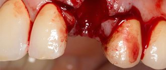
Exostosis of the oral cavity is the proliferation of bone and cartilage tissue in a certain area of the jaw row. Visually, it looks like a lump or nodule that tends to grow rapidly.
The sharp edges of the hardened cartilage tissue that forms exostosis can damage the mucous membrane covering it, leading to injury and severe pain, making it difficult to open the mouth and eat.
On the upper jaw, exostotic formations are most often localized in the area of masticatory elements on the outer or inner side of the row. When the lower jaw is affected, growths form at the base of the incisors, canines and premolars.
As a rule, cartilaginous growths have a benign structure, but there are known cases of their transformation into malignant neoplasms.
Symptoms of exostosis of the jaw
At the initial stages of development, the pathology usually goes unnoticed, as it manifests itself asymptomatically. It is often discovered when examining a patient for other dental diseases. The formation is clearly visible on an x-ray.
As the growths increase, characteristic symptoms appear. Their intensity depends on the clinical picture: location, shape, size.
By what signs can pathology be detected?
- the appearance of a tubercle or bump on the gums;
- sensation of a foreign object in the mouth, especially if the protrusion is located on the side of the tongue, at the bottom of the mouth;
- pain syndrome of varying intensity (occurs when injured by an acute growth of soft tissue);
- changes in the color of the mucous membranes in the area where the protrusion is located;
- jaw dysfunction if an osteophyte has formed near the jaw joint.
Unlike diseases of an infectious-inflammatory nature, it does not cause an increase in temperature, burning sensation, purulent abscesses, fistulas, and does not affect the tightness of the gums.
REASONS FOR THE APPEARANCE
Most often, the cause of exostosis is heredity. In this case, areas of slight growth of bone or cartilage are observed already in childhood.
If there is no growth and no discomfort, the specialist recommends regular monitoring without surgical intervention.
In addition to the genetic factor, exostosis can be caused by the following reasons:
- injury to the jaw rows, their fractures, causing chipping of bone tissue fragments with subsequent improper fusion;
- infectious inflammatory processes, as a result of which bone tissue atrophies and is destroyed;
- pathological structure of the jaw rows, resulting in the formation of curvature of bone tissue and the formation of growths;
- hormonal disorders, due to which the qualitative composition of the bone changes, causing its destruction and susceptibility to the formation of growths.
Dentists also note that very often the development of exostosis is a consequence of complex tooth extraction.
The reason for the growth of bone tissue in this situation is the lack of natural smoothing of the edges of the socket, improper fusion of bone tissue, or excessive trauma to the periosteum during the procedure.
Main reasons
Exostosis is the result of overgrowth of jaw tissue. Experts say that the main cause of this disease is a hereditary predisposition. We also list a number of indirect factors:
- Changes in hormonal levels.
- Careless tooth extraction, during which soft tissue was injured.
- Infection during tooth resection.
According to statistics, these formations appear only in the case of complex operations, when it is necessary to remove several units at once or a multi-rooted tooth.
SYMPTOMS
The initial stage of development of the disease is characterized by the absence of pronounced symptoms, therefore, at this stage, detection of exostosis is possible only during a dental examination.
A specialist will be able to determine the presence of an area of overgrown tissue and suggest the cause of its occurrence.
As the disease progresses, symptoms become more noticeable:
- on the gum there is a tubercle covered with a mucous membrane, which may look unchanged or be covered with small spines;
- the growth increases in size and can turn from a pea into a large lump, which causes discomfort and interferes with the full position of the tongue;
- painful sensations of varying intensity occur;
- the mucous membrane gradually acquires a rich pink tint;
- there is a violation of the patency of blood vessels located in the oral cavity;
- the degree of mobility of the lower jaw is impaired.
Signs
The main symptom is the presence of a dense tubercle near the place where the tooth was removed. At first it does not manifest itself in any way, except that it disturbs a little in the mouth, but again this depends on the location of the compaction. The lump is growing very quickly. When the maximum size is reached, the following signs are present:
- Aching pain that intensifies when eating.
- The cone takes on a bright scarlet hue.
- At maximum sizes, it interferes with the normal position of the tongue.
INDICATIONS AND CONTRAINDICATIONS FOR OPERATION
Dentists note that mandatory surgical intervention to eliminate exostosis is required in the following situations:
- with accelerated growth of the tumor;
- if the patient experiences severe pain and discomfort;
- during planning of further orthopedic treatment, which becomes impossible in the presence of bone outgrowths;
- with significant cosmetic defects of the facial area associated with an increase in growth.
When planning an operation, it is also worth paying attention to the functioning of the human body, in which removal of exostosis is prohibited.
Dentists name the following contraindications to the procedure:
- some blood diseases, including blood clotting disorders and increased sugar levels;
- diseases of the thyroid gland and adrenal glands;
- hormonal imbalance;
- periods of exacerbation of chronic diseases.
PREPARATION
Surgical intervention is the only option for removing a growth of bone or cartilage tissue from the surface of the jaw row, and therefore requires careful preparation to ensure a favorable outcome.
During the patient’s initial appointment, the dentist clarifies his complaints and conducts a visual examination of the oral cavity in order to identify visible pathologies. At this stage, a number of concomitant diseases are often identified: caries, gum or periodontal inflammation, which require treatment.
To determine the exact location of the growth, its shape, size and nature, the specialist sends the patient for an x-ray examination. The completed image is the basis for making an accurate diagnosis and drawing up a treatment plan.
At the stage of preparation for surgery, the dentist directs the patient to undergo tests, which can be used to identify additional diseases that impede the procedure. Thus, mandatory steps include checking blood for clotting and sugar levels, assessing the balance of hormones and the functioning of the adrenal glands.
At the preparatory stage, the specialist also decides on the option of administering an anesthetic drug.
Surgery to remove exostosis is often performed using local anesthesia, but in some situations general anesthesia may be required.
Before administering it, the specialist checks with the patient for the presence of an allergic reaction and previous experience with pain relief.
What to do if exostosis occurs after tooth extraction
Osteocartilaginous formations in the mouth can only be treated surgically, so if you suspect a pathology, you should consult a dentist. The doctor makes an accurate diagnosis after examining the patient: visual examination and radiography. Differential diagnosis of the nature of the lumps may be required.
Exostosis itself does not disappear, so the doctor decides whether the growth needs to be removed. If the protrusions are small, then the operation can be postponed indefinitely. In this case, you need to undergo regular examination at a dental clinic to monitor changes in tissues.
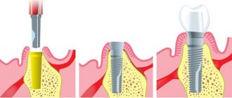
SURGICAL INTERVENTION
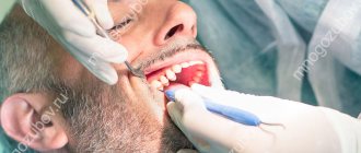
The procedure for removing exostosis begins with anesthetizing the area of the oral cavity that is to be operated on.
Before administering general anesthesia, the specialist determines whether the patient has an individual intolerance to the drug using a special test.
After administering the anesthetic, the specialist performs the following sequence of actions to remove exostosis:
- Disinfection of the oral cavity using special disinfectants, which eliminates the risk of infection of soft and bone structures during surgery.
- Making an incision in the gum tissue to gain access to the cartilage growth. To perform this operation, the dentist uses a scalpel of the appropriate size.
- Removing the build-up. Depending on the chosen method of the procedure, a dental chisel or a laser can be used at this stage, with the help of which the tumor is carefully cut off from the surface of the bone tissue.
- Sanding the treated area. To create a smooth surface and remove small particles of cartilage structure, which prevents recurrence of the disease in the future, the bone tissue is carefully ground using a dental drill. To avoid overheating of the bone, the specialist ensures timely cooling of the treated area.
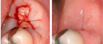
- Apply sutures to the excised area of gingival tissue and secure with a sterile bandage.
The duration of the operation to remove exostosis often does not exceed 2 hours. When using a laser, the time is reduced to 40-50 minutes.
Exostosis
Signs
Exostosis is a painless disease and may not manifest itself for a long time. And it is most often discovered by chance, for example, during radiography. But quite often it happens that exostosis can be felt. There are cases when exostosis grew to such a size that it was visible even to the naked eye.
Description
Typically, exostosis develops between the ages of 8 and 18 years. This disease appears especially often during puberty. It practically never occurs in children under 6 years of age.
Most often, exostoses appear in the upper third of the tibia, the lower third of the femur, the upper part of the fibula, the upper end of the shoulder and the lower end of the forearm bones. They can form on the scapula, collarbone, ribs, and quite rarely they can be found on the bones of the metatarsus and hand, and on the vertebrae. Exostoses do not form on the bones of the skull.
These formations can be of different sizes - from the size of a pea to the size of a large apple. There are cases where exostosis was the size of a child's head.
Their number can also vary from one to several tens and even hundreds.
Reasons for the formation of exostosis:
- inflammation;
- fracture;
- injury;
- infringement;
- infections (syphilis);
- abnormalities of the periosteum or cartilage;
- some endocrine diseases.
There are two types of osteochondral exostosis: multiple exostotic chondrodysplasia and solitary osteochondral exostosis.
You should not think that if exostosis does not cause discomfort, then it is safe. This disease has serious complications. The growth can put pressure on neighboring organs, causing their deformation and dysfunction. It can even deform bones. Another dangerous complication is a fracture of the exostosis leg. However, the most dangerous complication is the degeneration of exostosis into a malignant tumor. This occurs in approximately 1% of cases. Exostoses on the shoulder blades, femurs, pelvis, and vertebrae are most prone to this.
Diagnostics
The diagnosis is made based on the results of an x-ray examination. However, the outer cartilaginous layer of the exostosis is not visible on the x-ray, so it should be remembered that the size of the actual exostosis is larger than the study results would suggest. This is especially true for children, in whom the size of the cartilage layer can reach 8 mm.
It is necessary to differentiate this disease from bone tumors.
Treatment
Treatment of exostosis is only surgical. It is performed by an orthopedic traumatologist under local or general anesthesia. The choice of anesthesia depends on the size of the exostosis and its location. During the operation, the growth on the bone is removed, and its surface is smoothed.
The operation is now performed through a small incision. Often, if the exostosis was minor and the anesthesia was local, the patient can leave the hospital on the same day.
The prognosis is good. Usually, after removal of exostosis, a lasting recovery occurs.
Prevention
The only prevention of exostosis is regular examination and preventive examination. It is especially important to carry it out among children, since the formation of exostosis can cause improper development of the skeleton and will cause a lot of trouble in the future.
© Dr. Peter
REHABILITATION
Removal of exostosis is not a simple operation, so the recovery period most often does not exceed a week.
This time is enough for the gum tissue to recover after the cut, and for the bone to take on its natural shape.
To ensure an easy recovery period and to avoid postoperative complications, the patient should follow these rules:
- regularly take antibiotics and other medications prescribed by a specialist;
- avoid eating foods at too high or low temperatures;
- limit physical activity;
- stop smoking and drinking alcohol;
- exercise caution during daily hygiene procedures.
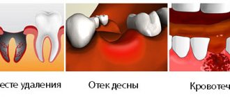
POSSIBLE COMPLICATIONS
Postoperative complications associated with the removal of exostosis are quite rare in medical practice, and according to the experience of dentists, they are associated with the patient’s failure to comply with the rules of the rehabilitation period.
The consequences of unscrupulous adherence to doctor’s recommendations include the following pathological conditions:
- Complete or partial divergence of the suture material. The cause of this phenomenon may be poor diet, smoking, or mechanical damage to the gums during brushing. To fix the problem, you should immediately consult a doctor, who will treat the damaged surface and re-stitch the stitches.
- Inflammation of the gums in the suture area. Due to poor oral hygiene, pathogenic microorganisms can accumulate in the wound area, the result of which leads to suppuration and swelling of the soft tissue. You can only get rid of this complication with the help of a specialist.
Rehabilitation after removal of exostosis
The wound heals completely in about 1 week. For better tissue healing and prevention of complications, it is necessary to comply with the doctor’s requirements.
During the recovery period, you should not exercise or overheat the body. It is prohibited to visit the bathhouse, sunbathe on the beach, or take a hot bath. Otherwise, bleeding from the wound may occur.
In order not to injure the damaged mucous membrane, you should not eat rough, spicy or sour foods. Food should be liquid or pureed, at a comfortable temperature. It is undesirable to smoke, as nicotine constricts blood vessels, which prevents rapid healing.
When brushing your teeth, you should avoid the operated area. The toothbrush should be soft. After eating, you should rinse your mouth with warm water. Daily rinsing with antiseptic solutions, for example, chlorhexidine, miramistin, is recommended. For these purposes, you can prepare decoctions of chamomile, sage, calendula, and oak bark.
In the first few days, pain and swelling of the tissues persist. To relieve acute pain, you can take analgesics. A cold compress will help relieve swelling. Ice or frozen product is wrapped in plastic wrap, cloth and applied to the cheek several times a day.
If the patient is in poor health or has chronic diseases, the doctor may prescribe antibiotics.
If you ignore the dentist’s requirements, complications may occur: infection and inflammation of the wound, bleeding, suture dehiscence.