From this article you will learn:
- what types of pulpitis are there,
- what distinctive features they have in relation to each other.
In dentistry, it is customary to divide pulpitis into several forms, the division of which is based primarily on the severity of symptoms. The classification of pulpitis accepted in Russia was developed at the Semashko Institute in 1989. The author of this classification is Platonov, and in accordance with it the following forms of this disease should be distinguished:
- acute pulpitis,
- chronic pulpitis,
- exacerbation of chronic pulpitis (in turn, each of these forms of pulpitis also has subtypes).
Why does chronic pulpitis develop?
The pulp is the soft tissue of the tooth; inside it there is a nerve and blood vessels that feed it. Like any organ, the pulp is characterized by a protective reaction to the invasion of infection - inflammation. In this case, swelling, suppuration and attacks of sharp throbbing pain occur. This is acute pulpitis.
Any infection can lead to inflammation of the pulp. Patients with deep caries, gum disease, sinusitis, osteomyelitis of the jaw, abrasion of teeth, and traumatic exposure of the pulp become ill especially easily.
The infection penetrates the dentin:
- through a crack in the root;
- with blood flow through vessels;
- from a carious cavity.
If the patient postpones going to the dentist, after 1-2 days, collagen fibers are produced in the pulp and the first stage of the disease begins - fibrous pulpitis.
Acute pulpitis can become chronic:
- As a result of the vital activity of pathogenic microorganisms: lactobacilli, staphylococci, streptococci. For example, after suffering from infectious diseases: rubella, influenza, ARVI.
- After a tooth injury: dislocation, fracture, incorrect treatment, or if the patient does not go to the dentist for acute pulpitis.
- When exposed to chemicals and dental materials.
Causes of fibrous pulpitis
Chronic fibrous pulpitis manifests itself when the coronal part of the tooth is opened, and the outflow of inflammatory exudate occurs through the carious cavity connecting to the pulp chamber.
Fibrous pulpitis can develop primarily, for example, in some people with low body reactivity. In this case, the disease develops independently in the closed cavity of the tooth.
Another reason for the occurrence of pulpitis is poor-quality treatment of deep caries: non-compliance with the rules of dental treatment, incorrect application of medicinal pads, etc.
Forms of chronic pulpitis
The disease causes severe changes in the structure and function of the tooth. Depending on the stage, it is customary to distinguish the following forms of chronic pulpitis:
Fibrous
In the initial stage, the inflamed cells of the neurovascular bundle degenerate and become thicker. The patient feels constant heaviness and swelling inside the tooth. The vascular bundle begins to respond to changes in ambient temperature with aching pain. This happens when a patient goes outside from a warm room in winter.
At this stage, the disease is reversible; with proper dental treatment, the dental nerve can be restored.
Ulcerative
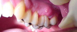
If the inflammation progresses, ulcerations appear on the pulp crown. This is the result of the proliferation of putrefactive bacteria in the pulp chamber. Dark putrefactive plaque in the recesses of the enamel is noticeable visually. The tooth begins to hurt when pressed, and there is a constant unpleasant taste in the mouth.
Hypertrophic
If, after acute pulpitis, the tissues not only increase in size, but also begin to divide, a polyp forms inside the tooth. The growth has a grayish-pink color and is detected during electroodontodiagnosis.
Due to the characteristics of the growth of dental tissues, this type of pulpitis occurs in patients under 30 years of age.
Concrete
In patients over 45 years of age, calcification salts are deposited in the dentin: denticles, petrificates. Their chemical composition causes irritation and then inflammation in the neurovascular bundle.
The symptoms of concrementous pulpitis are similar to trigeminal neuralgia: pain radiating to the temple and chin, aching pain at night.
Gangrenous
This is the last stage of chronic inflammation of the pulp, at which time the neurovascular bundle is completely destroyed. The tooth is deprived of blood supply and nutrition, darkens, and the putrid smell from the mouth increases.
The apical opening of the root is deformed. Gangrenous pulpitis most often leads to the development of acute periodontitis or the formation of granuloma at the root of the tooth.
Treatment of initial K04.00 (pulp hyperemia),
For initial pulpitis, conservative treatment is carried out.
Mostly preparations containing calcium hydroxide are applied to the bottom of the cavity, and then filled with permanent fillings, it is better to monitor after three months.
Clinical picture of acute K04.01 (according to MMSI acute focal) pulpitis
- Complaints: prolonged pain from all irritants, mainly at night. There are also spontaneous pains.
- The pain is clearly localized, the light intervals can last for several hours, and later these light intervals become shorter.
- When the chewing teeth (molars) are inflamed, pain during an attack can spread (radiate) to the ear, temple, and teeth of the opposite side (antagonist teeth).
- Inspection - deep carious cavity, a lot of softened dentin, which, when removed, can open the pulp chamber.
- Percussion is painless
- EDI – 25-40 or within normal limits
- Probing is painless
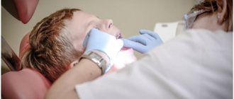
Inflammation of the pulp in children
Chronic pulpitis in children can develop immediately, bypassing the acute stage. This is possible due to:
- Features of the structure of milk teeth . They have a wide chamber within the pulp, small roots and a wide opening at the apex of the tooth root. Through it, infection from the tissues surrounding the tooth easily penetrates into the dentin.
- The child’s tendency to colds and ENT diseases. Pathogenic pathogens that cause frequent colds and runny noses easily infect the periodontal tissue. The infection then travels through the bloodstream to the neurovascular bundle.
- Frequent injuries and microtraumas that a child receives from falls and collisions. Pathogenic pathogens penetrate tooth tissue through microcracks.
In children's teeth, the infection spreads quickly - to the crowns of adjacent milk teeth and the rudiments of molars. This leads to the development of multiple caries.
This is why regular dental checkups for your child are so important. If your baby complains of pain, heaviness and discomfort in the tooth, an unpleasant odor appears from the mouth, or pain occurs when eating cold or hot food, be sure to consult a doctor. The health of baby teeth must be monitored just as carefully as permanent teeth.
Symptoms of chronic pulpitis
With different forms of the disease, its manifestations may vary. For example, with hypertrophic pulpitis there is almost no pain, but there is bleeding of the tooth root. The reason is the growth of foreign tissue in the tooth.
If inflammation is still accompanied by pain, with chronic pulpitis it has a characteristic feature - a delayed nature. The patient continues to feel pain even after the irritant is removed. Even with relative stabilization of the condition, a feeling of heaviness in the tooth remains.
Symptoms of chronic pulpitis:
- bleeding;
- sensitivity to cold, hot food and other external irritants;
- the tooth aches in attacks, including at night;
- there is an unpleasant taste in the mouth;
- difficulty chewing and biting food;
- bone tissue is sparse in the root zone;
- under the filling you can see a dark carious border;
- tooth enamel has changed color.
Clinical picture of initial K04.00
There is no history of spontaneous pain. When interviewed, it turns out that pain comes from various irritants, which quickly goes away after they are eliminated. A painful attack is provoked by cold and hot stimuli (temperature). Almost always the patient points to the causative tooth.
Pain from temperature stimuli quickly (within a few seconds) goes away. When talking with the patient, it turns out that the tooth has not hurt before.
- The tooth cavity is not opened.
- Percussion is painless.
- Probing is painful at one or more points.
- Electroodontometry - 10-12, and sometimes 20 microns (normally 2-6 microns).
- X-ray – no changes.
Diagnosis of the disease
Chronic pulpitis cannot heal on its own. The patient may experience acute pain, flux, and progressive inflammation at any time. Which, in turn, leads to the formation of a cyst, phlegmon, and fistula.
Any pain in the teeth and gums should be diagnosed immediately.
The dentist’s task when chronic pulpitis is suspected is to establish the form of inflammation and the nature of the process.
For this use:
- Probing and tapping the tooth. This way you can determine the level of pain sensitivity, and therefore the degree of damage to the nerve bundle.
- Electroodontodiagnosis (EDD) is an assessment of the sensitivity of the pulp to electric current. High sensitivity indicators indicate serious destruction of the vascular bundle, lower ones indicate its viability. This is the most accurate study of the degree of damage to the nervous system.
- X-ray. The image shows fibrous growths, calculus deposits and traces of deformation in the root canals.
In difficult cases, the doctor may prescribe a computed tomography scan of the tooth.
Forecast and prevention of fibrous pulpitis
It is very important to start treatment of fibrous pulpitis on time by seeking dental help. Exacerbation of fibrous pulpitis is quite painful and treating its advanced stage becomes increasingly difficult. A competent and timely approach to the treatment of fibrous pulpitis helps to eliminate pain and discomfort, preserve the tooth and prevent the spread of the pathological process to periodontal tissue.
Prevention of fibrous pulpitis consists of following hygienic rules for caring for the oral cavity, both personally and with the help of professionals. It is necessary to undergo regular dental examinations, keeping the condition of your own teeth under control, promptly treating caries and fibrous pulpitis.
What to do in case of exacerbation of chronic pulpitis
Chronic pulpitis is unpleasant because tooth pain can recur in any unfavorable environment. Exacerbation can be caused by:
- stress;
- influenza, ARVI, ENT diseases;
- hypothermia;
- injury, including minor.
Pain during exacerbation of chronic pulpitis affects the jaw and the trigeminal nerve area - temple, chin, cheek.
If your chronic dental disease has worsened or you have a toothache, make an urgent appointment with your dentist. You should not take painkillers in this situation - they can weaken the effect of the anesthetics that the doctor will use during treatment.
Treatment of complicated stages of acute pulpitis
Focal and diffuse pulpitis have their own varieties. The first has serous and purulent forms. Focal acute serous pulpitis often occurs as a complication of caries or during improper treatment of caries. It affects the root part of the pulp, which causes sharp attacks of pain. Diffuse serous acute pulpitis spreads to both the coronal and root parts of the pulp, causing severe swelling, which can develop into phlegmon.
Acute purulent pulpitis (pulp abscess), as the name suggests, is characterized by the accumulation of pus in the pulp chamber, throbbing pain, which can radiate to various parts of the jaw and head (which is why the patient often cannot identify the diseased tooth). For successful treatment, it is necessary to completely remove the pus from the canals, and in most cases the tooth is subsequently killed. Treatment of acute traumatic pulpitis is usually more difficult.
Treatment
Chronic inflammation of the pulp can be treated using a combination or surgery. The choice of method depends on the degree of destruction of the tooth nerve:
- The combined method helps at the initial stage, fibrous or hypertrophic form of pulpitis. All the doctor needs to do is remove the apical part of the nerve bundle and cover the stump with a special calcium-containing paste. Then install the seal. This way the tooth will retain its nutritional channel and functionality.
- With the surgical method (pulpectomy), the pulp must be completely removed. This method is justified for gangrenous pulpitis. The operation takes at least an hour and is performed under anesthesia. To do this, it is enough to visit the doctor once.
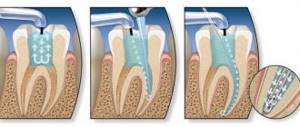
During a pulpectomy, the doctor’s sequence of actions is as follows:
- Preparation of the oral cavity, removal of filling remains and caries.
- Removal of the first part of the pulp in the tooth crown.
- Opening root canals with an ultra-fine bur, extracting nerve fibers. In modern clinics, doctors use a convenient and safe tool - an electronic endo-tip; it controls the depth and level of filling of the root canal.
- Treatment of canals using thin endodontic plates, disinfection of canals.
- Filling and filling the tooth cavity with a photo- or nanocomposite.
The doctor monitors the quality of the work performed using a control targeted image. The manipulation is considered complete if the canal is completely filled, the apical opening of the root is sealed, and there are no signs of possible perforation of the root canals.
Only treatment by a dentist will solve the problem of chronic pulpitis and you will no longer have to fear an acute attack of pain that may return at the most inopportune moment. If you put off visiting a doctor and take painkillers, keep in mind that this is harmful to your health and does not cure inflammation, but only leads to serious complications and more complex and expensive treatment.
Acute diffuse pulpitis
Usually it is the next stage of focal pulpitis (sometimes the peak form of chronic) and appears on the third or fourth day. With diffuse pulpitis, inflammation affects both the coronal and root parts of the pulp. The pain is paroxysmal and long-lasting, with periods of relief being rare. The pain is often radiating and radiates to various parts of the head. The fabric begins to become saturated with exudate.
Some sources also highlight acute fibrous pulpitis, but this is not entirely true. Fibrous pulpitis is a form of chronic; acute fibrous pulpitis is also not a completely correct term. It is a chronic complication of diffuse pulpitis. When gangrenous, extensive tissue necrosis occurs and the tooth often takes on a characteristic gray tint.


