Causes of the disease
Other diseases and bad habits can lead to the development of this disease, such as:
- viral and bacterial infections;
- nicotine abuse;
- weakened immune system;
- sinusitis and tonsillitis;
- ARVI;
- dental diseases.
Risk factors are:
- hypothermia;
- increased mental and physical stress;
- frequent stress.
The influence of factors often leads to accelerated tumor growth. Teeth that were previously filled or reconstructed are most susceptible to this disease.
What is a dental cyst
A dental cyst is a benign neoplasm ranging in size from 0.9 to 3 centimeters inside the bone tissue at the root of the tooth. The cyst looks like a “sac” that can be filled with fluid or pus.
The appearance of a dental cyst is a protective reaction of the body to inflammatory processes. The body attacks the affected cells, which begin to die, and in order to isolate these cells from the surrounding healthy tissues, the same “pouch” is formed around them.
A cyst can appear next to any tooth, however, most often the tumor is diagnosed in several places:
- On the front upper teeth.
- Above the teeth, which are located near the maxillary sinuses.
- Under wisdom teeth.
Symptoms
Many patients wonder what a dental cyst is and how to treat it. To find the answer, you need to understand the symptoms. As already noted, signs of the presence of a formation can be noticed only at a late stage. The symptoms of a dental cyst are as follows:
- pain in the jaw area that does not have a specific location;
- discomfort when chewing food;
- inflamed gums.
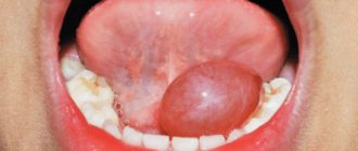
A tubercle is formed in the area of the tooth root, which subsequently increases in volume. The consequence of this process can be flux.
A cyst under a tooth can cause the following conditions:
- physical weakness;
- increased size of lymph nodes;
- increase in body temperature.
A serious danger is that the severity of pain with this disease is much lower than with other dental diseases. An extracted tooth with a cyst cannot be restored, so you must immediately consult a doctor if any discomfort occurs.
Jaw cysts
A primordial jaw cyst or keratocyst occurs in the area of the angle of the mandible or the third molar, in some cases it appears in the place where the tooth should have been. This cyst has thin fibrous walls, its inner surface is lined with squamous epithelium. Due to pronounced parakeratosis, its contents resemble cholesteatoma; odontogenic epithelium is found in their walls. Keratocysts are either single-chamber or multi-chamber; multiple cysts are often multilocular. However, they are combined with other developmental defects and often recur after removal.
A follicular jaw cyst or unerupted tooth cyst develops from the enamel organs of unerupted teeth. The cyst is localized in the alveolar edge of the jaws and is most often associated with the second premolar, third molar, canines of the upper and lower jaws. The walls of such jaw cysts are thin and consist of multilayered squamous epithelium that lines the cavity from the inside. Epithelial cells are changed and often flattened. Sometimes there are cells that produce mucus. In the cavity of the cyst there are one or several teeth, both formed and in their infancy.
Radicular jaw cysts account for 80% of all jaw cysts. Chronic periodontitis is usually preceded by a perihilar cyst, which develops from complex granulomas in the area of any teeth. Radicular cysts on the upper jaw are diagnosed twice as often as on the lower jaw. Typically, the diameter of cysts is from 0.5 to 2 cm, but there are cysts up to 3 or more centimeters in diameter. The multilayered epithelium that lines the inner surface of the cyst has no signs of keratinization; the cyst wall is fibrous, infiltrated with lymphocytes and plasma cells. During periods of exacerbation, the cyst becomes inflamed, its cells hyperplasia, which leads to the formation of network-like processes that are directed into the thickness of the wall. This symptom is characteristic only of radicular cysts.
Often these cysts become inflamed, and neutrophilic leukocytes appear in the infiltrate; if the epithelium is completely melted, then the entire internal surface of the cyst consists of granulations that can completely fill the cyst cavity. Radicular cysts often suppurate and cholesterol crystals and xanthoma cells are found in its walls. If radicular cysts are diagnosed in children, then areas of osteogenesis are found in the outer parts of the wall. According to their location, radicular jaw cysts can be adjacent, push aside, or penetrate into the maxillary sinus; in the latter case, the likelihood of developing sinusitis with exacerbation of inflammation of the cyst is quite high. Large cysts are complicated by bone destruction and thinning of the cortical plate; in jaw cysts of a dysontogenetic nature, the likelihood of developing tumors is much higher, although cell malignancy is not common.
Treatment methods
Once a cyst is detected, it is necessary to begin treatment as soon as possible. Depending on the type and severity of the disease, the treatment method will be determined: therapeutic, surgical or laser.
The therapeutic method is used for small diameter cysts. First, the doctor makes a hole in the tooth, through which the contents of the cyst are pumped out and the medicine is injected. The canals are treated with a disinfectant, after which a temporary filling will be installed. The treatment period can take from 1 to 3 months.
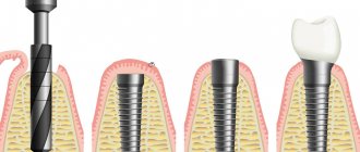
Most often, to treat a dental cyst, a surgical method is used with complete or partial preservation of the tooth. Less often, the tooth has to be removed completely.
There are three types of surgical treatment:
- Cystectomy. This is the most gentle type of treatment. To begin with, treatment and disinfection of the tooth canals is carried out, after which an incision is made in the gum, a hole is sawed out in the bone and the cyst is removed along with part of the root.
- Hemisection. This type of operation is similar to the previous one, only in addition to part of the root, part of the tooth crown is also removed, which makes this type of treatment more traumatic.
- Cystotomy. The simplest and least traumatic type of operation. In the area of the cyst on the gum, the doctor makes an incision through which the contents of the cyst are pumped out. Despite the simplicity of the operation, the recovery process will be long.
Laser cyst treatment is a new technique that allows you to quickly, sterilely and painlessly treat the affected tooth. During treatment, the fillings are removed and a laser is inserted into the canals to remove the cyst. Remaining liquid and other particles are removed using a pump.
Diagnostic examination
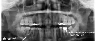
The leading method for identifying cystic systems is radiography. X-ray examination includes general images (frontal plane), local ones, focused on the affected tooth.
For small formations, an intraoral radiography of the jaws (orthopantomogram) is performed; large cystic systems are detected using an extraoral radiographic method.
The widespread use of orthopantomograms is due to the information content of panoramic images of the jaw rows. This method of examination allows you to accurately identify the location, the degree of growth of the formation, and the effect it has on neighboring healthy teeth. The negative characteristics of the procedure include a fairly high radiation dose, so an orthopantomogram is performed no more than once every 6 months.
Magnetic resonance imaging (MRI) is performed. During the procedure, the skull is scanned and a large number of pictures are taken with layer-by-layer images of tissues in different planes. The patient does not receive radiation; radiation exposure during this procedure is reduced to zero. There are a number of contraindications for MRI: it is not recommended during pregnancy, the procedure is contraindicated for patients with pacemakers or pumps.
How dangerous is a cyst?
At first, when a cyst forms, the child may not complain of discomfort, so the cyst increases in size without making itself felt. But at one point, a cyst on the gum of a child’s baby tooth may fester—then, when the shell of the cyst ruptures, all its contents will spill into the bone tissue, opening access to infection.
The child may experience swelling in the jaw and even facial asymmetry. Further spread of pus is fraught with serious consequences - odontogenic periostitis, destruction of the jaw bone, diseases of the digestive system, kidneys, liver, heart, bone marrow damage and the subsequent development of osteomyelitis. The disease may also be accompanied by a fistula on the child’s gum.
Photo of a tooth cyst in a child
Diagnostics
At the initial stage, it is almost impossible to detect a dental cyst, since there are no external signs of the disease. Patients often associate symptoms such as pain, discomfort, and swelling with other dental problems and delay visiting a doctor until the last minute. But even during an examination by a dentist, it is often not possible to detect a cyst, since the capsule is located in the tissues of the jaw and is inaccessible for visual inspection.
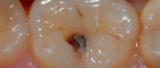
To make an accurate diagnosis, X-ray diagnostics or computed tomography are used. In these images, the cyst looks like a darkened elliptical or oval-shaped spot in the jaw tissue in the immediate vicinity of the tooth. Only through an image can one accurately determine the presence of a cyst in a patient.
Symptoms of a dental cyst
At the very beginning of cyst formation, there may be no symptoms. The presence of a neoplasm can be suspected by the occurrence of periodic pain when loading the affected tooth, the appearance of increased sensitivity to cold and hot foods and drinks, and soreness of the gums in the area of the affected tooth.
In addition, symptoms of a dental cyst may include:
- Changes in enamel color.
- The appearance of bad breath.
- A feeling of tightness in the area of the diseased tooth.
- Swelling of the soft tissue around the cyst.
- The appearance of a bump on the gum or palate. This symptom appears if the cyst reaches a sufficiently large size.
- It becomes impossible to eliminate the pain with painkillers.
If you have several symptoms, you should immediately consult a doctor for diagnosis and treatment.
Treatment of a cyst without tooth extraction
The success of treatment directly depends on how early it was detected. Therefore, dentists recommend that patients undergo regular preventive examinations and seek help at the first signs of illness. More recently, therapy necessarily involved tooth extraction. Of course, this approach did not suit either the doctors or especially their patients. Today, dentists' methods have changed significantly, and it is possible to get rid of a cyst without losing the beauty of your smile.
Treatment of the cyst can be surgical or conservative. The choice of therapy method depends on: the type, age of the patient, personal wishes and is determined for each patient.
Cost of treating a cyst under a tooth
The price of dental services for the treatment of cysts in our clinic is about 8,500 rubles. and depends on the following factors:
- type;
- complexity of the disease;
- method of treatment (surgical or therapeutic);
- use of complex equipment;
- expenses for surgical or dental supplies;
| Service | Price |
| Consultation | For free |
| Orthopantomogram OPTG of the jaw | from 1,100 ₽ |
| Cystectomy | 5 300 ₽ |
| Hemisection | 3 300 ₽ |
Look at the detailed price list for dental treatment at NovaDent dentistry in Moscow and the pain-free region.
If you have been diagnosed with a dental cyst, it should be treated by a qualified dentist. Many patients do not fully understand the danger of this disease. Photos of the cyst can be found on the Internet. From them it can be understood that this can cause the destruction of large areas of bone tissue. And this, in turn, poses a risk of jaw fracture and tooth loss.
Expert of the article you are reading: Svetlana Viktorovna Derevyakina Chief physician, doctor of the highest qualification category, therapist, periodontist, leading specialist of the NovaDent network
27 years
Clinical experience
Petrovsko-Razumovskaya
Verkhniye Likhobory
st. Dubninskaya, 27, building 1
+7
Free consultation with this specialist
Prevention is the best cure
You can detect a cyst in children in the early stages of formation on an x-ray, which is recommended to be taken every six months if the child has diseases such as chronic pulpitis or periodontitis. Cysts are most likely to appear in children aged 7 to 12 years, during the period when baby teeth are replaced by permanent teeth. The cyst forms mainly in the area of the first molars, most often on the lower jaw.
The child must be taught regular, and most importantly, high-quality oral hygiene. If you don’t do this and don’t take your child to the dentist, the consequences can be disastrous. It is also worth taking note of the following: firstly, it is possible and necessary to treat pulpitis and caries of baby teeth; secondly, severely affected by caries is an indication for the removal of a baby tooth, which should be carried out in a timely manner, otherwise they will become a source of infection and subsequently provoke the appearance of a cyst.
You, as a parent and person responsible for the health of your children, must monitor the condition of the entire oral cavity from the first erupted tooth of your child. Remember: the visible part is only 40% of the entire tooth, so you should not assume that its healthy appearance indicates the health of the entire tooth. Don’t forget about going to pediatric dentistry - any disease in the early stages can be treated much easier and more effectively.
Causes of cyst formation
The mechanism of development of a dental cyst is generally simple - inflammation is caused by viruses and bacteria entering the tissue through the bloodstream. The risk of developing a cyst is higher in people with various dental problems - untreated caries, periodontitis, periodontitis. People with reduced local immunity of the oral cavity are also more vulnerable to the disease; these also include patients suffering from chronic tonsillitis, sinusitis and other ENT diseases.
In addition, the cause of cyst development can be:
- congenital malformations of the jaw apparatus;
- injuries;
- complications after dental treatment;
- incorrect installation of dentures;
- pathological eruption of wisdom teeth;
- deviations in teething in children.
Weakened immunity, frequent colds, and smoking are additional factors influencing the development of the disease.