Sometimes patients ask the question: “edentia – what is it?” The term comes from the word “dent” - teeth, and the prefix “a” - without. Literally, “without teeth.” This means adentia is the partial or complete absence of teeth.
Even minor voids in the dentition lead to disruption of chewing function and degradation of the jaw bone. And this, in turn, is fraught with gastrointestinal diseases, metabolic disorders, and aesthetic defects.
This means that proper prosthetics are very important to eliminate adentia. And to prevent pathology - timely treatment of caries and periodontal diseases.
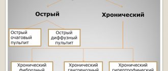
Classification of edentia
The basis for the classification of dental edentia is the etiological factor that provoked the disease, as well as the age period in which it first appeared.
It follows from this that edentulous teeth can be:
- congenital in nature (primary) - the formation of a tooth germ did not occur due to any reasons, most often pathological disorders of body functions;
- acquired character (secondary).
Depending on the age period of manifestation of adentia, it happens:
- temporary bite (manifests itself during the eruption of baby teeth);
- permanent bite (manifests itself during the period of change of milk teeth to permanent teeth).
Also, a special place in the classification of this phenomenon is occupied by a type of edentia called false edentia - characterized by the combination of several tooth crowns into one or a violation of the timing of teething.
Based on the lack of teeth in the jaw, the following types of edentia are distinguished:
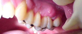
- partial - characterized by the absence of several teeth in a row;
- complete - absolutely all teeth are missing on the jaw.
Congenital partial adentia is said to occur when the patient is missing no more than 10 teeth (mostly located in the frontal area of the upper jaw). If the lack of teeth exceeds 10, then they speak of multiple adentia. Partial edentulism of the jaw of a congenital nature is diagnosed in the absence of no more than 15 teeth in one row.
Partial adentia of an acquired nature is determined by the class of dental defect:
- first class - all molars of the jaw are missing;
- second class - absence of all molars on one side of the jaw;
- third class - absence of the first molars of the jaws;
- fourth class - absence of teeth in the front part of the jaw.
However, the classification of acquired partial adentia does not end there because each class has several subclasses, and quite often a complex disease consisting of a class and a subclass is diagnosed.
According to location, adentia can be symmetrical or lack symmetry.
Complications
The human body usually “does not notice” the loss of one or two dental units, adapting through compensatory mechanisms. However, an increase in the number of voids in the jaw leads to impaired chewing function, deterioration of the periodontal condition, the development of inflammation of the temporomandibular joint, and subsidence of the jaw bone in empty areas.
This provokes diseases of the gastrointestinal tract, impaired absorption of food, changes in facial structure, and unclear diction. As for aesthetics, the absence of one tooth in the smile zone is enough for a person to feel psychological discomfort.
The closer to complete edentia, the more the described problems worsen. Biting and chewing more or less solid food becomes almost impossible. A person has to eat liquid and pureed food, which affects its taste and nutritional value. It is possible to develop hypovitaminosis and a lack of macro- and microelements.
The lips and cheeks droop, the tip of the nose droops, and the face “shrinks” at the bottom. The facial muscles atrophy, the face takes on an senile appearance. Speech changes - it becomes inarticulate. This leads to a decrease in the patient’s social contacts and provokes chronic stress.
Causes of edentia
Today, dentists cannot name the only reason why a person develops edentia. Some experts argue that the pathology is the result of follicle resorption. In turn, the follicle can resolve both against the background of various concomitant diseases, and in connection with inflammatory processes, as well as under the influence of toxins produced by pathogenic bacteria.
If a patient is diagnosed with adentia during the period of permanent dentition, then this is associated with a complication during the eruption of primary teeth, which was accompanied by a violation of the norms and timing of this process. Also one of the reasons is a person’s predisposition to pathology, which is hereditary. Disruption of the process of tooth germ formation may be associated with dysfunction of the endocrine system of the human body.
Another theory for the appearance of adentia (especially when the disease is congenital) is considered to be a violation of the formation of the fetus in the womb. During this period, the formation of all vital systems occurs, including the formation of teeth. If the fetus had any defects, they could interfere with the development of tooth buds, which led to adentia. Acquired adentia is the result of trauma to periodontal tissues, both as a result of blows or bruises, and as a result of poorly performed dental treatment. Carious disease can provoke the appearance of congenital adentia. If a diagnosis of adentia is suspected, only a comprehensive examination will help determine the cause of the pathology.
Why does the disease occur?
Primary adentia is characterized by the initial absence or rapid death of tooth germs.
The reason for this may be both a hereditary factor and a harmful effect that could be exerted on a woman’s body during the perinatal period, as a result of which the formation of the fetus was disrupted, which affected the development and growth of tooth germs. The formation of temporary teeth in the fetus occurs from 7 to 10 weeks, and permanent units are formed closer to 17 weeks. It should be noted that congenital adentia is extremely rare, literally in a few cases. The cause of this phenomenon is considered to be hereditary ectodermal dysplasia. Such a complex disease simultaneously affects the formation of the fetus as a whole, as a result of which problems are observed with the skin and hair, with the development of nails and many other tissues and organs of the human body.
Causes of destruction and resorption of tooth germs:
- endocrine disruptions;
- teratogenic factors;
- metabolic disorders in the fetus;
- past infectious diseases;
- ichthyosis;
- hypothyroidism;
- pituitary dwarfism.
Secondary adentia differs in that tooth loss occurs during a person’s life. The causes of edentia in this case are lifestyle, nutrition, oral care, and diseases acquired as a result of this. Sometimes surgical intervention performed incorrectly can have a harmful effect on the oral cavity. Any inflammatory processes can cause systematic tooth loss.
Diseases in which secondary adentia occurs:
- caries;
- periodontitis;
- pulpitis;
- periodontitis;
- injured enamel and structure;
- abscesses;
- deletion.
Symptoms of primary adentia
The congenital form of this pathology can occur both during permanent dentition and during the period of childhood teeth, when the occlusion is not yet fully formed. The cause of the disease has not been determined. During the dental examination, the specialist notes the complete absence of temporary teeth. Based on the results of the x-ray, the fact of the absence of tooth germs is established. The lower part of the face is comparatively smaller than normal size, which indicates an abnormal development of the jaw (incomplete). The bone bed has the shape of a small protruding tubercle above the gum, the tubercles of the upper jaw are indistinct, and the palate has a characteristic flattening.
Types of edentia
Teeth edentia is classified according to their number into partial (one or more teeth are missing) and complete (not a single tooth). According to the nature of development, primary and secondary are distinguished. In addition, there are different stages of development of adentia, for example, for secondary partial adentia, compensated (dispensary treatment of surrounding tissue), subcompensated and decompensated are observed. In the last two stages, adentia, the treatment of which is complicated or impossible, is subject to prosthetics in order to restore dentofacial functions.
Primary complete edentia of temporary occlusion
The main symptoms of the disease are the inability to bite off and chew food; therefore, the patient’s diet consists of eating only soft foods that do not require much effort to eat.
If we talk about symptoms from the speech apparatus, then there is a noticeable violation in the pronunciation of letters such as D, Z, T, L, Ch, N.
Another symptom is impaired respiratory function, which is associated with problems in the development of the nasal passages. The patient breathes through his mouth.
A characteristic sign of the disease is impaired hair growth on the head, eyebrows and eyelashes are completely absent, nails are either underdeveloped or (as well as hair) absent. By the time the baby teeth erupt, the infant fontanelle is not overgrown; often the frontal part of the face is much larger than during normal development.
Symptoms in the primary form
Adentia in children can be detected during the formation of a primary dentition or already with a permanent one. The specialist even notices the symptoms visually: in the absence of teeth and their rudiments, in most cases the structure of the facial skeleton is disrupted. The lower third of the face is unusually smaller in size, the palate is flat, the jaws are poorly developed, and the supramental fold is clearly defined. Developmental disorders can also be observed on the skull: for example, the bones of the maxillofacial apparatus are not fused, the fontanelle and the bones of the skull do not heal as expected. If the cause of the disease is anhydrotic ectodermal dysplasia, in parallel one can observe the absence of eyelashes and eyebrows, early aged skin, and damage to the mucous membranes.
Complete edentulism of the jaw of the primary form is characterized by the fact that the patient cannot eat normally: he is unable to bite and chew food, it is possible to eat only liquid or puree foods. Due to a violation of the structure of the skull, including underdevelopment of the nasal passages, the patient has mixed breathing, breathing simultaneously through the nose and mouth. The person also experiences difficulties in the functioning of the speech apparatus; he is unable to pronounce sounds that involve the teeth and tongue.
Primary partial adentia is characterized by the absence of a normal number of units in the dentition. Neighboring teeth shift into the area of the formed voids, the jaws are not always developed. The location does not matter: they can be outside the dentition, or creep on top of each other. Pronunciation is impaired (with frontal defects), and gingivitis may develop.
Symptoms of secondary adentia
Secondary adentia is characterized by the absence of the required number of teeth due to their damage by caries, inflammatory processes, treatment of various tumors and other reasons that led to tooth loss.
The disease of secondary complete adentia is manifested by the absence of teeth. In this regard, the lower jaw is unnaturally close to the upper jaw, and soft tissues retract, which manifests itself in the form of wrinkles and folds. The muscles that hold the jaw and are responsible for movement are either underdeveloped or completely atrophied. A patient with this disease cannot eat properly, eat solid food, speech and chewing functions are impaired.
Symptoms in the secondary form
Secondary adentia is not a congenital disease. It can be diagnosed when the first primary teeth are removed or observed for the first time in the presence of a permanent dentition. The difference from the primary form is that the continuity of the dentition is broken, and this occurs after the teeth have been formed
If the teeth are completely lost, the lower jaw gradually approaches the nose, soft tissues fall inward, folds and wrinkles appear on the skin. Losing all teeth can lead to the development of exostoses, bony protrusions that cause pain. Over time, nutrition becomes disrupted and speech changes.
The way you chew food also changes. In the process of living with partial edentia, the remaining teeth may shift, as a result of which the load on them during chewing is distributed differently. At the same time, there is no load at the sites of the holes, destruction of bone tissue occurs. Teeth may wear out, pain may appear, reactions to the temperature of food and drink may develop, and diseases may develop. Displacement of the jaw with significant loss of teeth can occur up to subluxation or dislocation of the joint between the jaw and temple.
Multiple absence of teeth is very noticeable in older people due to hollow cheeks. But the change in the oval of the face is not determined by the years, but by the number of teeth and how the tissues adapt to new conditions.
The loss of large numbers of teeth seriously complicates life. If teeth are missing, a person smiles less often, his behavior and adaptation in society changes. Poor chewing of food leads to stomach diseases.
Partial secondary adentia
Characterized by displacement or divergence of formed teeth. The main effort associated with chewing food falls on existing teeth, which gradually leads to the destruction of soft tissue in those places where teeth are missing.
A complication of this disease is the pathological wear of formed teeth, and their sensitivity is also noted. Closing the jaws is accompanied by pain. Teeth react sharply to chemical and thermal irritants, periodontal pockets form, and there is a risk of dislocation or subluxation of the lower jaw.
In a patient with partial secondary adentia, the symmetry of the face is disturbed, the nasolabial folds become overly pronounced, and the corners of the mouth unnaturally droop. If the front teeth are missing, then the impression of jaw retraction is created.
Symptoms of adentia in children and adults
Diagnosis of this disease takes a long time. But there are a number of symptoms indicating its presence. It is especially important to take them into account when determining dental edentia in children. These factors include:
- reduction of jaws (both or just one);
- signs of retraction of the cheeks and lips;
- deformation of the alveolar processes;
- the appearance of wrinkles and folds approximately in the places where the twos are located;
- manifestations of muscular atrophy of the oral cavity;
- change in jaw angles.
All these symptoms can lead to the formation of a malocclusion. Malocclusions, in turn, cause certain shifts of the teeth and the formation of gaps in the jaw. In some cases, growths may form that affect the appearance and make chewing difficult.
Complete secondary adentia can be caused by caries, periodontitis, or surgical intervention. The doctor may also notice scars from injuries that led to tooth loss. Another symptom that explains the loss of masticatory organs is the presence of cancer.
It is worth noting that there is a steady downward trend in the age of edentulous dental patients. Poor ecology leads to the fact that young people are increasingly faced with this problem.
Diagnosis and detection of the disease
Edentia is a serious problem, and to solve it it is very important to promptly and accurately identify the presence of pathology and its type.
The first stage of diagnosis is to conduct a visual examination. Based on the results obtained, the doctor determines whether adentia is primary or secondary.
Then it is necessary to take an X-ray of the jaw, especially if the initial examination showed that the disease is congenital. This can only be confirmed with an x-ray, in which a specialist will check for the presence of dental follicles or identify signs of abnormal development.
When identifying the causes of adentia in children, at the initial stage of diagnosis it is necessary to find out the structure of the root system and find out how things are under the gums. Therefore, in the treatment of childhood adentia, the method of panoramic radiography is used. It allows the doctor to obtain a detailed picture, including information about the structure of the bone tissue of the alveolar processes.
Why is timely diagnosis so important?
Timely detection of adentia is of such high importance for one serious reason. The fact is that before starting any therapeutic actions, it is necessary to diagnose and eliminate all existing pathologies.
First of all, these include inflammatory infections of the oral cavity. During treatment, it is recommended to preserve the roots of the teeth as much as possible, since the absence of roots can lead to the loss of the entire masticatory organ. In addition, it is necessary to carry out complete disinfection of the oral cavity.
If the doctor suspects the development of acquired adentia, it is necessary to find out the cause of the disease as soon as possible. This is due to the fact that some pathologies can lead to complications during prosthetics. To eliminate these risks, the dentist checks for the following symptoms during the examination:
- exostoses (benign growths on bones consisting of bone or cartilage tissue);
- unremoved roots covered with mucous membrane;
- tumors and inflammatory processes;
- infections of the oral mucosa.
Types of adentia and their diagnosis
General symptoms indicating the presence of the disease were discussed above. But there are also a number of signs characteristic of different types of adentia. Their knowledge can also help to identify pathology in a timely manner.
Primary complete edentia
The rarest type of disease and at the same time the most serious. With primary complete adentia, the patient does not even have the rudiments of teeth and their roots. This pathology leads to serious consequences, namely:
- facial symmetry is disrupted;
- the correct formation of the alveolar processes of the upper and lower jaws is difficult;
- the secretion of mucus and saliva in the mouth slows down, which can cause additional inconvenience to the patient.
One of the symptoms of primary complete adentia may be paleness of the mucous membranes. The absence of baby teeth in children can also be easily determined by palpation. If such signs are present, it is necessary to urgently take appropriate measures.
Primary partial edentia
It is expressed in the congenital absence of some teeth in the patient. If partial adentia is detected in a child, the x-ray will show gaps in the root system. It should be noted that the absence of wisdom teeth is not considered edentulous.
As the disease develops, diastemas and tremata are formed - spaces between the chewing organs. In simple terms, the teeth begin to “creep”: if you look at a photo of edentia in children, you will notice large gaps between the teeth.
Primary partial adentia can be symmetrical or asymmetrical. In the first case, there are no paired teeth - doubles or fangs. Asymmetrical adentia means the absence of teeth in different places of the jaw.
Secondary complete edentia
This type of disease is diagnosed when it is acquired. Its symptoms can be observed on all jaws or only on one. Both primary and permanent teeth may be missing. Secondary complete adentia is diagnosed when all the chewing organs have fallen out or needed to be removed due to another disease.
One of the main unpleasant consequences of this pathology is jaw deformation. It becomes closer to the nose, soft tissues begin to retract. Also, this type of disease is characterized by muscle atrophy, leading to disruption of the shape of the entire jaw or one or both alveolar processes.
In addition, the loss of all teeth immediately leads to everyday problems - the patient will not be able to chew independently, and will also begin to swallow some sounds.
Secondary partial edentia
This is the most common type of disease, in which several masticatory organs are missing from the dentition. The loss of canines, twos or other teeth often leads to increased sensitivity of bone tissue. This is due to the gradual wear of the side walls of adjacent teeth.
With this complication, the patient faces discomfort, due to which he gradually stops eating solid food, which is difficult to chew. Both children and adults face this problem.
Possible consequences of edentia
Tooth loss is a serious disease that can lead to various unpleasant consequences, including the development of other serious diseases. Among them are the following:
- impaired jaw development, which leads to difficulty pronouncing various sounds;
- the patient is forced to exclude solid foods from his diet, which causes problems with intestinal motility and disruption of the gastrointestinal tract;
- development of persistent psychological discomfort. The patient’s self-assessment is distorted and depression may develop;
- in the complete absence of teeth, the jaw develops abnormally and can put pressure on any area of the head, thereby provoking the appearance of temporal tumors.
Diagnosis of edentia
Dental pathology such as adentia, the treatment of which is necessary to normalize the basic vital functions of a person, requires a high-quality, complete diagnosis, during which it will be possible to establish the main cause of the pathology and develop a treatment plan.
The process of diagnosis and subsequent treatment involves consultation with specialists such as a periodontist, surgeon, orthopedist, implantologist and orthodontist.
The initial diagnosis is established based on the collection and thorough analysis of the patient’s complaints, as well as the clinical picture of the disease. The dentist also draws up a chronological picture of the disease, in which the main role is given to comparing the patient’s age and the number of teeth, as well as the nature of their absence.
Dentists can make an accurate diagnosis based on a spot X-ray examination of the oral cavity. If all teeth or a significant number of them are missing, then a panoramic x-ray is performed; a computed tomography scan of the TMJ and its x-ray can also be performed in combination.
Based on the results of an X-ray examination, the doctor can determine whether there is a pathology in the development of the tooth germ, the level of coverage of the root by the gum, the presence of tumors inside the jaw, and identify inflammatory processes.
In order to draw up a treatment plan, an impression and subsequent production of a model of the patient’s jaw will be required. Performed individually for each clinical case.
What can be the complications of adentia?
This pathology requires mandatory consultation and treatment with a dentist. Partial or complete loss of teeth can significantly reduce the quality of life and health problems arise.
Without dental restoration, a person faces the following difficulties:
- Inability to eat solid foods.
- Limiting the diet, as well as poor chewing of food, leads not only to gastrointestinal disorders, but to vitamin deficiency and metabolic diseases.
- Losing more than half of your teeth significantly increases the stress on the remaining teeth. This leads to their abrasion, thinning and fragility.
- Due to jaw deformation, pathological changes in the oral mucosa occur.
- Periodontal disease causes pain when eating and brushing.
- Impaired diction provokes psychological discomfort and limitation of communication.
There is no need to wait for complete destruction of the dentition, since in this case longer and more expensive treatment will be required. If you have problems with your teeth, contact your dentist for a diagnosis.
Method of treating edentia
An acquired disease is treated by installing dentures; the ideal option is to install implants on pins, which is associated with better distribution of pressure on the gums and soft tissues. Using this method, you can select the required implant size as correctly as possible and fill the interdental gap with it.
Complete edentia of an acquired nature is also treated using prosthetics, in which removable systems are installed. If fixed systems are installed, then primary dental implantation is necessary as a basis for dentures.
Congenital dental adentia in children must be treated at the age of no later than four years, with complete removable plate dentures installed, which must be replaced every two years. If the patient is diagnosed with partial primary adentia, then partial plate dentures are installed.
It is necessary to ensure that while wearing dentures, stomatitis and bedsores, as well as allergies to dyes, do not develop. Treatment begins only after professional hygienic cleaning of the oral cavity.
Treatment methods
At the moment, the pathology is incurable, since humanity has not yet learned to grow new teeth. But there are many methods for replacing lost teeth with dentures and completely restoring aesthetic and chewing function.
In general, dentures are divided into removable, fixed and partially removable.
The fixed type includes bridge-like structures (“bridges”) and prosthetics on implants.
Removable ones are plates made of acrylic, nylon or a clasp structure made of metal and plastic.
Bridges
This is a chain of artificial crowns welded together that rest on your own teeth.
Prosthetics using this technology are performed if it is necessary to fill a void of no more than four units in a row (at most five), otherwise the “bridge” will lose stability. Also, the supporting units must be strong enough, since the chewing load will fall on them.
The disadvantage of this technology is the need to grind down the supporting teeth for crowns, and often their depulpation.
Prosthetics on implants
This is a relatively new technique, which is based on the ability of bone to fuse with titanium.
The prosthesis is a structure made of a titanium implant (usually in the form of a screw), an abutment (adapter), and an artificial crown made of metal ceramics or zirconium dioxide.
The titanium implant is surgically implanted into the jaw and takes about six months to integrate with the bone. After engraftment, the implant performs the function of a root, and thus the anatomical structure of the tooth is recreated.
After successful prosthetics, such a prosthesis begins to fully take on the chewing load. This means that the jawbone does not degrade, the chewing process remains completely physiological, and the adjacent teeth remain intact.
Contraindications and limitations of this method are based on its invasiveness and the possibility of implant engraftment.
Removable structures
These are solid plates made of acrylic or nylon, which represent a gum-colored base and artificial teeth.
A removable denture can be partial or complete.
Partial is used for partial edentia and is attached with hooks or clasps to the patient’s own teeth. The load during chewing is distributed over the entire prosthetic bed. Such “removers” change diction and taste perception, as they block the palate, and therefore require a long period of getting used to. This is especially true for acrylic dentures.
Partial removable dentures also include clasp systems. A clasp is an arch with crowns, bases and artificial teeth soldered onto it. This prosthesis is lightweight and requires almost no getting used to, since it does not cover the palate, but it has a number of clinical limitations.
Prosthetics with complete edentia
This is the most difficult task for an orthopedist, since in this case atrophic changes in the jaw occur, which means loss of landmarks in the height and shape of the lower part of the face. Therefore, the doctor needs to determine the size and shape of the prostheses so that they optimally restore the oval of the face. The orthopedist also solves the difficult task of securing prostheses.
The classic method of prosthetics is complete removable structures that are attached using a vacuum method, that is, they are suctioned to the palate. For reliability, it is possible to use specialized pastes and adhesives. However, the pullers on the lower jaw still hold much worse.
With the advent of implantation, removable, partially removable and non-removable complete prosthetics using implants became possible.
Fixed prosthetics
This is a bridge-like structure, in which the role of supporting teeth is performed by implanted dentures. The combination of supporting and “hinged” teeth can be different, but the more supports, the more reliable the bridge and the more physiologically the load is distributed.
Such prostheses are made of metal ceramics, zirconium dioxide and aluminum oxide. They are quite expensive, especially zirconium and ceramic.
Removable prosthetics on implants
With a limited budget, an alternative can be conditionally removable and removable prosthetics supported by implants, which provides reliable fixation of the prosthesis in the jaw, excellent aesthetics, and a minimum period of adaptation. Its cost is significantly cheaper than non-removable structures.
Some of the techniques:
- All on 4 and All on 6. From English this translates as “all on four” and “all on six.” Four or six implants are implanted into the patient’s jaw, into which, instead of abutments, fasteners are placed to attach an acrylic prosthesis. In this case, the chewing load is taken on by the implants, so for better distribution and reliability of the design, it is better to make a prosthesis on six supports. This is a conditionally removable structure. The patient cannot remove it himself, but the dentist can do this in the office.
- Bar prosthetics . Just like the previous case, two, four or six implants are implanted into the patient’s jaw. A metal beam is installed on their tops, on which the acrylic prosthesis is fixed by snapping. A person can remove this prosthesis himself, but it is not recommended to do this often so as not to break the locking structures.
Which method will be chosen depends on the clinical case and financial capabilities of the patient.
Adentia: disease prevention
Prevention of pathology must be carried out even while the child is in the womb. To do this, it is necessary to create ideal conditions for the development of the fetus and eliminate as much as possible the influence of unfavorable factors that provoke the development of defects.
Acquired edentia can be avoided only under the supervision of a dentist: for this you should undergo regular examinations, perform oral hygiene procedures, and perform prosthetics if a tooth has been lost. It is also important to carry out disinfection measures in the oral cavity, which will not allow pathogenic bacteria to generate pathogenic processes that can affect the normal functioning of the teeth.
Treatment
For rational planning of prosthetic therapy, complete edentulism is classified in detail. There are three types of edentulous upper jaw (according to Schroeder) and four types of edentulous lower jaw (according to Keller). The mucosa of the prosthetic beds was also classified according to the degree of atrophy into four classes (according to Supple). For the treatment of partial primary adentia, in accordance with the patient’s age, a pre-orthodontic trainer is used with registration at the dispensary. Adentia, the treatment of which begins at an early age in children (when permanent teeth erupt), is aimed at promoting normal eruption and preventing deformation. Typically, orthodontic preparation is carried out before therapy and restoration of the dentition during this period.
To restore the integrity of the bite, an adhesive bridge, prosthetics and implants are used, but this approach is only adequate if one or more teeth are missing. Treatment of patients with a complete form of secondary adentia is a multifaceted task, where restoration of bite functionality, prevention of pathologies and complications, and leveling of psychological and emotional consequences are carried out simultaneously. Prosthetics includes a number of activities preceding the production of a prosthesis - inspection, planning, preparatory procedures, actual production, fixation, adaptation, patient instruction and control.
Modern method of implantation
In case of complete or partial loss of teeth, the dentist can offer a modern option for implantation - “all on four”. It involves installing a prosthesis on 4 implants, which are implanted in the area of 2 and 5 teeth. This treatment option has few contraindications and is excellent for those who value comfort and reliability of the design.
Restoration of the lower row of teeth on 4 implants
Features of implantation using the “all on four” technique:
- only 4 implants need to be implanted, and not 8-12, as with other restoration options;
- installation of the prosthesis is possible immediately after fixation of the implants, that is, the technique involves immediate loading;
- there is the possibility of such treatment even with insufficient bone tissue thickness;
- A permanent prosthesis is placed, which with proper care can last a lifetime.
This modern technique is now being practiced more and more often, and many note its advantages over others. In addition, you can save money on it, because you only need to buy 4 implants.
But implantation has its contraindications:
- General absolute. Pathologies of the circulatory system, diabetes mellitus, arthritis, tumors, tuberculosis, immunodeficiency, central nervous system pathologies, sexually transmitted diseases and AIDS.
- General relative. Poor level of oral hygiene, defects of the alveolar process, age under 16 years, alcoholism.
- Local. A short distance to the nasal sinuses, a tendency to poor hygiene, insufficient bone tissue.
- Temporary. Acute period of infectious disease, traumatic sports, pregnancy, rehabilitation after surgery, drug addiction.
The dentist may refuse implantation for other reasons. These may be intolerance to anesthesia, being under stress, exhaustion of the body, general surgical contraindications, and some mental disorders.