There is a whole complex of pathologies in which a person develops a malocclusion due to the fact that the upper and lower rows of teeth do not meet in the anteroposterior plane.
The condition when the upper part protrudes forward is called prognathia (in other terminology, prognathic or distal occlusion). And if the lower half of the jaw is in front, the doctor diagnoses progeny.
These anomalies can be combined with each other, causing their symptoms to overlap.
Reasons for development
Prognathia develops for many reasons, which usually appear in infancy and early childhood. These provoking factors include:
- disruption of the function of normal breathing through the nose due to its constant congestion, as the jaw begins to stretch forward against the background of the cessation of its development in width;
- development of diseases of the jaw apparatus, especially during artificial feeding;
- the habit of sucking a finger, lower lip, gnawing stationery objects, excessive addiction to nipples contribute to the protrusion of the lower row of teeth;
- the jaw grows incorrectly if the child walks with his head down as a result of some kind of postural disease;
- malocclusion of baby teeth or their early removal due to caries;
- previous rickets and other diseases affecting bone tissue.
The disease can also manifest itself in certain disorders, inflammatory processes, insufficient intake of fluoride and calcium at the stage of development in the womb. Moreover, it is transmitted from parents to children.
Prognathia (distal occlusion)
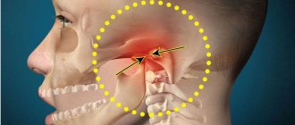
Prognathia (distal bite) refers to sagittal malocclusion and is characterized by inconsistency in the size, shape and position of the upper and lower jaws in the sagittal direction (Fig. 284). The degree of sagittal displacement is determined by the orbital (frontal) plane.
Some authors call this malocclusion prognathia due to the anterior position (protrusion) of the upper jaw in relation to the lower jaw, while others call it distal occlusion, since the lower jaw is located distal to the upper jaw.
The term “distal occlusion” was introduced by Licher. Brückl, Reichenbach, Korkhauz and others do not use the term “prognathia”. They designate its various clinical forms as a narrowing of the jaws with a close or fan-shaped arrangement of the upper front teeth or refer to it as a deep blocking (overlapping) bite. They use the term “distal bite” only when the lower jaw is distal.
Prognathia (distal occlusion) is a fairly common anomaly that occurs during the period of primary, mixed and permanent dentition. The causes of prognathia (distal bite) are varied. These include intrauterine and neuro-humoral factors, disruption of the functional balance of muscles, artificial feeding, diseases of early childhood (especially rickets), inflammatory processes of the jaws, impaired nasal breathing, bad habits, early removal of baby teeth.
Prognathia may be caused by excessive development of the upper jaw or upper dental and alveolar arches, underdevelopment of the lower jaw or lower dental arch, distal position or displacement of the entire lower jaw with its dentition with an overdeveloped or normal upper jaw. The relationship of the lateral teeth in the sagittal direction is characterized by the fact that the medial buccal cusp of the upper jaw closes with the lower one of the same name or lies in the gap between the second premolar and the anterior buccal cusp of the first molar. However, this sign is not constant. In the transversal direction, there may be normal overlap of the lower teeth by the upper teeth, and unilateral or bilateral linguo-occlusion may also be observed.
Teleradiographic studies by A. El-Nofeli and I.K. Irgenson established that with prognathia there is a discrepancy between the size of the upper dentition and the size of the base of the upper jaw, i.e., the apical base. With prognathia, there may also be a mesial or distal location of the upper jaw in the facial skeleton, and the latter may have a different size (normal, underdeveloped, overdeveloped). There is a decrease in the length of the body of the lower jaw and shortening of its branches. The severity of prognathia depends on the discrepancy between the size of the apical base of the upper and lower jaw.
There are various clinical forms of prognathia. As an independent anomaly, prognathia is rare. Most often, it is combined with anomalies in the position of individual teeth, an open or deep bite, and narrowing of the jaws, which in turn aggravate prognathism.
Based on data from teleradiographic studies, A. El-Nofeli identified two forms of distal occlusion: dental distal occlusion and skeletal. Dental distal occlusion is characterized by an abnormal arrangement of teeth and an abnormal shape of the dentition with the correct ratio of the bones of the facial skeleton and the bones of the skull. Skeletal distal occlusion is caused by morphological deviations of the facial skeleton and various options for the location of the upper jaw in the skull in combination with dental anomalies.
According to Engle, prognathia has two subclasses. With the first, there is a narrowing of the upper dentition with deviation of the frontal teeth forward (Fig. 284, a), with the second, an oral inclination of the upper and lower front teeth (Fig. 284, b). L.V. Ilyina-Markosyan also adheres to the division of prognathia into two forms.
The wide variety of clinical forms of prognathia and all possible combinations of its various symptoms cannot be classified into just two forms. However, the two mentioned forms of prognathia should be considered the most pronounced - the main forms of this anomaly.
Classification and characteristics
Prognathia is classified depending on the position of the jaw into several types:
- the first form involves insufficient development of the lower jaw against the background of normal growth of the upper jaw;
- the second is characterized by excessive formation of the upper jaw or upper row of teeth, while the lower remains in its normal position;
- in the third form, the symptoms of the two listed above are combined;
- the sign of the fourth is only a slight “squeezing” forward of the upper front teeth, usually ones and twos.
Pathology can also acquire:
- true;
- false;
- and dual form.
The true form of the anomaly is characterized by pronounced signs:
- The patient's middle facial region protrudes significantly forward. This shortens the upper lip and creates tension in the muscle tissues of the mouth.
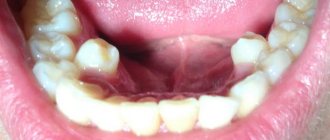
- The upper dentition extends beyond the oral cavity, biting the lower lip. In this case, the person cannot completely close his mouth.
- The pathology is characterized by a change in the position of the upper teeth: the gap between the incisors is noticeably expressed, crowding of the teeth or their fanning out may occur.
Article:
What exactly should you do if the baby has prognathia : the chin is absolutely not “strong-willed”, the lower jaw “crawled” back, and the upper teeth, like those of a rabbit, protrude forward?
Exercises for the lips and lower jaw - myogymnastics, are carried out in parallel with breathing exercises and wearing a vestibular plate (with the visor pointing down). Naturally, during myogymnastics the plate should not be in the baby’s mouth. The exercises are done 2-3 times a day, first for 5 minutes, then for 10-15 minutes, sitting in front of a mirror. Do not take the whole complex at once to work with your child. Start with one or two exercises. When you get good at them, master the next ones, but don’t forget to repeat the previous ones. And it would be a good idea to visit a speech therapist before starting self-study at home. 1. Exercise “Smile”. The lips smile naturally, the teeth are clenched in a “fence”, i.e. The upper teeth “stand” above the corresponding lower ones. The front teeth are clearly visible. Maintain this position for at least 10 seconds (the time lengthens from activity to activity).
2. Exercise “Tube”. The teeth are tightly clenched. Pull your lips forward with a tube and hold in this position for at least 10 seconds (the duration gradually increases).
3. Alternate exercises “Smile” - “Pipe” at the count of “one-two” at least 10-15 times. Teeth are clenched all the time! Take your time! Fix one or the other position of the lips.
4. Exercise “Fish” . The teeth are clenched tightly, the lips are extended forward (see exercise “Tube”). The lips open and close (as a fish does with its mouth in an aquarium).
5. Exercise “Funnel ”. Move the lower jaw forward, open the mouth slightly, and open the teeth. On the count of “one”, pull your lips forward with a “horn”; on the count of “two”, pull your lips inside your mouth, tucking them behind your teeth (the lower jaw is still pushed forward!). Do at least 10 repetitions.
6. Exercise “Annoyance”. Bite your upper lip with your lower front teeth at least 10-15 times. Hold this position for 5-10 seconds. The lower jaw is pushed forward as much as possible.
7. Exercise “Timpani”. The lips are slightly curled behind the teeth inside the mouth. Slap them one against the other, making a characteristic patting sound. The lower jaw is pushed forward as much as possible. Do this at least 10 times.
8. Exercise “Snorting horse ”. Relax your lips and snort, like a horse does. Move the lower jaw forward as much as possible. Do this at least 10 times.
9. Exercise “Deadbolt”. Move the lower jaw forward as much as possible. Move it from side to side at least 10-15 times with maximum amplitude.
10. Exercise “Hide and Seek”. a) “Hide” the upper lip by biting it with the lower front teeth. Only the lower lip is visible, it stretches up towards the nose. Hold in this position for at least 5 seconds (the time to complete the exercise increases from session to session);
b) “Hide” the upper and lower lips, drawing them into the mouth and slightly squeezing them with the front teeth. The lower jaw is pushed forward as much as possible. Hold in this position for at least 5 seconds (the time to complete the exercise increases from session to session).
11. Exercise “Plane” . “Cross” the lower central incisors along the upper lip at least 10-15 times. The lower jaw moves forward and “works” energetically.
How to eliminate progeny?
Correcting progeny (the lower jaw protrudes unnaturally) can be difficult. But regularly wearing a vestibular plate (with the visor pointing upward) and performing articulation exercises (myogymnastics) help cope with this malocclusion.
The child needs to imagine that his teeth and lips are “stubborn doors” that always strive to “skew.”
1. Articulation exercise “Aligning the doors” : a) open your mouth slightly, then, slowly moving the lower jaw back, place the edges of the lower central teeth on the edges of the upper ones, hold this position for 5-10 seconds;
b) open your mouth, then, moving the lower jaw back, compress your lips tightly, slightly biting them with your teeth, hold for 5-10 seconds.
What should you do if one door leaf prevents the other from closing? That's right, take a plane and remove all the “unnecessary”!
2. Exercise “Plane” : “Cross” the upper central teeth along the lower lip at least 10-15 times. The lower jaw moves back and actively “helps” in this work.
3. Exercise “Rabbit”: a) bite the lower lip with your upper front teeth at least 10-15 times. Then grab your lower lip with your upper teeth and hold this position for 5-10 seconds. The upper front teeth are clearly visible during this exercise.
b) try to reach the upper part of the chin with your upper incisors and also lightly bite it. Fix this low position of the upper jaw on the chin and hold it for 5-10 seconds. The upper front teeth are clearly visible.
And now he will play hide and seek with his lips and teeth.
4. Exercise “ Hide and Seek ”: “hide” the lower lip behind the upper front teeth and the upper lip, biting it. In this case, only the upper lip is visible, it seems to hang over the chin, but the upper teeth are not visible. Hold in this position for at least 5 seconds (the time increases from session to session.
With the help of articulation exercises and plates, the child acquires useful skills that help him cope with the pronunciation of difficult sounds, and he develops a correct bite. But you have to be prepared for the fact that wearing the plates and producing sounds will last for several years. This will require enormous patience from parents and children.
Diagnostic criteria
Diagnosis of abnormalities in the development of the jaw bones is carried out by external examination, as well as by examining the oral cavity by a dentist or orthodontist and by taking x-rays.
The formation of a violation will be indicated by:
The photo shows pronounced prognathia of the first type
- protruding upper jaw and its incorrect position at an angle relative to the base of the skull;
- pathologically round face;
- formation of a deep mental fold;
- inability to close your mouth;
- protruding lower lip;
- fan-shaped arrangement of teeth.
Depending on the individual characteristics and form of the disease, prognathic bite has the following symptoms:
- the patient cannot bite food due to protruding incisors;
- speech disorders occur;
- difficulty breathing through the mouth;
- With an incorrect bite, the oral cavity is seriously reduced, which is why a person loses the ability to chew food thoroughly.
How to treat distal bite (prognathism)?
Among the treatment methods, three main ones can be distinguished: two of them involve classical treatment with the help of orthodontic devices, and one is applicable in more complex cases. Let's start with it.
Orthognathic surgery
When, according to the doctor's testimony, classical, more conservative methods of correcting the bite are powerless, orthognathic surgery can be used.
In fact, this is an operation in which the entire jaw bone is corrected. At the same time, the shape of the face changes. Depending on the type of prognathia (upper or lower jaw), the bone either lengthens or shortens. In the case of a distal bite (defect of the upper jaw), a kind of “lengthening” of the lower jaw is carried out, which ultimately leads to the closure of the teeth, a correct bite and a beautiful silhouette.
Some nuances:
- orthognathic surgery is performed only in extreme cases and has a number of contraindications
- After the operation, treatment with braces is usually prescribed.
Treatment of malocclusion with surgery
Correcting an overbite with braces
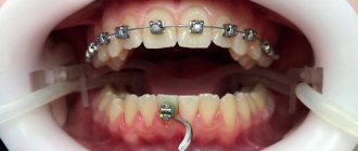
If the degree of prognathia is beyond the control of braces, correction of the bite with the help of braces is prescribed. Depending on the patient’s preferences, these can be classic metal braces (the most inexpensive option), or more aesthetic ones - sapphire or ceramic structures.
Treatment with braces can last two, three or even four years. If we talk about the lower bar, large bite defects can be corrected in 6-8 months.
Correction of prognathism using aligners or clear aligners
Aligners and mouth guards for the treatment of occlusion are a relatively new trend in the dental services market, but have already gained respect among patients. In terms of their capabilities, they are practically not inferior to braces, but at the same time, the treatment is much more “inconspicuous”.
Transparent aligners are not visible on the teeth, and can simply be removed while eating. This greatly simplifies the process of hygiene and dental care during the treatment of distal occlusion (it is known that in the case of braces, oral care, especially after eating, is not a simple procedure).
Material tested by DI experts
Defect correction
Frenkel apparatus
With true prognathia, correction of the patient's condition is accompanied by certain difficulties. In particular, when a patient develops a permanent bite, its correction by orthopedic methods is no longer possible. In this case, you cannot do without surgery.
Therefore, it is extremely desirable to detect the initial symptoms of pathology in a child in time and wean him from bad habits that accompany the disease. To do this, the orthopedist corrects the bite of baby teeth using orthodontic devices.
Actively used:
- activator tools that stimulate the growth of the lower jaw and prevent the development of the upper jaw;
- compression bandages that stop bone growth;
- use of the Frenkel apparatus, which gives the jaw an anatomically correct appearance.
- sanitation of the mouth and nasopharynx.
At the same time, during therapy, parents make sure that the child does not put his fingers or any foreign objects into his mouth.
The method of surgically correcting the bite involves cutting the tissue and directing the jaw to the side at the required angle so that the patient's upper and lower rows of teeth come together.
Then the bones are splinted in the correct position and grow together within 1.5-2.5 months. Next, there is a period of rehabilitation, after which the patient, in most cases, is restored to a normal bite and takes on the appearance of an absolutely healthy person.
Possible consequences
If prognathia of the upper jaw is not removed at school age (up to 14-15 years), then an adult may face a lot of complications:
- first of all, an external facial defect causes serious psychological trauma to the patient, which leads to the formation of an inferiority complex and problems in his personal life;
- secondly, chewing functions are impaired, which can also affect the state of the digestive organs;
- Another manifestation of pathology undiagnosed in childhood are deviations in the area of speech, most often a lisp;
- If the teeth are not positioned correctly, caries develops faster, and any careless movement can cause pain.
Thus, prognathic bite is a rather serious maxillofacial pathology that requires timely treatment by an orthodontist.
What is the cause of prognathia?
About 70% of children have some degree of distal malocclusion. What is the reason for such a common pathology? Dentists believe that genetics are to blame. The child inherits from the parents the shape of the teeth, the size of the jaw, as well as abnormalities of the masticatory apparatus.
But there are other factors that contribute to the formation of a prognathic bite:
- negative effects during intrauterine development - inflammatory diseases, lack of calcium and fluoride;
- chronic ENT diseases - if the nose is stuffy, the child begins to breathe through the mouth, which leads to narrowing and stretching of the upper jaw bone;
- bad habits - prolonged use of a pacifier, the habit of chewing pencils and other objects provoke displacement of the lower dentition;
- poor posture - incorrect position of the body leads to a constant tilt of the head forward, this disrupts the growth of the jaws;
- premature loss of baby teeth - removal of a temporary tooth ahead of schedule (for example, due to caries or crown fracture) causes displacement of adjacent crowns, so there is not enough space for the eruption of a new tooth.

