Vertical movement of the lower jaw.
The initial position of the lower jaw when opening the mouth is the state when the lips are closed. In this case, there is a gap of 2-4 mm between the dentition of the lower jaw and the upper jaw. This state is called a state of physiological rest. The movement of the lower jaw in the vertical plane occurs when opening and closing the mouth, due to the active contraction of the muscles: - depressors (mylohyoid, geniohyoid, anterior belly of the digastric muscle) - levator (proper masticatory muscle, temporalis, medial pterygoid muscle). The amplitude of mouth opening is strictly individual. On average it is 4-5 cm.
Phases of lowering the lower jaw.
1. With a slight lowering of the lower jaw (quiet speech, drinking), the articular heads in the infero-posterior part of the joint rotate around a horizontal axis passing through their centers. 2. With a significant lowering of the lower jaw (loud speech, biting), the hinge rotation in the infero-posterior part of the joint is joined by sliding of the articular heads together with the discs forward along the circumference of the articular surface. The result is a combined movement of the articular heads, in which the point of contact of two convex articular surfaces moves. 3. With the maximum lowering of the lower jaw, the sliding of the heads is delayed at the apex by the tension of the articular capsules, articular ligaments and muscles, and one hinge movement continues in the joint. The trajectories of movement of the lower teeth are concentric curves with a common center in the head of the lower jaw. They, just like the axis of rotation of the head, can move in space.
Unreducible displacement of the articular disc
Sometimes clicking in the temporomandibular joint occurs for months or even years before the joint suddenly seizes. At a certain point, the disc simply does not slide back onto the condylar process of the mandible. Opening the mouth becomes difficult and painful; Chewing is also very difficult. This condition is called “closed mouth blocking.” When this situation occurs, the clicking of the temporomandibular joint stops. Such blocking can be very short - a fraction of a second - or longer, for example, minutes or hours.
Unreducible displacement of the articular disc. The disc dislocates completely forward when the mouth is closed and does not slide back onto the condylar process when the mouth is opened
Sagittal movements of the lower jaw.
The forward movement of the lower jaw is carried out by bilateral contraction of the lateral pterygoid muscles. The movement of the head of the lower jaw is divided into 2 phases: 1- the disc together with the head slides along the surface of the articular tubercle; 2- the sliding of the head is accompanied by its articulated movement around its own transverse axis. The distance that the head of the mandible travels when it moves forward is called the sagittal articular path. This distance is on average 7-10 mm. The angle formed by the intersection of the line of the sagittal articular path with the occlusal plane is called the angle of the sagittal articular path. According to Gisi, it averages 33º. With an orthognathic bite, the protrusion of the lower jaw is accompanied by sliding of the lower incisors along the palatal surface of the upper ones. The path taken by the lower incisors when moving the lower jaw forward is called the sagittal incisal path. The angle formed by the intersection of the line of the sagittal incisal path with the occlusal plane is called the angle of the sagittal incisal path. On average, its value is 40-50°. Bonneville's three-point contact. When the lower jaw is advanced to the position of anterior occlusion, contact of the dentition is possible only at three points. Two of them are located on the distal cusps of the second and third molars, and one on the anterior teeth.
BIOMECHANICS OF MOVEMENTS OF THE LOWER JAW
Biomechanics is the science of human and animal movements. It studies movement from the point of view of the laws of mechanics inherent in all mechanical movements of material bodies without exception. Biomechanics studies objective patterns revealed during examination.
Studying the movements of the lower jaw allows you to get an idea of their normality, as well as identify disturbances in their manifestations in the functioning of muscles, joints, closure of teeth and the condition of the periodontium. The laws on the movements of the lower jaw are used in the design of devices - occluders and articulators. The lower jaw is involved in many functions: chewing, speech, swallowing, laughter , etc., but for orthopedic dentistry its chewing movements are of greatest importance. Chewing can be performed normally only when the teeth of the lower and upper jaws come into contact (occlusion). The closure of the dentition is the main property of chewing movements.
The human lower jaw moves in three directions: vertical (up and down), which corresponds to opening and closing the mouth, sagittal (forward and backward), transversal (right and left). Each movement of the lower jaw occurs with simultaneous sliding and rotation of the articular heads. The only difference is that in one case, hinge movements predominate in the joints, and in the other, sliding movements.
Vertical movements of the lower jaw. Vertical movements are made due to the alternating action of the muscles that lower and raise the lower jaw. Lowering of the lower jaw occurs with active contraction of m. mylohyoideus, m. geniohyoideus, etc. digastricus, provided that the hyoid bone is fixed by the muscles lying below it. When closing the mouth, the lower jaw is raised by contracting m. temporalis, m. pterygoideus medialis with gradual relaxation of the muscles that lower the lower jaw.
When the mouth is opened simultaneously with the rotation of the lower jaw around an axis passing through the articular heads in the transverse direction, the articular heads slide down and forward along the slope of the articular tubercle. With maximum mouth opening, the articular heads are positioned at the anterior edge of the articular tubercle. In this case, different movements take place in different parts of the joint. In the upper section, the disc slides down and forward along with the articular head. In the lower one, the articular head rotates in the recess of the lower surface of the disc, which for it is a movable articular fossa. The distance between the upper and lower rows of teeth in an adult at maximum opening is on average 4.4 cm.
Sagittal movements of the lower jaw. The forward movement of the lower jaw is carried out by bilateral contraction of the lateral pterygoid muscles, fixed in the fossae of the pterygoid processes and attached to the articular capsule and articular disc. The forward movement of the mandible can be divided into two phases. In the first phase, the disc, together with the head of the lower jaw, slides along the articular surface of the tubercles. In the second phase to sliding
Section I. Orthopedic treatment of patients with complete loss of teeth
the head is joined by its articulated movement around its own transverse axis passing through the heads. These movements are carried out simultaneously on the right and left. The greatest distance that the head can travel forward and down along the articular tubercle is 0.75-1 cm. When chewing, this distance is 2-3 mm.
The distance that the articular head travels when the lower jaw moves forward is called the sagittal articular path. Sagittal articular path
characterized by a certain angle.
It is formed by the intersection of a line lying on the continuation of the sagittal articular path with the occlusal (prosthetic) plane. By the latter we mean a plane that passes through the cutting edges of the first incisors of the lower jaw and the distal buccal cusps of wisdom teeth, and in their absence, through similar cusps of the second molars. The angle of the articular sagittal path,
according to Gysi, is on average 33°.
The path taken by the lower incisors when moving the lower jaw forward is called the sagittal incisal path.
When the line of the sagittal incisal path intersects with the occlusal plane, an angle is formed, which is called
the angle of the sagittal incisal path.
Its size is individual and depends on the nature of the overlap. According to Gysi, it is on average 40-50°.
Transversal movements of the lower jaw.
Lateral movements of the mandible result from unilateral contraction of the lateral pterygoid muscle. So, when the jaw moves to the right, the left lateral pterygoid muscle contracts, and when it moves to the left, the right muscle contracts. In this case, the articular head on one side rotates around an axis running almost vertically through the articular process of the lower jaw. Simultaneously
It is the head of the other side, together with the disc, that slides along the articular surface of the tubercle. If, for example, the lower jaw moves to the right, then on the left side the articular head moves down and forward, and on the right side it rotates around a vertical axis.
Angle of the transversal articular path (Bennett's angle).
On the side of the contracted muscle, the articular head moves downward, forward and somewhat inward.
Its path during this movement is at an angle to the sagittal line of the articular path. Otherwise it is called the angle of the lateral articular path.
On average it is 17°. On the opposite side, the ascending ramus of the mandible moves outward, thus becoming at an angle to its original position.
Transverse movements are characterized by certain changes in the occlusal contacts of the teeth. As the lower jaw shifts to the right and left, the teeth describe curves intersecting at an obtuse angle. The farther the tooth is from the articular head, the blunter the angle. The most obtuse angle is obtained at the intersection of curves formed by the movement of the central incisors. This angle is called the angle of the transversal incisal path, or Gothic.
It determines the range of lateral movements of the incisors and is equal to 100-120°. When lateral movements of the jaw are made, it is customary to distinguish between two sides: working and balancing. On the working side, the teeth are set opposite each other with cusps of the same name, and on the balancing side - with opposite cusps, i.e. The buccal lower cusps are set opposite the palatal cusps.
In prosthetic dentistry, one of the unresolved problems is the problem of articulation. The solution to this problem should be understood as studying a wide range of issues related to
Chapter 6. Concept of stability of prostheses
related to the biomechanism of interaction in the human dental system in normal and pathological conditions, and the development on this basis of progressive modern methods of prosthetics.
The only criterion that determines the correct articulation of artificial teeth is the presence of multiple and unhindered sliding of teeth during the chewing movements. This feature, on the one hand, ensures uniform distribution of chewing pressure, stability of dentures, increasing their functional value, and on the other hand, prevents the occurrence of pathological changes in the soft and hard tissues of the bed.
Creating correct articulation of dentures is impossible without establishing those elements that, under physiological conditions, provide dynamic contacts between teeth. The most widely used methods for constructing artificial teeth are based on the theory of balancing and the spherical theory.
Balancing theory
(joint theory). The main requirement of the classical balancing theory, the most prominent representatives of which are Gysi and Hanau, is the preservation of multiple contacts between the dentition of the upper and lower jaws in the phase of chewing movements. According to Gysi, chewing movements occur cyclically, in a “parallelogram”. The preservation of the cusp and incisal contacts is the most important factor in this theory, the authors of which believe that the inclination of the articular path gives direction to the movement of the mandible and that this movement is influenced by the size and shape of the articular cusp. According to the requirements of Gysi's theory, it is necessary:
• precise determination of the articular path;
• recording of the incisal path;
• determination of the sagittal compensation curve of the line;
• determination of the transversal compensation curve of the line;
• taking into account the height of the cusps of the chewing teeth.
At the end of the 19th century. Bonneville noted 3-point contact as a cardinal sign of physiological articulation of the dentition. With anterior occlusion, tooth contact is possible at three points: one of them is located on the front teeth, and two on the distal cusps of the second or third molars. Some authors consider a full-fledged masticatory apparatus only from the point of view of this contact, both qualitatively and quantitatively. Others believe that only when making prosthetics for toothless jaws, it is necessary to strictly observe the principles of articulatory balance and the laws of multiplicity of contacts in order to obtain maximum effectiveness of the prostheses. Hanau analyzes the articulation system and especially emphasizes the difference between the position of the dentures in the articulator and in the mouth, due to the lack of elasticity of the tissues.
From a whole series of articulation laws, Hanau identified 5 main factors, calling them the articulation five:
• inclination of the articular path;
• severity of the compensation curve;
• inclination of the reference plane;
• inclination of the upper incisors;
• height of the cusps.
All these factors are subject to change. There is an inverse relationship between the quantities. For example, increasing the curvature of the compensation curve changes the inclination of the incisors and vice versa.
A.I. Pevzner (1934) and other authors criticize the theories of Gysi and Ganau, believing that a food bolus between the teeth when
Section I. Orthopedic treatment of patients with complete loss of teeth
| contact with the upper ones. Given that the fangs are on a turn, Gisi recommended installing them without contact with the antagonists. Staging according to Gysi using the mandibular tubercle method, “tubercle” method. In an effort to maximize the stability of the prosthesis on the lower jaw, Gysi recommends setting the orientation plane from the line of the canine cusps, then parallel to the Camper line, passing at a height of 2 mm below the upper lip and connecting with the apexes of the alveolar cusps of the mandible. Premolars and the first molar are installed along the found orientation plane. The second molar is placed on the leveling plane. Taking into account the type of bite and the initial shape of the occlusal surface of the teeth is an important factor determining the success of orthopedic treatment. Therefore, when placing artificial teeth, it is necessary to take into account the relationship of the alveolar processes of the upper and lower jaws in the central occlusion. Principles of teeth placement according to Hanau. Hanau’s technique is built in accordance with the principles of articulation set forth in Gysi’s theory, the main of which is the principle that determines the dominant role of the temporomandibular joint in the movement of the lower jaw. The relationships established by Ganau between the 5 articulatory factors are summarized by him in the form of several laws: 1. With an increase in the inclination of the articular tubercles, the inclination of the occlusion plane increases. 2. As the inclination of the articular tubercles increases, the inclination angle of the incisors decreases. 3. As the inclination of the articular tubercles increases, the height of the tubercles increases. |
When biting and chewing, it separates the dentition and thereby disrupts the balance just at the moment when the need for it is greatest. This is the main drawback of the method of constructing artificial dentition in accordance with the theory of balancing
The design of rational prostheses for toothless jaws is a complex biomechanical problem, and its solution must be constructed in accordance with the laws of mechanics. This means that the installation of artificial teeth should be based on requirements that satisfy the existing principles of biostatics and biodynamics of the masticatory apparatus.
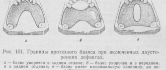
Anatomical setting of teeth according to Gish
consists of installing all the teeth of the upper jaw within the prosthetic plane parallel to the Camper line, passing at a distance of 2 mm below the upper lip.
In its second modification,
the so-called step setting, Gysi proposed, taking into account the curvature of the alveolar process of the lower jaw in the sagittal direction, to change the inclination of the lower parts of the jaw. By using a "stepped" arrangement, Gysi's goal was to increase the stabilization of the lower jaw prosthesis.
The third, most common placement of teeth according to Gysi,
consists in establishing the chewing teeth along the so-called leveling plane. The leveling plane is the average value in relation to the horizontal plane and the plane of the alveolar process. According to this technique, the lateral teeth of the upper jaw are placed as follows: the first molar touches the plane only with the buccal cusp, the remaining cusps and all the cusps of the second molar do not touch the leveling plane. The lower teeth are placed in the den-
Chapter 6. Concept of stability of prostheses
4. With increasing depth of the sagittal occlusal curve, the inclination of the occlusion plane of the prosthesis decreases.
5. As the degree of curvature of the sagittal occlusal curve increases, the inclination angle of the incisors increases.
6. As the inclination of the occlusion plane of the prosthesis increases, the height of the cusps decreases.
7. As the inclination of the occlusal plane increases, the inclination of the incisors increases.
8. As the inclination of the occlusion plane increases, the height of the cusps decreases.
9. As the inclination of the incisor angle increases, the height of the cusps increases.
To ensure all of the above points in their mutual connection, it is necessary, as Ganau believed, to use an individual articulator.
According to the Hanau method, when installing a lateral tooth, it is necessary to check the degree of individual overlap of the teeth, ensure tight, uniform contacts between the teeth in a state of central occlusion (creating a balanced occlusion), as well as smooth sliding of the cusps of the teeth and their multiple contacts on the working and balancing sides (creating a balanced, “balanced” articulation of teeth).
Spherical theory.
A common requirement of numerous theories of articulation is to ensure multiple sliding contacts between artificial dentitions during the chewing phase. From the point of view of fulfilling this general requirement, the spherical theory of articulation developed in 1918 by Monson should be considered the most correct. The spherical theory of articulation most fully reflects the spherical properties of the structure of the dental system and the entire skull, as well as the complex three-dimensional rotational movements of the lower jaw. Prote-
zitting on spherical surfaces provides:
• articulatory balance in the phase of chewing movements (Gusi);
• freedom of movement (Hanau, Hylteb-randt);
• fixing the position of central occlusion while simultaneously obtaining a functional impression under chewing pressure (Gusi, Keller, Rumpel);
• formation of a tuberculate chewing surface, eliminating the formation of shedding moments that disrupt the fixation and stabilization of dentures (see Table 6.1).
Therefore, prosthetics on spherical surfaces are rational for the prosthetics of toothless jaws, the use of plate dentures in the presence of single natural teeth, the production of splints for periodontal disease, for the correction of the occlusal surface of natural teeth in order to create correct articulatory relationships with artificial teeth on the opposite jaw and targeted treatment for joint diseases . Proponents of the spherical theory first of all note that it is easier to place artificial teeth on spherical surfaces.
Extraoral method of recording the central relationship of the jaws (according to Git).
This method was proposed in the 1920s. After determining the height of the lower part of the face and designing the occlusal plane, a small pin is strengthened in the center of the upper wax ridge, extending beyond the lips in a vertical downward direction. A metal platform covered with a thin layer of wax is fixed on the lower roller. The pin must touch the surface of the record. The patient is asked to make lateral movements of the jaw until he gets tired. An angle is drawn on the plate
Biomechanics of the lower jaw
Table 6.1
| Articulatory theories of dentition construction | Gysi's theory | Monson theory | Hanau's theory | Balancing theory | Spherical theory |
| Basic provisions | The inclination of the articular path gives the direction of movement of the lower jaw, which is influenced by the size and shape of the articular tubercle | Complex movements of the lower jaw are determined not by articular paths, but by the surfaces of the dental cusps, which give directions to these movements | The theory is similar to Gusi's theory. He analyzes the articulation system and especially emphasizes the difference between the position of the dentures in the articulator and in the mouth due to the lack of tissue elasticity | Takes into account: 1) the angle of inclination of the sagittal composite path; 2) the angle of inclination of the sagittal incisal path; 3) the angle of inclination of the transversal articular path; 4) the angle of inclination of the transversal incisal path; 5) the angle of inclination of the cusps of artificial teeth; 6) the angle of inclination of the occlusal curves; 7) directions of the occlusal plane | Provides: 1) articulatory balance in the phase of chewing movements (Gusi); 2) freedom of movement (Hanau, Hyltebrandt); 3) fixation of the position of the central occlusion with simultaneous obtaining of a functional impression (Sapozhnikov); 4) formation of a tuberous chewing surface (Sapozhnikov) |
| Determining factors | 1. Accurate determination of the articular path. 2. Recording the incisal path. 3. Determination of the sagittal compensation curve. 4. Determination of the transverse compensation curve of the line | 1. The inclination of the articular path. 2. Depth of the compensation curve. 3. Tilt of the orientation plane. 4. Inclination of the upper incisors. 5. Height of the mounds |
Chapter 6. Concept of stability of prostheses
approximately 120° (gothic angle). The placement of the pin at the apex of the angle will indicate the central position of the lower jaw in relation to the upper jaw.