V-Line surgery – to make your face oval!
A massive, wide, heavy jaw looks impressive in men, but does not adorn women. An elongated, angular or square lower part of the face does not correspond to the generally accepted canons of femininity, according to which facial features should be graceful and chiseled.
On the contrary, triangular, oval and intermediate V-shaped face shapes (oval in the form of a “heart”) have been considered a sign of aristocratic origin for centuries. The V-line of the jaw gives a soft, youthful charm to the features, since anatomically the lower third of a child's face is triangular in shape, which is the structure we associate with youth.
Ideal proportions of the lower third of the face
The lower jaw consists of a horseshoe-shaped body and ascending branches, which are directed towards the temples and are located at an angle to the body of the jaw. If this angle exceeds 125°, the lower third of the face has an oval or triangular shape; with a less flat angle, the jaw looks square or round. The angle of the lower jaw is not only an anatomical, but also an age-related feature: in young children it is 150-160°, and the figure decreases with age.
The ideal proportions of the lower part of the face were calculated back in the Renaissance: a horizontal line is drawn from the middle of the interval between the lips and chin; when it intersects with the ascending branch of the lower jaw, it should form an angle of 100-110°. Such proportions create a “v-line”, that is, when viewed from the front, the line of the lower jaw resembles the Latin letter “V”.
Types of bite
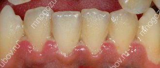
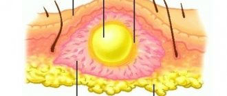
What is a bite? This is the relative position of the upper and lower jaws, taking into account their correct position when fully closed. With a correct bite, the upper row of incisors should cover the lower row by only one third. The upper incisors must be in clear contact with the same incisors of the lower jaw. With a correct bite, there are no gaps between the teeth.
In orthodontics, there are several types of bite, each of which can only be diagnosed by an orthodontist upon examination.
Reshaping the lower jaw with V-Line surgery
The possibilities of modern surgery make it possible to change the shape of the lower third of the face and bring it closer to the desired oval or triangle.
Depending on the initial shape of the lower jaw, V-Line plastic surgery (Jaw Reduction, V-Line Mandibular Contouring) may include:
- changing the contour of the lower jaw by resection of the corners
- straightening the edge of the jaw body using grinding
- narrowing of the chin using T-osteotomy (excision of a small section of bone tissue in the center or pushing it down and forward)
Mandibuloplasty is performed under general anesthesia and lasts an average of 1.5 hours. In the vast majority of cases, intraoral access is used in the retromolar region (behind the last teeth) and along the lower arch of the vestibule of the oral cavity (under the lower front teeth), which does not leave marks on the face. The period of postoperative hospital stay is 1 night.
The active rehabilitation period lasts 10-14 days: during this period it is necessary to use a face mask, there are some restrictions on food. The face recovers completely after surgery within 2 months.
V-Line bone grafting does not disrupt the structure of the dentition and is easily tolerated. The operation is indicated for patients aged 20-25 years with fully formed bone tissue.
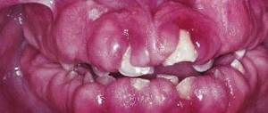
Crooked front teeth in adults
The main reasons why crooked front teeth appear in adult patients:
- Tooth extractions and long-term lack of integrity of the dentition. As soon as at least one lateral or front tooth is missing, immediately (after 3-6 months) displacement of the teeth adjacent to the defect begins. And the antagonist tooth begins to move vertically into the territory of its distant brother. After 2-3 years, the tilt and displacement of the teeth worsen and prosthetics or orthodontic treatment are required.
- The eruption of wisdom teeth, with a lack of space in the jaws, leads to crowding and then curvature of the front teeth. In this case, it is necessary to remove the eighth teeth in time before the front teeth begin to shift.
- Bad habits. This is holding pens and pencils between the teeth, biting off nails with the front teeth, etc. Quite often, this leads to chipping of the enamel or displacement of the incisors.
Best results with 3D planning
The V-Line operation is designed to create a feminine and aristocratic oval face, an elegant small chin, and a more youthful appearance by changing the jaw line.
The VIP Studio virtual plastic laboratory makes it possible to 3D model the ideal proportions and desired shapes before surgery. Based on computed tomography and light scanning data, an accurate three-dimensional model of the face, head and neck is formed. The surgeon and the patient jointly plan the scope of the intervention; based on the model, calculations are made that are used during the operation.
Thanks to the VIP Studio virtual plastic technology, which Dr. Guryanov uses in his work, the result of the operation is as close as possible to the client’s expectations.
Diagnosis and treatment of patients with deformities of the lower jaw in the angular area
The problem of diagnosing disproportion in the lower zone of the face and treating patients with this pathology, which is expressed in the form of a protruding angle of the lower jaw and causes an aesthetic problem for many people, especially females, is considered. A set of diagnostic methods is presented, which makes it possible to differentiate types of pathology into groups to facilitate the choice of treatment methods. Treatment methods that allow obtaining high aesthetic and functional results are presented.
The increase in the number of patients turning to a surgeon to change their face shape is due to increased migration, as well as the desire of women to have more graceful, sophisticated and at the same time proportional facial contours.
Progress in maxillofacial surgery has made it possible to consider the issues of diagnosis, planning and treatment of patients with deformities of the facial skeleton from different angles. However, a trend has emerged that cannot be ignored due to the growth of urbanization and the expansion of plastic surgery capabilities. A number of deformations have been identified, the treatment of which is aimed, rather, at the aesthetic aspects of facial anatomy, rather than at functional disorders of the dentofacial system. These are the so-called facial disproportions, which are characterized by deviations from the norm in the sizes of the lower, middle and upper zones of the face, both in the sagittal plane and in the transversal and vertical.
One of these deformations is the protruding angles of the lower jaw, which, according to the pathogenetic mechanisms of development, are related to hypertrophy of the masticatory muscles.
L. Whitaker first reported the possibility of reducing the width of the lower third of the face using osteotomy of the outer cortical plate of the angle of the mandible along with the masseter muscle. After Legg's first report of m. hypertrophy. masseter, many authors began to use partial myectomy m. masseter W. Adams and J. Converse proposed correcting this pathology surgically, using resection of m. masseter, and ostectomy of the angles of the lower jaw either externally or intraorally. Yang and Park, S. Baek et al. also proposed surgical classification and treatment, and divided patients into three groups, followed by surgical correction of the angular contours only. But the protruding angles of the lower zone of the face must be considered as a whole, from the point of view of the harmony of the entire face, and correction of not only the angles of the jaw, but also muscle hypertrophy, deformation of the chin, using a differentiated approach.
After analyzing foreign literature, we came across a huge variety of terms that can be used to designate disproportion in the lower zone of the face, characterized by wide and enlarged angles of the lower jaw. In the international classification, the term “hypertrophy of the masticatory muscles” is used, which characterizes changes in the bone tissue of the lower jaw. Since most of such work was carried out by scientists from Asia, where resection of the angles of the lower jaw is a popular operation, we decided to use their terminology, namely to call such a disproportion the protruding angles of the lower jaw, since this term most accurately characterizes it and we do not use this term in the domestic literature met.
Without dwelling on the description of the anatomy of the angle of the lower jaw, the parotid-masticatory region, which is well covered in the medical literature, it should be noted that manifestations of disproportion are caused by both the increase in the angle of the lower jaw (usually against the background of underdevelopment of the chin of the lower jaw), and by a change in the shape of the angle in the transversal and sagittal planes.
Most publications describe various methods for treating protruding mandibular angles. However, the authors did not conduct a thorough scientific study of diagnostic methods, did not differentiate the diverse forms of this pathology and did not determine the exact indications for the use of certain treatment methods, which was the reason for discussing this urgent problem of reconstructive plastic surgery.
facial surgery.
Material and methods
Over the past 10 years, 30 patients have come to us with protruding angles of the lower jaw. We spent at
They provide surgical and non-surgical correction of facial contours in order to improve its aesthetics and solve the psychosocial problems of patients. The age of the patients ranged from 19 to 42 years, there were 40 women and 6 men.
Preoperative diagnostics included the following examination methods: clinical examination, anthropometry, clinical photography, multislice computed tomography (MSCT), electromyography (EMG) of the masticatory muscles, ultrasound of the masticatory muscles themselves.
Clinical photometry was carried out using a semi-professional Nikon D500 SLR camera (focal length 1.5 m). Patients were photographed in frontal, profile, semi-profile and axial views. Particularly valuable results were obtained after 3D processing of data obtained using a multislice computed tomograph (Fig. 1).
Rice. 1. Three-dimensional construction of the facial skull.
This provided objective visualization, volumetric-spatial representation of the intervention area and made it possible to assess the volume of the upcoming correction. We obtained digital computed tomography data using MSCT performed on a SIEMENS Sensation 16 device. They were used to construct a three-dimensional model of the facial skull in the preoperative and postoperative periods, as well as to plan the possible results of the upcoming treatment.
Functional studies of the masticatory muscles are also important. Many of the patients who came to us with hypertrophy of the masticatory muscles themselves complained that, especially in the afternoon and at night, they noticed discomfort, expressed in tension and fatigue in the area of the angles of the lower jaw, as with one of the symptoms of bruxism. Patients suffering from bruxism identified by neurological examination were excluded from the study.
All patients with protruding mandibular angles underwent EMG recording the force of jaw compression. EMG was performed using the SINAPSIS device in the preoperative period, as well as in the early and late stages of the postoperative period. EMG made it possible to record and evaluate bioelectrical processes in the masticatory muscles, correlate the recorded values with the values obtained during a clinical examination, with MSCT and ultrasound data, and also outline a treatment plan. In addition, studying the electrical biopotentials of muscles helps to prevent such complications as weakness of chewing functions in the postoperative period, i.e. EMG data of the masticatory muscles objectively show the maximum volume of myectomy.
The above examination methods were used in the preoperative, postoperative periods and in the long term after treatment. Analysis of data from all examination methods obtained at the preoperative stage allowed us to classify patients based on the morphological features of the deformities:
Group 1 - mild deformity with protrusion of the corners in the posteroinferior direction, i.e. in the sagittal plane, which reveals the facial profile;
2nd group - noticeable protrusion of the corners not only in the sagittal plane, but also in the lateral (main deformity);
3rd - there are pronounced features of the 2nd group in combination with hypertrophy of m. masseter; 4th group - the deformation is based only on hypertrophy of m. masseter
Of our 30 patients, 6 were assigned to group 1, 11 to group 2, 9 to group 3, and 4 to group 4.
Patients of the first 3 groups were treated surgically, patients of the 4th group received botulinum toxin injections
type A in a dose of 20 units. into the chewing muscles themselves.
Operation techniques
All operations were performed under general anesthesia with endotracheal intubation. We only used
intraoral surgical access. The incision of the mucous membrane began in the retromolar region 1 cm above the last lower molar and then continued along the lower buccal-alveolar fold of the vestibule of the mouth to the projection of the 1st lower molar. Then you should go to the periosteum, covering the area of the bone of the lower jaw behind and downward from the last lower molar. The periosteum is dissected and exfoliated along with the fibers attached to it m. masseter to the area required to open the entire angle of the lower jaw. In patients of the 1st group, marginal resection of the mandibular angle with bending osteotomy was then used, in patients of the 2nd group - sagittal splitting ostectomy of the mandibular angle, in patients of the 3rd group - sagittal splitting ostectomy of the angle and partial resection of the masticatory muscles themselves (Fig. 2) .
Rice. 2. Patient A. before (left) and 6 years after surgery (right).
During myectomy, the internal fibers of m. were resected along the sagittal plane. masseter The volume of bone and muscle resection was determined according to data obtained after calculations and processing of the results of the preoperative examination, and was agreed with the patient. After removing the excess mandibular angle, the so-called contouring of the angles was carried out, as a result of which it was possible to obtain the maximum natural shape of the mandibular angle (Fig. 3, 4).
Rice. 3. Patient E. before surgery.
Rice. 4. Patient E. after surgery.
In the early surgical period (days 15–20) and in the long term after surgery (1–5 years), we used the same examination methods as before treatment.
According to an analysis of the results of treatment of patients with protruding angles of the lower jaw: in 25 out of 30
the person achieved a satisfactory result, the square contours of the lower area of the face were corrected
more refined, the face became proportional, acquired an oval shape, and symmetry was preserved. In 1 patient, asymmetry persisted for 1 month, associated with hematoma and asymmetrical edema that occurred in the early postoperative period. After 1 month, the asymmetry gradually disappeared spontaneously; no special re-surgical correction was required. All patients in group 4 achieved partial correction; the effect lasted an average of 3-4 months, after which additional botulinum toxin injections were required (Fig. 5).
Rice. 5. Patient R. before (left) and after (right) treatment with botulinum toxin.
Conclusion
The results of studies conducted in 30 patients with protruding angles of the lower jaw, the complex of diagnostic methods we developed and the distribution of patients into clinical groups depending on anatomical and anthropometric indicators turned out to be effective, which contributed to obtaining good aesthetic and functional results.
Authors:
Doctor of Medical Sciences F.H. Nabiev, asp. K.V. Filippov, asp. P.V. Libin