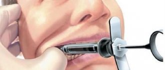
Technique of torusal anesthesia
1. The patient opens his mouth as wide as possible (Fig. 101, B). Injection site: the intersection point of a horizontal line drawn 5 mm below the chewing surface of the third upper molar with the groove formed by the lateral slope of the pterygomaxillary fold and the cheek. 2. The syringe is located on the first lower molar (or even on the second lower molar), the needle is directed perpendicular to the plane of the mandibular ramus.
Rice. 101. Torusal anesthesia according to M.M. Weisbrem (S.N. Weisblat, 1962). A - diagram of the location of nerves in the area of the mandibular eminence: 1 - buccal nerve; 2 - lingual nerve; 3 - inferior alveolar nerve; 4 - mandibular eminence (torus); 5 - a horizontal line drawn 5 mm below the chewing surface of the third upper molar (reaches the torus). B — position of the needle injection, position of the syringe during torusal anesthesia
3. The needle is advanced into the soft tissue to the bone to a depth of 15-20 mm, an aspiration test is performed, 1.5-2 ml of anesthetic is injected, anesthetizing the inferior alveolar and buccal nerves. 4. Having advanced the needle a few millimeters, 0.3-0.5 ml of anesthetic is released to anesthetize the lingual nerve. Anesthesia occurs within 3-5 minutes. Note : In the last 3 years, using modern strong anesthetics, we have not advanced the needle to anesthetize the lingual nerve (point 4). When 1.5-2 ml of anesthetic is injected near the bone, the anesthesia zone covers both the inferior alveolar, buccal and lingual nerves. 5. Anesthesia zone : the same tissues as with mandibular anesthesia, as well as: mucous membrane and skin of the cheek, gums of the lower jaw from the middle of the second premolar to the middle of the second molar on the vestibular side. 6. Complications are the same as with mandibular anesthesia.
Section 7. COMPLICATIONS OF LOCAL INJECTION ANESTHESIA
7.1. LOCAL COMPLICATIONS
Injection anesthesia may be accompanied by local or general complications.
Local complications of injection anesthesia are presented in table. 1.
Table 1.
Local complications of injection anesthesia
| Complication | Causes | Prevention | Treatment |
| Damage to a vessel by a needle (more often with infraorbital or tuberal anesthesia) | Violation of anesthesia technique | Advancement of the needle following the flow of anesthetic solution | Squeeze the vessel for several minutes using a tampon. After 2-3 days, resorption therapy is prescribed |
| Needle breakage | Violation of anesthesia technique | Needle quality control. Compliance with the rules of anesthesia | If the end of the needle is visible, the fragment is removed on an outpatient basis. If the end of the needle is not visible, the fragment is removed in the hospital by dissecting the tissue under radiographic control |
| Damage to the nerve trunk (pain, paresthesia) | Violation of anesthesia technique | Advancement of the needle following the flow of anesthetic solution | Prescribe analgesics, vitamins (group B), physiotherapeutic procedures |
| Damage to the fibers of the internal pterygoid muscle, contracture of the lower jaw | Violation of needle advancement technique. Using curved needles | Careful control of the advancement of the needle along the inner surface of the jaw branch to the mandibular foramen. Prohibition on the use of deformed needles | Prescribe analgesics, physiotherapeutic procedures, mechanotherapy |
| Post-injection pain, swelling | Damage to the periosteum. Rapid administration of anesthetic. Using an expired anesthetic | Compliance with the rules of anesthesia. Slow administration of the anesthetic; exclusion of subperiosteal administration of anesthetic. Prohibition on the use of expired drugs | Prescribe physiotherapeutic procedures, analgesics (according to indications), antihistamines |
| Paresis of facial muscles | Violation of anesthesia technique. Introduction of a large dose of anesthetic for extraoral anesthesia at the tubercle of the upper jaw and for mandibular anesthesia | Compliance with the rules of anesthesia. Accurate dosing of anesthetic | Does not require treatment, goes away after the anesthetic wears off |
| Diplopia (double vision) | Spread of anesthetic into the infraorbital canal and paresis of the extraocular muscles | Compliance with the rules of anesthesia. Accurate dosing of anesthetic | Does not require treatment, goes away after the anesthetic wears off |
| Tissue necrosis (the first sign is sharp pain at the beginning of solution administration) | Incorrect administration of a non-isotonic solution (alcohol, calcium chloride, etc.) | You should read the name of the drug on the capsule before administering its contents to the patient. | Stop administering the drugs immediately! The tissues are generously infiltrated with a weak anesthetic solution [0.25% procaine (Novocaine♠), lidocaine, etc.] and, if possible, they are widely dissected and drained. If necessary, hospitalize the patient |
Control questions
1. List the local complications of injection anesthesia.
2. Name the cause of damage to the vessel by the injection needle.
3. What preventive measures must be taken to avoid damaging the vessel with an injection needle?
4. What are the first actions of a doctor when a vessel is injured by an injection needle?
5. Name the reasons for an injection needle breaking off during local anesthesia.
6. What preventative measures must be taken to avoid breaking the injection needle during local anesthesia?
7. What should be done if an injection needle breaks off during local anesthesia?
8. List the causes of damage to the nerve trunk from an injection needle.
9. What preventive measures should be taken to avoid damaging the nerve trunk with an injection needle?
10. What should be done in case of injury to the nerve trunk with an injection needle?
11. Name the reasons for damage to the fibers of the internal pterygoid muscle by an injection needle.
12. What preventive measures must be taken to avoid damaging the fibers of the internal pterygoid muscle with an injection needle?
13. What should a doctor do if the fibers of the internal pterygoid muscle are injured by an injection needle?
14. Name the causes of post-injection pain or swelling.
15. What preventative measures should be taken to avoid post-injection pain and swelling?
16. What is the doctor’s tactics in case of post-injection pain or swelling?
17. Name the reasons for paresis of facial muscles after injection anesthesia.
18. What preventive measures must be taken to avoid paresis of facial muscles?
19. What is the doctor’s tactics in case of paresis of facial muscles?
20. What are the causes of diplopia after injection anesthesia?
21. What preventive measures should be taken to avoid diplopia during injection local anesthesia?
22. What should be done if diplopia occurs?
23. Name the causes of tissue necrosis after injection anesthesia.
24. What preventive measures must be taken to avoid tissue necrosis?
25. What should a doctor do if tissue necrosis develops during injection anesthesia?
Technique of “high” anesthesia of the inferior alveolar nerve
Carrying out standard mandibular anesthesia does not always provide sufficient anesthesia of the lower jaw, which can be explained by the presence of additional innervation. The teeth of the lower jaw can receive additional innervation from the hypoglossal, auriculotemporal and upper cervical nerves. The hypoglossal branch leaves the trunk of the inferior alveolar nerve approximately 1 cm above the mandibular foramen and therefore cannot be switched off using conventional techniques. Branches from the auriculotemporal nerve extend to the pulp of the lower teeth through openings in the ramus of the lower jaw. This innervation is blocked by “high” anesthesia according to GAGow-Gates and JOAkinosi.
Features of local anesthesia for double socket nerve
Mandibular anesthesia, as the main method of anesthetizing teeth in the lower jaw, has been known since the end of the nineteenth century. The very principle of its implementation has remained unchanged, but the technique has undergone a number of changes.
Thus, classical mandibular anesthesia is performed in the area of the mandibular foramen (Halsted method, 1885; Fischer, 1911; Egorov). “High” methods of blocking the inferior radial nerve have become more modern, during which an anesthetic depot is created in the area of the torus of the lower jaw (Weisbrem, 1940), the notch of the lower jaw with the patient’s mouth closed (Vizirani, 1960, - Akinozi, 1977), and the articular process mandible (Gow-Gates, 1973). Such a variety of methods makes us think that there is a problem in carrying out local conduction anesthesia of the teeth of the lower jaw. According to a number of researchers, mandibular anesthesia is ineffective in 10-15% of cases [12].
One of the reasons for its ineffectiveness is the anatomical variations in the structure of the mandibular nerve and canals [13].
There are studies confirming that mandibular anesthesia is in most cases ineffective when the patient has a double mandibular canal. It was noted that after its implementation in this group of patients, the pain sensitivity of the dental tissues remains, at the same time, there is a feeling of numbness of the lower lip and chin on the corresponding side of the anesthesia [7, 8, 12, 13, 16, 17].
Anatomy of the inferior alveolar nerve
The mandibular nerve is a continuation of the third branch of the trigeminal nerve and is located in the thickness of the lower jaw along the length from the mandibular to mental foramina. According to a number of authors, the mandibular nerve along its entire length forms three main branches: the retromolar branch, the molar branch, and the incisive branch [1-3]. Throughout its entire length, the mandibular nerve can run as one main bundle, but can be divided into two [4] or even three branches [5].
In terms of prevalence, double inferior alveolar canal occurs in less than 1% of the population. Thus, according to Grover PS and Lorton L., when studying 5000 orthopantomograms, a double lower alveolar canal was observed in 4 cases (0.08%) [7]. According to other authors, a study of 6000 OPTG revealed double mandibular canals in 57 cases (0.95%) [6].
Classification of double mandibular canals
Having studied 3612 OPTGs, Notje et al. identified the following variations in the structure of the double mandibular canal: type 1 - two canals that begin through a single opening; type 2 - branch of the small canal at the level of the second and third molars; type 3 - two canals begin from different foramina, but unite at the level of the molars [6]. Heasman proposed a classification of the position of the canal in relation to the roots of the teeth: 67.7% - type 3 (average), 15.6% - type 2 (high), 5.2% (low), type 4 (mixed) - 11.5 % [8].
The most famous is the Langlais classification of double canals of the lower jaw (Fig. 1): type 1 - bifurcation at the level of the third molar (0.367%), type 2 (0.517%) - bifurcation within the jaw ramus or beyond, then union, type 3 - mixed (0.03333%): on the one hand type 1, on the other - type 2, type 4 (0.03333%) - two channels that begin with different holes [9].
Rice. 1. Classification of double mandibular canals according to Langlais.
Formation of a double channel
Of course, the double alveolar nerve is an anatomical variation in the structure of the jaws, which is formed during the prenatal period of development. Johansson et al. found that the development of the mandibular nerve proceeds from three independent canals, which then combine into one canal. [10]. Chaves et al., studying preparations of the jaws of prenatal children, found that the mandibular canal is divided into three independent highways that innervate a certain group of teeth. Thus, the first canal approaches the rudiments of the incisors and canines, the second - to the premolars, the third - to the molars of the lower jaw. This fact largely explains why, in primary adentia, the lesion affects a certain group of teeth on each side of the lower jaw [11].
During the development of the lower jaw, intramembranous ossification, three independent canals with nerve trunks are combined into a canal with the nerve of the same name. Currently, there is no consensus on the reason for the formation of the double mandibular canal. However, it can be assumed that changes in the mineralization of the jaws during the developmental stages of the child may prevent the approximation of the main branches of the mandibular nerve, which leads to the development of several independent canals.
Tactics of local anesthesia for double inferior alveolar nerve
Before starting dental treatment, it is necessary to conduct an additional X-ray examination, such as orthopantomography. It allows you to evaluate the position of the mandibular foramen in relation to the occlusal surface of the molars, the number of canals, and their topography according to Langlais.
For anesthesia of the teeth of the lower jaw with a double mandibular canal, “high” methods of anesthesia are recommended (torusal, Gou-Gates, Vizirani-Akinozi). Of course, it is not advisable to do OPTG for each patient before mandibular anesthesia, but it is worth thinking about performing it in cases where the patient himself complains about the “ineffectiveness” of pain relief during dental treatment. It is safe to say that there is a group of patients with an unfavorable history in relation to local anesthesia, for whom X-ray diagnostics, up to computed tomography, are actually indicated to identify the reasons for the ineffectiveness of local anesthesia.
For anesthesia of the teeth of the lower jaw with a double mandibular canal, “high” methods of anesthesia (torusal, Gou-Gates, Vizirani-Akinozi) are recommended. They ensure the creation of an anesthetic depot above the mandibular foramen, thereby resolving the issue of an anomaly in its position with a double mandibular canal.
If the doctor prefers classical mandibular anesthesia, its effectiveness should be assessed after an average of 10 minutes. If the patient notes a feeling of numbness of the chin, but there is no anesthesia of the pulp, the anesthetic depot has been created correctly and there is no need for repeated mandibular anesthesia. Repeated mandibular anesthesia should be carried out using the “high” type and only if the patient does not have a feeling of numbness in the chin and lower lip. As an additional method of pain relief, it is recommended to carry out intraligamentary or intraosseous anesthesia, which is guaranteed to lead to anesthesia of a specific tooth.
Clinical case
Patient V., born in 1982, complained of periodic pain in tooth 37. Objectively: 38 - partially erupted with a mesioangular position. DS: Dystopia 38. During the examination, an OPTG was performed, which revealed a double mandibular canal, presumably starting with one mandibular foramen, then running separately along the entire length of the ramus and body of the mandible and ending with two foramina (type 2 according to Langlais) (Fig. 2, 3). The patient was recommended to remove 38. From the anamnesis, it was found that during previous dental interventions there were often problems with pain relief.
For the operation of removing tooth 38, mandibular anesthesia was performed by palpation (1.8 ml Ubistesin 1:100000 3MESPE) and buccal infiltration anesthesia (0.5 ml Ubistesin 1:100000 3MESPE). 5 minutes after anesthesia, the patient noticed a feeling of numbness in the edge of the lower lip on the left. During the creation of surgical access and partial odontopreparation, patient 38 experienced pain. As an additional anesthesia, intraligamentary anesthesia (0.8 ml Ubistesin 1:100000 3MESPE) was performed, the tooth was removed painlessly.
Discussion
In this clinical example, the patient received standard anesthesia used for the extraction of lower jaw teeth. However, in this case, there was no complete anesthesia of the pulp 38. The authors of the article suggest that this problem is associated with the presence of a double mandibular canal in the patient. The double mandibular canal contains both nerves and vessels responsible for the blood supply to the mandible [6].
It is believed that the main canal, which opens at the level of the occlusal surface of the molars and has a larger diameter, always contains the mandibular nerve. The additional canal, which has a smaller diameter, in most cases starts from the main mandibular foramen. There are double mandibular canals, which begin with two independent openings, while the additional opening can be located either above the main one, closer to the base of the articular process, or below the main one, in the distal part of the jaw branch.
The main canal, which opens at the level of the occlusal surface of the molars and has a larger diameter, always contains the mandibular nerve. According to the literature, the additional canal of the lower jaw may contain branches of the mylohyoid nerve (n. myelohyoideus), which is closest in topographic-anatomical relation to the mandibular nerve . The mylohyoid nerve is not amenable to anesthesia during standard mandibular anesthesia due to the fact that the anesthetic cannot diffuse through the sphenomandibular ligament, through which the mylohyoid nerve passes [14].
According to Wilson, this is explained by the fact that the mylohyoid nerve branches from the mandibular nerve 14.7 mm above the mandibular foramen, i.e., this zone may be outside the range of action of the local anesthetic solution [15]. Lew et al. described a clinical case of ineffective local anesthesia of the mandibular teeth in a patient with a double mandibular canal [13], DeSantis et al. described a similar case [17], in this article the authors also encountered this problem. At the same time, there is evidence of the effectiveness of mandibular anesthesia in patients with a double mandibular canal [16].
Conclusion
A double mandibular canal may cause failure of mandibular anesthesia. This is largely due to the dislocation of the additional mandibular foramen and the innervation of the teeth from the mylohyoid nerve. Additional x-ray diagnostics (OPTG, CT) allows us to find out the reason for the ineffectiveness of anesthesia of the lower jaw. When identifying double mandibular canals using an x-ray, you can determine their topography, choose the technique of conduction anesthesia and the planned location for creating a local anesthetic depot.
LITERATURE
- Oikarinen VJ. The inferior alveolar artery. SuomHammaslaToim 1965;61(Suppl 1):1–131.
- Poirot G, Delattre JF, Palot C, Flament JB. The inferior alveolar artery in its bone course. SurgRadiolAnat 1986;8:237–244.
- Zoud K, Doran GA. Microsurgical anatomy of the inferior alveolar neurovascular plexus. SurgRadiolAnat 1993;15:175–179.
- Northje, C.J.; Farman, A.G.; Grotepass, FW Variations in the normal anatomy of the inferior dental (mandibular) canal: a restrospective study of panoramic radiographs from 3,612 routine dental patients. Brit. J. oral Surg., Edinburgh, v. 15, p. 55-63, 1977.
- Auluck, A.; Keerchilatha, MP Trifid mandibular nerve canal. Dentomaxillofac. Rad., London, v. 34, no. 4, p. 259, Jul., 2005.
- Northje, C. J.; Farman, A.G.; Joubert, JJV The radiographic appearance of the inferior dental canal: an additional variation. Brit. J. oral Surg., Edinburgh, v. 15, p. 171-2, 1977.
- Grover, PS & Lorton, L. Bifid mandibular nerve as a possible cause of inadequate anaesthesia in the mandible. J. Oral Maxillofac. Surg., 41:111–9, 1983.
- Heasman, PA Variation in the position of the inferior dental canal and its significance to restorative dentistry. J. Dent., Chengtu, v. 16, no. 1, p. 36—7, Feb., 1988.
- Langlais, R. P. Broadus, R. Glass, B. Bifid mandibular canals in panoramic radiographs. Journal of the American Dental Association (1985), Vol 110, Issue 6, 923-926.
- Johansson CS, Hildebrand C, Poulsen B (1992). Anatomy and developmental chronology of the rat inferior alveolar nerve. AnatRec 234:144–152.
A complete list of references is in the editorial office.
Gau-Gates Method
The technique of this anesthesia is very complex and consists of creating an anesthetic depot near the head of the articular process of the lower jaw. 1. The patient opens his mouth wide. 2. The doctor draws a mental line from the corner of the mouth to the depression (pit) near the tragus of the ear. This is the plane in which the needle will advance. 3. The needle is inserted from the side of the opposite mandibular canine and directed through the medial palatal tubercle of the upper second molar located on the side of the anesthesia. 4. The needle is injected into the mucous membrane (the needle injection site is much higher than with conventional mandibular anesthesia) and deepened into the soft tissue towards the bone (to contact with the head of the articular process) (Fig. 102 A).
Fig. 102. Needle position for “high” blocking of the inferior alveolar nerve. A - according to the Gau Gates method. B - according to the Akinosi method.
5. The needle is slightly pulled back, an aspiration test is performed, and after injection, the entire contents of the syringe enter the soft tissue near the articular process. 6. The patient should keep his mouth open for several minutes until signs of anesthesia of the lower jaw appear.