Pathological occlusion is a violation of the relative position of the dentition, observed when the upper and lower jaws are completely closed. Today, most people have a different bite than normal. However, as a rule, such violations are not critical and do not require correction.
In turn, serious anomalies in the structure of the bite can lead to disruption of the functions of the dentofacial apparatus, other organs and systems of the body, and cause morphological and aesthetic disruptions. Such violations require timely detection and correction using modern orthodontic structures and devices.
Types of pathological bite
Modern orthodontics identifies five key forms of pathological occlusion:
- mesial (mandibular bone protrudes significantly forward);
- distal (the front part of the upper row of teeth protrudes significantly forward);
- deep (mandibular incisors are overlapped by upper incisors by more than ½ length);
- open (in this case, part of the teeth on both jaws cannot close, forming a gap);
- cross (one or both sides of one of the jaws are underdeveloped).
Some researchers include in this classification such disorders as diastema and dystopia. Dystopia is a pathology of occlusion in which individual teeth are located outside their standard position in the dentition. In turn, the term diastema refers to such a violation of the position of the teeth, in which a gap is formed between adjacent central incisors, having a width of up to 6 mm.
Prevention
Malocclusions are various types of deviations from the normal arrangement of the dentition relative to each other. Such deviations can appear both in adults (for example, after the eruption of wisdom teeth or as a result of injury), and in children during the period of growth and formation of the dental system.
The severity of an abnormal bite can vary significantly - depending on the severity of the pathology, grades I, II and III are distinguished. However, even fairly minor malocclusions sometimes create very serious problems for a person’s normal life, ranging from psycho-emotional ones to problems with eating.
What are malocclusions?
Orthodontists use Engle's classification in their practice. He identified 3 types of bite, depending on how the first molars (that is, the so-called molars) close together.
The first class according to Engle is considered the norm of occlusion, a kind of standard that the orthodontist tries to achieve if there are any deviations from the normal relationship of the teeth. It was revealed that it is the closure of teeth according to the first Angle class that is the most physiological for the entire human dental system.
Today, orthodontists classify occlusion deviations in the lateral part of the teeth as sagittal anomalies, and deviations in the anterior part of the dentition as vertical malocclusions.
Malocclusions also include such pathologies when, with normal closure of the teeth, the following defects are present in the lateral region:
The median diastema is the gap between the first incisors of the upper jaw. In the early mixed dentition (from 2.5 to 4.5 years), diastema is a normal physiological condition when the frenulum of the upper lip passes between the temporary central incisors. During normal development, during the eruption of the lateral incisors and canines, this gap closes, and the attachment of the frenulum is displaced and woven into the mucous membrane of the upper lip. In some cases, the cause of a diastema may be the presence of a supernumerary tooth in the area of divergence of the central teeth of the upper jaw (this pathology can be identified by the results of an x-ray examination).
Teeth crowding – this malocclusion occurs when the size of the teeth and dental arches do not match. Approximately 60% of European children have some degree of dental crowding. In this situation, the loss of a permanent or temporary tooth can cause adjacent teeth to move into the area of the defect to fill the void. Crowding of the lower teeth during adolescence is mainly due to the eruption of wisdom teeth and the pressure they place on the dentition.
Trema – spaces between teeth. It is important to understand that in a mixed dentition, the presence of three is a normal phenomenon, caused by the fact that the baby teeth move apart and prepare space for the permanent larger teeth. Tremas can appear with microdentia - small sizes of the teeth themselves. In any case, the child’s parents should pay attention to such spaces between the teeth, as food gets clogged in them, which, if hygiene is poor, can lead to caries and gum inflammation.
Transposition or dystopia of teeth - these similar terms refer to the eruption of a tooth in an unusual place for it. There are several reasons for this phenomenon. For example, this may be an abnormal position of the tooth germ due to a hereditary factor, pathologies of the fetus during pregnancy, illness of the mother in the first stages of pregnancy, birth trauma of the child, the use of forceps during obstetrics, etc. The cause of dental dystopia may be another - lack of space in the dentition causes them to erupt outside the dental arch: in the cheeks, in the lips, causing injury to the child when chewing and forming a source of inflammation, because sometimes it is quite difficult to reach such a tooth when brushing.
Let's take a closer look at the following malocclusions.
Distal bite.
Distal occlusion is the most common malocclusion pathology among the European population. Many people associate its occurrence with the nature of the food we eat - we began to eat more soft food, and therefore the need for chewing and effort disappears. The lower jaw decreases in size, no longer moves forward as much, and the upper jaw prevails over the lower jaw. Distal occlusion is a class II anomaly according to Engle's classification.
In the distal bite, two subclasses are distinguished depending on the inclination of the maxillary incisors.
Class II, subclass I - the upper incisors are inclined towards the upper lip. The reasons for the formation of this phenomenon may be the habit of thumb sucking, prolonged sucking of the pacifier, the habit of placing the tongue between the teeth, as well as hyperactivity of the muscles of the upper lip and the orbicularis oris muscle.
Facial signs of this type of occlusion are a concave profile, unclosed lips, and compensatory pulling of the lower lip forward and upward. Sometimes there are cases of excessive activity of the lower lip (for example, with the habit of biting the lower lip), then the upper incisors move forward, and the lower ones fall back from the normal position.
Class II, subclass II - the upper incisors are inclined towards the palate. A provoking factor may be the habit of biting the upper lip, as well as the infantile, that is, childish type of swallowing with tension in the muscles of the lips and cheeks. In such cases, when examining the patient, the lips are closed, the lower lip is thickened, and a deep fold is visible on the chin.
Distal occlusion is often accompanied by speech disorder, inability or difficulty biting food, difficulty breathing, as well as pain and dysfunction in the temporomandibular joint.
Mesial bite
Compared to the distal bite, with the mesial bite the opposite situation is observed - when the upper jaw lags behind the lower jaw in size. This is the third class of malocclusion according to Angle's classification.
The reasons for the development of mesial occlusion may be:
- birth injury;
- early removal of upper teeth;
- genetic predisposition - for example, the child inherited a massive lower jaw from his father and a small upper jaw from his mother.
Often, with this anomaly of occlusion, one can see a phenomenon called dentogingival compensation: the teeth on the upper jaw are crowded, while on the large, forward lower jaw they are located evenly, there may be gaps between them (three).
Facial signs of mesial occlusion: a convex profile, a noticeably protruding chin, recession of the upper lip and protrusion of the lower lip.
Mesial occlusion contributes to the development of disorders of the temporomandibular joint - due to the anterior position of the head of the upper jaw in the articular fossa, there is a constant stretching of the TMJ ligaments, tension in the temporal and masticatory muscles, and possible development of pain when eating, as well as headaches. Sometimes patients complain of injury to the upper lip by the teeth of the lower jaw while eating.
Open bite.
An open bite is a non-occlusion of the teeth in the anterior region, which causes a gap to form between them. Normally, the upper incisors should overlap the lower incisors by one third of the crown size. With an open bite, there is no overlap at all or minimal.
The following types of open bite are distinguished:
- anterior open bite – there is no overlap in the anterior part of the dentition with the lateral teeth closed;
- lateral open bite - when the teeth in the anterior part overlap, the lateral teeth do not close.
Among the reasons for this anomaly are described:
- hereditary factor;
- mouth breathing - in this case, the child needs to consult an ENT doctor, because it is important to understand why the child breathes through the mouth. Perhaps there was an injury and there is a curvature of the nasal septum, or the presence of adenoids. Sometimes a weakened immune system and frequent colds can also make it difficult for a child to breathe through the nose;
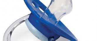
- habit of thumb sucking, prolonged sucking of pacifiers and other objects;
- infantile type of swallowing and the habit of inserting the tongue between the teeth;
- congenital malformations – cleft of the alveolar process of the lip and palate;
- endocrine disorders;
- tumors of the maxillofacial region.
Facial signs of an open bite: the mouth is half-open, but if it is possible to close the mouth, then the face is tense.
Patients complain of the inability to fully bite and swallow food, and often experience a lisp.
There are 3 degrees of severity of open bite depending on the size of the vertical gap: I degree - up to 5 mm, II degree - from 5 to 9 mm, III degree - more than 9 mm.
They also pay attention to which teeth meet in the lateral sections.
Deep bite.
A deep bite is one in which the upper teeth excessively overlap the lower teeth. Sometimes the lower teeth rest with their cutting edges on the mucous membrane of the palate, then they speak of a traumatic deep bite.
Possible causes (etiology) of deep bite:
- early loss of chewing teeth (due to injury or complications of caries leading to their removal, or their primary absence - adentia);
- violation of nasal breathing;
- incorrect type of swallowing;
- speech dysfunction;
- bad habit of sucking various objects;
- violation of the timing of teething, especially in the lateral parts of the dentition;
- early wear of temporary teeth.
As in the case of open bite, three degrees of deep bite are also distinguished, depending on the severity of the anomaly (that is, on the amount of overlap of the lower dentition with the upper one).
Facial signs of deep bite:
- turning the lower lip outward;
- expressiveness of the chin fold;
- shortening of the lower third of the face (sometimes doctors use the term “bird face”).
As a rule, with this malocclusion, patients complain of difficulty biting and chewing food, and pain in the temporomandibular joint often occurs, and headaches are possible. Very often a speech defect occurs - patients speak through their teeth.
Crossbite.
As the name suggests, in a crossbite, the teeth intersect each other when they meet.
With a crossbite, there is a discrepancy in the size of the jaws in the lateral region. Orthodontists classify this type of bite as a transversal anomaly, and the pathology can be unilateral or bilateral.
Crossbite occurs in both the anterior and posterior regions.
With a lateral type of bite, orthodontists distinguish the following types of this anomaly:
- when the lower jaw is displaced towards the tongue - lingual crossbite;
- towards the cheek – buccal crossbite;
- and towards the palate - palatal crossbite.
Causes of the anomaly:
- bad habits (listed above);
- trauma or damage to the jaw, including birth trauma;
- application of forceps during obstetrics;
- absence of individual teeth;
- disorders of the temporomandibular joint (TMJ) – ankylosis, habitual dislocation of the joint, underdevelopment of the joint on one side;
- indestructibility of the surfaces of baby teeth;
- violation of the sequence and timing of teething.
Frequent complaints from patients and parents:
- the presence of an aesthetic defect with a noticeable discrepancy in the size and position of the jaws;
- difficulty eating;
- violation of sound pronunciation;
- gum disease due to possible injury during chewing and speech;
- problems with the gastrointestinal tract.
As a rule, vertical malocclusions are combined with anomalies in the sagittal direction.
Causes of pathological bite formation
Any malocclusion occurs under the influence of the following factors:
- injuries received during childbirth;
- bad heredity;
- a number of diseases suffered by a woman during pregnancy;
- chronic diseases of the nasopharynx;
- mistakes when feeding a baby with a bottle or breast;
- multiple carious lesions of teeth;
- formation of mouth breathing;
- inadequate, unhealthy diet;
- relative lack of fluoride or calcium;
- exchange failures;
- injuries of the dental system;
- the child adopting an incorrect position during sleep;
- pacifier abuse at an early age;
- habit of sucking collar, finger, toys;
- incorrect posture;
- rickets;
- uneven wear of temporary teeth;
- prolonged replacement of temporary teeth with molars;
- long before they are replaced with permanent ones, removal of temporary teeth.
Distal occlusion - what is it?
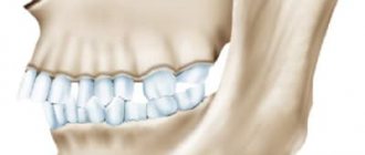
Distal bite (prognathia) is a type of anomaly when the teeth of the lower row or the entire jaw are significantly shifted back, which does not allow for the correct functioning of the entire dental system. Often the chin is smaller and looks underdeveloped, which is why the lower part of the face appears to be slanted - in common parlance this profile is called a “bird face”. From photographs taken before and after treatment of distal occlusion, one can assess the degree of distress and how much the anomaly deforms facial features.
The photo shows the profile of a person before and after treatment
The cause of the problem may also be the opposite situation, when the upper row of teeth is too forward, and the jaw protrudes significantly above the lower one. Often the pathology in question is complicated by deep occlusion. In such cases, orthodontic experts diagnose a deep distal bite.
Malocclusions: consequences and complications
Unfortunately, if left untreated, malocclusion can become one of the factors provoking the development of a number of pathological processes in the body. In particular, malocclusion can be a consequence of:
- changes in the principles of functioning of the dental system;
- increasing the load on some teeth and weakening it on others;
- accelerated abrasion of the enamel layer of teeth;
- noticeable increase in tooth sensitivity;
- changes in the shape of the face, loss of its symmetry, appearance of aesthetic defects, disturbances in facial expressions;
- speech dysfunction;
- increasing the risk of injury to the tongue, cheeks, gums;
- increasing the risk of periodontal disease;
- TMJ damage;
- the appearance of inflammatory processes;
- problems arising during prosthetic and implantation procedures;
- the appearance of night apnea;
- respiratory dysfunction;
- the occurrence of diseases of the nasopharynx, digestive organs, and respiratory tract.
What are the dangers of distal bite:
- inharmonious profile (“bird-like” face), small chin
The contour of the soft tissues of the face directly depends on the position of the teeth: if the upper teeth protrude, and the lower ones, on the contrary, are pushed back or crowded, then the profile will look ugly. The posterior position of the lower jaw entails a posterior position of the chin. In severe cases of distal malocclusion, the face appears bird-like.
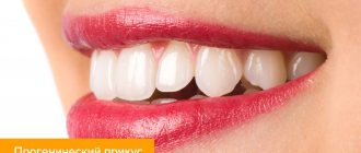
- protruding upper front teeth
Without being in a bite, in most cases the upper front teeth protrude forward strongly, sometimes a person cannot calmly close his lips
- crowded teeth
Due to the narrowing of the jaws with this pathology, there is a lack of space for teeth, which entails their crowding.
- deep bite and reduction of the lower third of the face
The lower jaw is in the posterior position (and/or the upper in the anterior position), causing the upper teeth to completely overlap the lower ones. A deep bite entails a reduction in the lower third of the face - this makes the person look older and the profile looks inharmonious. Often there is such a deep bite that the lower incisors injure the upper gum in the palate.
- difficulty breathing
ENT organs are directly related to the dentofacial apparatus. The maxilla is a bone that anatomically forms the floor of the nasal airway. The lower jaw, together with nearby soft tissues, influences the volume of the laryngopharyngeal space. The more posterior the jaw is, the smaller the space, which makes breathing more difficult.
- problems with posture, curvature of the spine
The jaw bones are closely connected to the musculoskeletal system. Malocclusion can be both a cause and a consequence of spinal problems.
- tooth wear
If there is a lumpy closure of the teeth, the enamel wears off faster and the teeth wear out.
- diction defects
Teeth are involved in the pronunciation of many sounds: for example, the sounds “t”, “d”, “f”, “v”, “s”, “z”, “sh”, “zh” and others. Misaligned teeth or a bad bite makes it difficult or impossible to pronounce certain sounds.
- pain in the joint (temple area), as well as clicking, crunching, etc.
Often the cause of pathology of the temporomandibular joint is a distal bite: the cartilage in the joint wears out under increased load, which gives the corresponding symptoms.
– high risk of injury to the front teeth
In the distal bite, non-occlusion of the upper and lower anterior teeth is most often observed (when viewed in profile). Left without underlying support, the upper front teeth cannot withstand the shock load - chips of the tooth crown, or even root fractures, occur.
- chronic inflammation of the lips (cheilitis)
If, due to a bite, the lower lip is constantly bitten, it becomes inflamed, chapped, cracked, and bleeds. Therefore, the cause of cheilitis is not always a lack of vitamins or windy weather.
- difficulty biting food
If the front teeth cannot be placed “joint to joint,” the function of biting food suffers.
The main stages of correcting malocclusion
Orthodontics deals with the correction of pathological occlusion. In this case, the therapy program includes several successive stages.
- Diagnostic measures. To make a correct diagnosis and prepare a treatment program, doctors may need radiovisiography, orthopantogram or CT data. The resulting images help to assess the severity of changes that have occurred in the structure of the dental system.
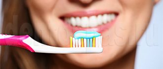
- Dental sanitation of the oral cavity (professional hygiene, treatment of caries).
- Fight against accompanying pathological processes.
- Installation of braces or other corrective structures.
- Retention measures that allow you to consolidate the results obtained during orthodontic treatment (wearing dental retainers).
Correcting a bite is a difficult, lengthy and expensive procedure that requires the patient to have great patience and a responsible approach to compliance with all medical instructions. However, only in this way can one achieve preservation of the aesthetics of the dentition and prevent the development of a number of pathological processes.
How long does treatment last?
How long it will take to correct a distal bite depends on the age of the person, the severity of the problem, and the type of corrective equipment used. For schoolchildren, the situation can return to normal within six months to a year. Adults most often have to see an orthodontist for several years.
There is no need to worry about the long treatment period. The most important thing is to cure the existing dental disease. After all, its consequences can be the most unfavorable.
Probable causes of pathology development
If the external signs of the problem are similar in all patients, then the reasons for the development of the anomaly can be very diverse. Let's consider the most common prerequisites leading to the formation of malocclusion in children and adults:
- problems with posture,
- genetic inheritance,
- serious injuries to the face and skull,
- abnormal jaw sizes as a congenital pathology,
- abnormal narrowing of the jaw shape,
- large or small sizes of incisors,
- disorders in skeletal bone development,
- acute deficiency of fluoride and calcium in the body,
- too little breastfeeding time, when the lower jaw is underdeveloped,
- underdeveloped chewing apparatus due to lack of hard food in the diet.
Often the problems can be hereditary.
The problem in question is quite serious, because it is more difficult to treat than others. An integrated approach is required, and often even braces alone will not help correct the situation. As a rule, if the pathology is clearly expressed, it is necessary to resort to surgical treatment protocols.
Treatment of malocclusions
Modern methods of orthodontic treatment include mechanical and functional means. Mechanical systems, in turn, are divided into removable (plates, aligners, etc.) and non-removable systems (braces) for correcting the bite.
Removable appliances are mainly used in children with primary or mixed dentition. For permanent occlusion, a non-removable technique (bracket system) is used. Braces are metal or ceramic plates that are attached to the teeth from the outside or inside. They are connected by a wire made of a special composite metal, which applies even pressure on the teeth and returns them to the correct position. Traditional records perform the same function. All these orthodontic devices allow not only to correct the bite, but also to put crooked teeth in place.
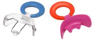
For children 7–10 years old, preventive devices are used - trainers, which affect the muscles of the maxillofacial area and tongue. They can stimulate or slow down jaw growth, change the width of the palate and the shape of the jaw bones, helping to align teeth during their eruption. This device does not require round-the-clock wearing; the child wears it only at night and does not experience any complexes due to problems with diction and appearance.
Rational and timely treatment leads to normalization of the bite and straightening of the teeth.
How to prevent the problem - preventive measures
Obviously, the problem can only be prevented in childhood. But for this, parents need to take note of the following preventive recommendations:
- If you have a choice, preference should be given to breastfeeding,
- the introduction of solid food into the diet should be gradual but timely,
- it is required to regularly visit specialists - not only the dentist, but also other specialized doctors, including for the prevention of chronic pathologies associated with the respiratory system,
- parents should monitor the child’s posture and, if there are visible deviations, immediately begin to correct them under the supervision of an appropriate specialist,
- You will also have to wean your baby off the pacifier and thumb sucking habit in time.
Thumb sucking in childhood can cause malocclusion in the future.
Distal bite is a fairly common problem, but this does not mean that the abnormality does not require close attention and appropriate treatment. Alas, the defect will not disappear on its own, and prolonged ignoring of the pathology will sooner or later lead to more serious consequences not only for the oral cavity, but also for the body as a whole.
- According to WHO.
Nota Bene!
It is never too late to treat malocclusions, but it must be remembered that the sooner treatment begins, the better the result.
Expert: Alesya Aleshkina, dentist
Prepared based on materials:
- Kurlyandsky V. Yu. Orthopedic dentistry. – M.: Medicine, 1977.
- Persin L. S. Orthodontics. – M., 1999.
- Guide to orthopedic dentistry / ed. A. I. Evdokimova. – M., 1974.
- Handbook of Dentistry / ed. A. I. Rybakova and G. M. Ivashchenko. – M., 1977.