How to avoid blisters on the roof of your mouth?
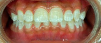
Doctors consider the oral cavity to be a mirror image of the entire body, signs of infection and disturbances in the functioning of all internal organs. The presence of blisters on the palate is not only unpleasant, but also very dangerous, as they are a gateway for infection.
In addition, they can be evidence of a severe pathology in the body, and their inaccessibility (unlike skin rashes) makes its diagnosis and treatment very difficult.
Blisters on the roof of the mouth can bother both children and adults, causing pain and discomfort. They need to be dealt with only with the help of a specialist.
Burn of mucous membranes
If you consume too hot liquid or food, a burn to the mucous membrane may occur.
There are 3 stages of damage:
- Redness of the tissue appears.
- A watery, transparent bubble appears on the roof of the mouth.
- Dying and rejection of burned tissues.
For mild to moderate burns, the oral cavity should be rinsed with antiseptic solutions; anti-inflammatory gels can be applied to the damaged areas. Before healing, you should avoid eating irritating foods so that the blister does not open and an ulcer forms on the roof of your mouth.
When to see a doctor
The appearance of a blister on the roof of the mouth is often a symptom of a dangerous pathology in the body and always requires contacting a specialist. Many possible reasons causing its appearance necessitate testing to establish the correct diagnosis.
First of all, you should contact a general practitioner (pediatrician), who, after a thorough examination, will refer you to a dermatologist, ENT specialist or oncologist.
If blisters appear not only on the palate, but also on other areas of the mucous membrane or skin, you should consult a dermatovenerologist. If your throat is affected (swelling and pain), then you have a sore throat and should be treated by an ENT specialist.
What not to do with blisters
The first thing that should not be done is to ignore the symptoms, since possible infections or cancer can cost the patient his life. Blisters on the palate always pose a danger in the form of infection entering the body and the development of a palate abscess.
What is prohibited to do during treatment:
- warm the throat so as not to increase suppuration;
- remove rashes yourself;
- If ulcers appear, the mouth should not be disinfected with alcohol-containing products.
How to make calendula decoction at home, watch this video:
Remember that you should not self-medicate, because very often it is possible to cure the underlying disease only with the help of antibiotics prescribed by a doctor.
How to avoid their reappearance
It is necessary to perform preventive procedures:
- carry out oral hygiene daily;
- regular visits to the dentist (once every six months);
- support immunity with vitamins;
- lead a healthy lifestyle;
- Healthy food;
- Monitor your blood sugar levels regularly;
- quit smoking;
- avoid spicy and too hot foods so as not to injure the mucous membranes.
It is important to follow all the doctor’s instructions and not interrupt treatment, otherwise you will not be able to avoid the recurrence of blisters.
A small number of rashes can be easily eliminated, and if the disease starts, the unpleasant consequences will not take long to appear. Strengthen your body and you will quickly defeat all bacteria and viruses.
Herpetic stomatitis
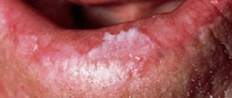
The disease is caused by the herpes virus, and blisters with cloudy liquid appear on the palate, tongue, inside of the lips, cheeks, and there is a burning and itching sensation in the mouth. The nasolabial triangle may also be affected. Before the bubbles appear, patients feel unwell, body temperature rises, mucous membranes hurt and itch, and regional lymph nodes become inflamed. The rashes are usually multiple and can merge into one large lesion.
After some time, the blisters on the oral mucosa spontaneously open. In their place, erosions remain; when an infection occurs, inflammation can develop and ulcers can form. According to the severity, herpetic stomatitis can be mild, moderate and severe.
Treatment is aimed at suppressing the herpes virus. Patients are prescribed regular treatment of the oral cavity with antiseptic agents, and anti-inflammatory and painkillers are applied to the affected areas. Immunomodulators, vitamins and antiviral medications are taken internally.
Drug treatment
The existence of a huge number of treatment methods does not mean that it is possible to quickly and easily eliminate both the blister itself and the pathology that caused it.
You can alleviate your condition a little on your own, and then only for a while.
The main treatment should be comprehensive and should be prescribed by a doctor. Medicines (sprays and ointments) purchased at the pharmacy help relieve pain, burning and itching. This especially applies to drugs that contain an antibiotic:
- rinse your mouth with an antibiotic solution;
- Apply the required amount of ointment (Vishnevsky, Tetracycline or Levomikol) to a cotton swab;
- Carefully treat the blister and the skin around it with ointment;
- wait until the product is absorbed and dry (leave your mouth open and tilt your head back slightly);
- The blister should be treated every 5 hours, after consulting with your doctor;
- can be treated with iodine, hydrogen peroxide or potassium permanganate (a very weak solution);
- Irrigating the mouth with Chlorhexidine and rinsing with Iodinol will help get rid of the infection;
- pharmaceutical tablets (for resorption): Gramidin, Septolette, etc. help get rid of infection in the oral cavity.
However, they should be used very carefully so as not to damage the palate. If blisters appear (without headache, cough or fever), you must urgently contact a specialist to diagnose and determine the exact cause of the disease.
Professional treatment includes:
- rinsing with antiseptics;
- use of anti-inflammatory drugs;
- anesthesia;
- antibacterial therapy;
- use of antiviral and antifungal drugs;
- treatment with antihistamines (for allergies).
In addition to prescribed medications, the patient must follow certain medical recommendations.
Treatment with folk remedies
The use of folk remedies to treat blisters on the roof of the mouth perfectly complements medication treatment. Let's look at the most effective of them:
- Soda solution: 1 tsp. dissolve soda in 1 tbsp. water (warm). This remedy relieves inflammation and has an antimicrobial effect.
- A decoction of oak bark will help remove the infection and also relieve redness: 1 tbsp. l. bark pour 1 tbsp. boiling water, put on fire and simmer over low heat for about half an hour, then cool. Rinse your mouth with the resulting decoction 5-6 times a day.
- Aloe compresses: remove the skin from 1 leaf of the plant and knead thoroughly, wrap the pulp in gauze and apply to the blister for 20 minutes.
- A decoction of calendula helps relieve itching from a blister on the roof of your mouth and is also an excellent antiseptic. Use for rinsing.
- Goldenseal infusion: 2 tsp. plants pour 1 tbsp. boiling water, leave for 2 hours and use for rinsing.
- Decoctions of rose hips, propolis and chamomile provide an excellent anti-inflammatory effect. In addition, they are all absolutely safe.
To treat blisters, you should stop the underlying pathology, and not hope that the disease will go away on its own. If treatment is not started in time, the situation will only get worse.
Dühring's dermatitis herpetiformis
This is a skin disease caused by intestinal dysfunction. Patients develop painful blisters on the skin and mucous membranes of the mouth. External signs are very similar to those of herpes.
The rashes come in different sizes and types: they can be tense with clear liquid, covered with a crust, or have the form of a papule. Their appearance is preceded by general malaise, chills, itching, and burning. The bubbles are most often localized on the hard palate, cheeks, and mouth. The disease is chronic, so relapses occur periodically.
After 3 days, the blisters on the mucous membrane in the mouth open and form erosions. After another 3 days, the wounds heal, leaving an inflamed area or a small scar in their place.
The disease can develop at any age, but most often affects men 30–40 years old. For treatment, sulfone drugs, vitamins, antihistamines, corticosteroids, and a special diet are prescribed.
Pathohistology of pemphigus
At the onset of the disease, intercellular edema of the epithelial layer and destruction of intercellular bridges in the lower parts of the germ layer appear. As a result of the loss of communication between epidermocytes, cracks are formed first, and then blisters , predominantly of suprabasal localization. The basal cells, although they lose contact with each other, remain attached to the basement membrane. The cavity of the bladder, as a rule, contains round acantholytic cells with large hyperchromatic nuclei and pale cytoplasm. During the healing process, proliferation of the papillae and elongation of the epithelial outgrowths are noted. The infiltrate consists of eosinophilic granulocytes, plasma cells and lymphocytes.
In other words, the basis of the pathological process in pemphigus is acantholysis - a disruption of the connection between the cells of the spinous layer as a result of disruption of the desmosome - tonofilament complex, resulting in the formation of acantholytic cells, which are characterized by a large nucleus. The cytoplasm of the cells is strongly basophilic and pyroninophilic, which is due to the high content of RNA in it. In acantholytic cells there is a high level of protein metabolism and oxidative processes (exceeding the metabolism of spinous cells), which indicates their secretory activity. It is assumed that acantholytic cells originate from the basal and lower rows of spinous cells.
Vesical vascular syndrome
In people suffering from hypertension or cardiovascular diseases, a dense blister may appear in the mouth on the cheek, soft palate, or tongue. It looks like a single red bubble that stays in the mouth for up to 2 days. This manifestation is called bladder syndrome. The cause of blisters is the rupture of small blood vessels in the mouth when blood pressure rises.
After perforation of the bladder, erosion is formed, which epithelializes after 3–5 days. When infected, suppuration occurs and a deep trophic ulcer is formed.
Vesicovascular syndrome is most often observed in women over 40 years of age. Treatment is carried out under the supervision of a cardiologist.
Treatment options for inflammation and pain in the palate
Some causes of inflammation of the palate are quite dangerous, so treatment should begin immediately after diagnosis. Treatment tactics depend on why the mucous membrane lining the upper part of the oral cavity is inflamed.
In case of mild mechanical or thermal damage to the palatal tissues, you can rinse your mouth with a solution of soda and salt or tinctures and decoctions of herbs: chamomile, sage, oak bark. To relieve pain caused by a person burning the oral mucosa while tasting a dish, it is enough to rinse your mouth with cold water or a soda-salt solution.
If the palate is quite inflamed, you should resort to the use of local anti-inflammatory and analgesic medications. To quickly remove pain and itching, you should supplement traditional drug therapy, and also eliminate the risk of infection in small scratches and sores.
To get rid of inflammation of the palate, you should treat not only the symptoms, but also the disease that provoked its appearance. For example, if you have a sore throat, you need to take antibiotics; the therapeutic course can last 1–2 weeks. Fungal diseases are treated only with antifungal drugs in the form of ointments, sprays and gels. Viral - antiviral. All infectious pathologies indicate a weakened immune system, and therefore require a therapeutic course to strengthen it.
If the palate is inflamed due to dental caries or pulpitis, treatment by a dentist will be required. A person will not be able to get rid of the disease until his teeth are treated, since caries is a constant source of infection in the mouth.
Essential medicines
Wounds and ulcers formed due to inflammation of the upper palate should be treated with antiseptics, for example, Rotokan, Chlorhexidine or Furacilin. The following drugs fight fungal inflammation:
- Viferon, Pimafucin and Nystatin ointments.
- Clotrimazole cream.
- Nizoral.
- Borax with glycerin.
If the roof of your mouth hurts too much, you can make warm infusions for rinsing with chamomile, propolis, eucalyptus or oak bark at home. But it is better to purchase an anesthetic in the form of a spray at the pharmacy: Hexoral, Lidocaine Asept or Benzocaine. Dental gels such as Cholisal, Lidochlor, Kamistad have mild analgesic properties. Their main function is to relieve inflammation, and their secondary function is anesthesia.
If inflammation of the palate, accompanied by swelling, is caused by viral agents, antiviral medicinal sprays Miramistin, Lugol, as well as Acyclovir ointment are suitable. If the palatal tissues are swollen due to viral stomatitis, you can use folk recipes for medicines that can be easily prepared at home. The main ingredients in them are sea buckthorn and rosehip oils, propolis tincture.
Erythema multiforme exudative
An inflammatory disease of the mucous membranes and skin is called erythema. The acute course is manifested by the formation of blisters, papules, and blisters in the mouth. The course of the pathology is long-term with the occurrence of periodic
relapses. The rashes are most often localized on the inside of the lips, cheeks, tongue, soft palate, and floor of the mouth.
Before the appearance of blisters, patients complain of general malaise, fever from 37˚ to 38˚, burning in the mouth, aches throughout the body. Afterwards, hyperemic spots appear, in the center of which a bubble filled with serous fluid forms. Painful sensations are constantly present. Patients cannot talk or eat.
The blisters break open after a few days, and in their place erosions, covered with a fibrous coating, form. When wounds become infected, inflammation occurs, the ulcers become covered with a yellow-gray coating, which is also found on the teeth and tongue. Regional lymph nodes become inflamed and salivation increases.
An exacerbation lasts 2–3 weeks, healing of erosions occurs in 7–10 days without tissue scarring. Treatment consists of taking disensitizing, anti-inflammatory drugs, and vitamins. Antiseptic treatment of the oral cavity and erosions is carried out locally. Severe forms of erythema are treated in a hospital setting under the supervision of a physician.
Pemphigoid (non-acantholytic pemphigus)
It is believed that autoimmune processes play an important role in the pathogenesis of the disease. All types of pemphigoid are characterized by the absence of acantholysis in epithelial cells, subepithelial location of blisters, severe inflammation, absence of acantholytic cells, and negative Nikolsky's sign. The symptom of subepithelial perifocal detachment may be positive.
Pemphigoid is benign, the general condition of patients suffers little, and the prognosis for life is favorable.
Bullous pemphigoid (pemphigoid bullosa) usually occurs in people over 60 years of age. The skin and mucous membranes of the mouth, nose, and genitals are affected. In approximately 10% of cases, the disease begins with damage to the oral mucosa.
The clinical picture is characterized by the formation of subepithelial large tense blisters on erythematous or unchanged epidermis and oral mucosa. Bubbles 0.5-2 cm in diameter are most often localized in the inguinal folds, lower abdomen, and flexor surfaces of the upper extremities.
The oral mucosa is affected in approximately one third of patients with bullous pemphigoid. Against the background of edematous and hyperemic oral mucosa, tense blisters with serous or hemorrhagic contents with a diameter of 5-20 mm appear. They persist for several hours, sometimes days, then erosions covered with fibrinous plaque form in their place. Unlike acantholytic pemphigus, erosions epithelialize after 10-15 days without scarring or atrophy, but can reappear after a certain time. The favorite localizations of blisters in non-acantholytic pemphigus are the border of the hard and soft palate and cheeks. Rashes on the oral mucosa are almost painless; on the skin they are accompanied by itching, burning, and pain.
Sometimes the pathological process can be localized on the gum, while the mucous membrane of the gingival margin on the vestibular surface is hyperemic, swollen, and bleeding. Nikolsky's sign in this case is often positive, but no acantholytic cells are detected. The course of the disease is persistent and long-term, with periodic remissions.
Over time, the severity of bullous pemphigoid weakens and it may disappear spontaneously.
Pathohistological changes are characterized by the formation of subepithelial blisters. In the dermis and the lamina propria of the mucous membrane, swelling and a massive infiltrate, consisting mainly of eosinophilic granulocytes, are observed.
Dühring's dermatitis herpetiformis is characterized by a polymorphism of skin rashes accompanied by burning and itching. Lesions of the mucous membranes are uncharacteristic. There is eosinophilia in the blood and exudate of the blisters.
Exudative erythema multiforme, unlike bullous pemphigoid, occurs acutely, with fever and malaise. Mostly young people are affected. With exudative erythema multiforme, blisters are located on the edematous, hyperemic mucous membrane of the mouth and skin. With treatment, the process resolves quickly.
The occurrence of bullous toxicoderma, in contrast to bullous pemphigoid, is usually associated with taking medications. In addition, rashes on the oral mucosa and skin during bullous toxicoderma are polymorphic (erythema, vesicles, blisters) against the background of general disorders of the body (fever, weakness, malaise).
- Cicatricial pemphigoid (pemphigus cicatricans)
In the oral cavity, blisters measuring 0.2-1.5 cm appear on hyperemic or unchanged mucous membranes. The most typical localization is the soft palate, uvula, tonsils, and buccal mucosa. The blisters are tense, have a dense covering, with serous or hemorrhagic contents. When the blisters are opened, deeply located erosions of a meat-red color are formed, not prone to peripheral growth. A characteristic feature is the ability to form blisters in the same places, resulting in cicatricial changes in the mucous membrane, leading to the development of adhesions and strictures.
When blisters are localized on the conjunctiva of the eyes, cicatricial changes can develop, leading to its wrinkling, limited mobility of the eyeball, cicatricial deformation of the lacrimal canals, ulceration of the cornea and blindness.
Damage to the red border of the lips by cicatricial pemphigoid can contribute to the development of microstomia. Nikolsky's symptom is negative. No acantholytic cells are found in fingerprint smears.
On the skin, blisters are most often localized on the scalp, face, limbs, and inguinofemoral folds. Since blisters appear in the same places, after their elimination smooth atrophic scars remain.
- Benign nonacantholytic pemphigus
The clinical features of the course are almost no different from non-acantholytic pemphigus. The blisters are tense, with a dense covering, serous or serous-hemorrhagic contents (3-10 mm in diameter), located subepithelially on the unchanged or slightly hyperemic oral mucosa, without the phenomena of acantholysis. In some patients they occur almost continuously, in others - with remissions from several days to several weeks. A few hours after the appearance of the blisters, they open with the formation of slightly painful, rapidly epithelial erosions. Subjective complaints of patients are reduced to a feeling of slight burning and tightening of the mucous membrane. After 1-2 weeks, erosions epithelialize without leaving a scar.
Pemphigus
A flaccid transparent bubble has appeared in the mouth, what is it? This may be a manifestation of an autoimmune pathology - pemphigus. It most often affects people over 50 years of age. There are several types of disease:
Pemphigus vulgaris is manifested by the formation of complete blisters, candidiasis may occur, and a white cheesy coating will appear. A specific smell appears in the mouth. Later the rash spreads throughout the body.- Pemphigus foliaceus. After opening, the blisters become covered with lamellar crusts, under which new rashes constantly form. A thick layer of crusts forms. If infection occurs, patients feel deteriorating sharply and their body temperature rises. The mucous membranes are rarely affected.
- Pemphigus vegetans. When the blisters open, erosions covered with crusts form. Folding of the tongue may occur.
- Erythematous pemphigus. The first rash appears in the center of the face and takes the shape of a butterfly. The blisters become covered with crusts, which, when rejected, leave scars. Later the disease takes a generalized course.
Pemphigus is a dangerous disease, it can be benign and malignant, and therefore requires immediate treatment from a dermatologist and dentist.
Etiology and pathogenesis of pemphigus
The etiology of pemphigus is not fully understood.
The infectious (viral) theory of pemphigus is popular, but so far it has not been possible to isolate the virus, and there is also no evidence of the contagiousness of this disease.
The theory of neurogenic origin was based on the constant detection of degenerative changes in the nervous system during pathological studies, but the primacy of these changes has not been proven. They are apparently secondary, arising as a result of a sharp metabolic disorder.
There are also theories of endocrine and toxic origin.
A major role is currently assigned to autoimmune mechanisms of disease development. This theory is based on the detection in the blood of patients with pemphigus of circulating antibodies of the IgG type, which are related to the intercellular substance of the spinous layer of the epidermis; the amount of antibodies depends on the severity of the disease.
Studies by A.G. Avtanuilov and N.A. Mashkilleyson showed that in pemphigus, the DNA content in the nuclei of acantholytic cells is increased, and there is a direct connection between the increase in the level of nuclear DNA and the severity of the disease, which apparently leads to a change in the antigenic structure of these cells and the production of IgG autoantibodies against them.
According to N.A. Mashkilleyson, an important role in the pathogenesis of pemphigus belongs to changes in T- and B-lymphocytes, and if responsibility for the activity of the pathological process lies with B-lymphocytes, then the number and functional state of T-lymphocytes determine the occurrence and course of the disease.
Among the conditions for the development of the disease, damage to the mucous membrane, emotional trauma, and blood changes also occur.
Epidermolysis bullosa
This is a genetic pathology that affects newborn children. The disease has several forms (simple, borderline, dystrophic), the clinical manifestations depend on this. With any of its types, thinning of the skin and mucous membranes is observed; with a minor injury, a transparent bubble with liquid can form in the mouth, on the palate, or in any part of the body.
First, a tense blister filled with cloudy fluid appears on the affected area in the mouth. After opening it, painful erosions and ulcers form, and candidiasis may occur. After healing of deep wounds, the tissues scar and lead to deformation of the mucous membranes and malocclusion.
The pathology can affect any internal organ, skin, bones, eyes, hair and nails. Unfortunately, the pathology is incurable.
Vegetating type of pemphigus
The vegetative type of pemphigus is benign. Patients can feel well for many years. Bubbles are located around the holes, as well as in the area of skin folds. When the bubbles open, they reveal erosions, at the bottom of which soft vegetations appear that have a fetid odor. Such vegetations are covered with serous-purulent or simply serous fluid. Pustules can be seen along the contour of the neoplasms, so pemphigus vegetans must be distinguished from the chronic form of pyoderma. Nikolsky syndrome will be positive only near the affected areas of the skin, however, in the terminal stages, pemphigus vegetans is similar to pemphigus vulgaris in its clinical manifestations.
Diagnosis of pemphigus
An important diagnostic sign of pemphigus vulgaris is a positive Nikolsky sign . There are 3 methods. One Nikolsky symptom does not confirm the diagnosis of pemphigus; it is also observed in medicinal stomatitis. In this regard, the diagnosis of pemphigus must be confirmed by cytological studies - detection of Tzanck cells. Acantholytic cells are round and smaller than normal cells of the spinous layer, the nucleus is large relative to the entire cell, its diameter is 1/3-1/2 or more than the diameter of the cell, the nucleus is loose with 1-6 lighter nucleoli; the cytoplasm is two-layered - light (perinuclear zone) and dark blue (peripheral).
With pemphigus vegetans, the cytological picture is identical to that with pemphigus vulgaris. However, in seborrheic and pemphigus foliaceus, multinucleated cells are not found, acantholytic cells are found in smaller numbers, are monomorphic, and often have an oval and irregularly triangular shape.
You should also use the immunofluorescent method, which allows you to detect in the blood serum of patients with pemphigus antibodies to the intercellular substance of the spinous layer of the epidermis (with indirect RIF) of the IgG type and IgG deposits in the area of the intercellular substance and the membrane of the cells of the spinous layer (with direct RIF).
Differential diagnosis of pemphigus and diseases that make up the group of blistering dermatoses is based on symptoms associated with the localization of blisters in relation to the epithelium. With pemphigoid, the blisters are located subepithelially, so they have a thicker cover than with pemphigus and last a longer time, and therefore, when examining the oral mucosa in such patients, blisters with transparent contents are observed, which is impossible with pemphigus.
Erosions formed at the site of blisters with pemphigoid are usually located on a slightly hyperemic base, their surface is often covered with fibrinous plaque.
With pemphigus vulgaris, the oral mucosa around the erosions is not externally changed, and the erosions themselves can be covered with a collapsed bladder cover, which is very easily removed with a spatula. Acantholytic cells and a positive Nikolsky sign distinguish pemphigus vulgaris from non-acantholytic pemphigus.
For pemphigus, RIF allows one to determine deposits of immune complexes containing IgG in the area of the membranes of spinous cells and the cementing substance between them. With indirect RIF, circulating IgG is determined.
In non-acantholytic pemphigus, these same immune complexes are also found in the basement membrane area.
A blistering rash as a manifestation of an allergy to drugs helps to distinguish between anamnesis (medication intake) and relatively rapid healing after discontinuation of the causative drug.
The blisters are located subepithelial. There is no acantholysis, Nikolsky's sign is negative, acantholytic cells are absent. Allergy tests clarify the diagnosis. Blisters in the bullous form of lichen planus also occur under the epithelium, but there is no acantholysis. Around the bladder or in other areas of the mucous membrane, typical papules of lichen planus can be observed.
Diagnostic methods
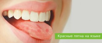
Rashes may indicate the presence of a pathology such as ordinary stomatitis or candidiasis. However, often such a symptom indicates dangerous diseases occurring in the human body. It is for this reason that when a rash appears on the palate, it is imperative to undergo a thorough diagnosis and not self-medicate.
The patient examination includes the following activities:
- General examination of the areas of the rash and examination of the medical history. In addition, the presence of any additional symptoms must be clarified.
- Laboratory research. Based on the results of a general blood test, you can find out the cause of the rash and make an accurate diagnosis. An increase in the number of leukocytes or erythrocyte sedimentation rate indicates an inflammatory process or an infectious disease. Against the background of an allergic reaction, an increase in the number of eosinophils is noted.
- Analysis of images of the contents of the rashes themselves. In fungal pathology, micelles of pathogenic fungi can be seen when examined under a microscope. Signs of cancer are the presence of atypical cells in the samples.
Based on the results of the studies, the doctor may prescribe additional tests for HIV infection or syphilis, as well as a thorough dermatological examination.
We recommend watching the video:
What you can do on your own
If red spots are detected in the oral cavity, it is recommended to immediately consult a doctor and not self-medicate. It is possible to make an accurate diagnosis and select effective therapy only after laboratory tests and determination of the cause of the disease.