Non-carious lesions of teeth is a summary concept used mainly in the domestic medical literature. In foreign dentistry, other terms are used, for example, “pathology of non-carious teeth,” and each pathology or disease is considered separately.
As the name of this concept suggests, non-carious lesions of hard dental tissues refer to all lesions, diseases, damage to enamel, dentin, cement that are non-carious in nature, i.e. without the participation of bacterial microflora. Non-carious lesions and diseases can have extremely diverse clinical signs, causes and patterns of development. They can be hereditary (therefore genetic studies and consultations are necessary), congenital, acquired, they can be limited to one single tooth or generalized (affect all dentitions), they can affect different areas in a certain chronological order, depending on the stage of development of the dental system.
Unfortunately, despite the long period of research into non-carious lesions of dental tissue, this group of dental diseases has not been studied fully enough; there are often no uniform descriptions of clinical pictures, possible complications and prognoses of their development. This significantly complicates making an accurate diagnosis, choosing the right treatment and prevention.
Causes of development and prevalence
Statistics on the average prevalence of non-carious dental lesions are very contradictory. But they are believed to be the second most common after caries, and their share is 1-2% of the population (but according to some studies, the share of such diseases that affect tissues even before teething reaches 5-14%). There is evidence that the incidence of such non-carious pathologies as wedge-shaped defects and hyperesthesia is highest in women aged 45-65 years.
The causes of non-carious lesions can be very diverse and are not well understood. In general, the common cause is considered to be impacts (including failures) of various types that disrupt amelogenesis, dentinogenesis, and mineralization processes.
Non-carious lesions of teeth classification
The classification of non-carious lesions is characterized by quite a wide variety, and there are no generally accepted views on it. In foreign practice, classification is found mainly only according to chronological criteria (before teething and after teething). Summarized data on the classification of non-carious lesions can be presented in the following table.
| Classifying features | Type or type of non-carious lesion |
| General changes in hard dental tissues | Hypoplasia (has the greatest significance). Underdevelopment (in terms of quantity) of dental tissues, hereditary or acquired. Hyperplasia. Redundancy of hard dental tissues, hereditary or acquired. Dysplasia . Violation of the proper maturation and differentiation of hard dental tissues. Sometimes hypoplasia and hyperplasia are considered special cases of dysplasia. Necrosis . Destructive destruction of hard dental tissues without the participation of bacterial microflora (unlike caries). Sclerosis . Increased and pathological compaction of hard dental tissues. |
| Clinical nature of the lesion process | Hereditary dysplasias (for Stainton-Capdepont disease, amelogenesis imperfecta and dentinogenesis) Abnormalities and lesions dental tissues in some congenital pathologies (Hutchinson-Fournier teeth, congenital porphyria, osteopetrosis, hemolytic anemia, ectodermal dysplasia). Increased or decreased abrasion of dental tissues. Pathologies associated mainly with the influence of external factors (microdentia, tetracycline teeth, local hypoplasia, wedge-shaped defects, erosion, fluorosis, traumatic lesions). Hyperesthesia . Increased sensitivity of teeth. |
| Terms of formation and development of the disease | Before teething , i.e. during the period of tooth formation (for example, hyperplasia and hypoplasia, some forms of fluorosis, hereditary and acquired dysplasia, amelogenesis imperfecta, dentinogenesis, osteogenesis, drug-induced dysplasia, hypophosphatesia). After teething (for example, pathological abrasion or wear of teeth, wedge-shaped defects, erosive lesions, some forms of fluorosis, hyperesthesia, trauma). Sometimes this also includes some types of discoloration and staining of teeth. |
Treatment of fluorosis
Fluorosis
– damage to tooth enamel, accompanied by the appearance of white, yellow, brown spots on it and destruction of the tooth structure. A chronic disease that does not allow for long delays in treatment and an indifferent attitude towards the destructive process. Fluorosis is a systemic pathology that manifests itself on the teeth.
Causes of the disease: excess fluoride in the human body. The main source of this element is drinking water, so most often the cause of enamel fluorosis is an increased dose of fluoride in the composition of consumed liquids. Obviously, fluoride is found not only in water, but it is absorbed from food in much smaller quantities.
Types of fluorosis
Fluorosis can be divided into two types: endemic (associated with high levels of fluoride in drinking water) and professional (occurs in people who spend a lot of time in enterprises and premises where the fluoride content in the air exceeds the sanitary standard). The concentration of fluoride in water should not exceed 1.5 g/ml. Most often, enamel is affected in children whose skeletal system and enamel are not yet fully formed and who live in an area of water with a high fluoride content. For adults there is a limit - 5-6 g/ml.
Stages of fluorosis and its treatment
- The initial stage of fluorosis is characterized by the formation of white spots on the vestibular surface of the teeth. Treatment is carried out by bleaching, remineralization, fluoridation, and grinding of the affected areas.
- Fluorosis up close! The initial stage, which manifests itself in chalky white spots, smooth to the touch, sometimes light yellow spots.
- The streak form is characterized by the appearance of small white dots and streaks on the teeth. Treatment consists of eliminating the root cause, using filters or bottled drinking water. There are special filters that can be used to reduce the concentration of fluoride in water. Dental treatment is carried out in several ways depending on the manifestation of the disease: whitening, restoration of damaged enamel structure with the help of remineralizing agents, filling defects.
- The spotted form is distinguished by the appearance of light yellow spots. Upon visual examination, the shine and smoothness of the affected enamel is noted. The affected area increases, small spots merge into large ones and cover several teeth at once. The treatment consists of carrying out measures that are used in line form. For significant defects, the doctor may consider it advisable to install veneers.
- The chalky-mottled form provokes the appearance of dull spots on the surface of all teeth, as well as the appearance of dark brown pigmented spots, yellowing of the enamel with the formation of multiple small specks and spots. Treatment is carried out using artistic restoration or installation of veneers.
- The erosive form is characterized by the formation of significant dental defects. Round yellow or brown lesions appear on the enamel, and tissue loss is observed. With such manifestations of fluorosis, remineralization and bleaching will be ineffective. Treatment is carried out by filling with composite materials, installing veneers or crowns.
- The destructive form is the most difficult, as there is rapid destruction of the tooth structure, disruption of the shape of the crowns, fragility of the teeth, and possible chipping and cracks. Multiple aesthetic defects are accompanied by dysfunction and cause pain. Treatment of the destructive form is carried out using therapeutic and orthopedic methods. If possible, teeth are restored using composite materials through restoration. Significant defects and chips of enamel require the installation of crowns or dentures.
Prevention and treatment of non-carious lesions of hard dental tissues
Non-carious diseases are characterized by great diversity and only general principles of their prevention and treatment can be indicated.
Prevention of non-carious dental lesions is aimed mainly at increasing the body’s overall resistance, strengthening the dental structure and includes vitamin therapy and taking medications with microelements, adjusting the diet to optimize calcium salts, and treating concomitant diseases, including during pregnancy.
Treatment of non-carious dental lesions depends on the type of disease and its stage, but almost always begins with remineralizing therapy. In the acute period, temporary filling is performed with ionomer cements, and in a gentle manner, with minimal preparation. This is preparation for further functional filling with composite materials (elimination of factors of functional overload of the dentition) and aesthetic restoration. In general, modern methods make it possible not only to prevent further development of lesions, but also to ensure the full functioning of the dental system.
Request a consultation
Destruction of tooth enamel
Necrosis of tooth tissue
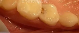
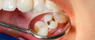
- this is a non-carious destruction of the structure of the enamel and dentin of teeth due to the influence of negative endogenous and exogenous factors, it is considered a very difficult and serious illness, which often ends in a serious deterioration or even loss of chewing function. It should be noted that it has become more common in recent years than 10-15 years ago.
Let's take a look at the most common forms of necrosis of hard dental tissues.
Chemical (acid) necrosis
It can develop as a result of exposure to various acids (acidic products) on dentin and enamel. Tooth decay can go unnoticed, since the enamel is not damaged in the early stages. This happens either as a result of a negative production factor (which is a high concentration of acids and various other substances within the workplace), or due to frequent or constant exposure to acid-containing products, medications, and drinking. The enamel softens and gradually breaks down, exposing dentin, which is a fairly soft tissue.
Getting carried away with drinks like Coca-Cola increases the prevalence of this pathology and complete tooth destruction. Further, pain may occur when exposed to temperature and chemical stimuli. As dentin loses enamel, it becomes dark.
Its treatment depends on the degree of manifestation of chemical necrosis of teeth. When teeth are completely destroyed, implantation is performed. Thus, the scope of therapeutic measures in the initial forms can be limited to stopping or maximally reducing the effect of chemical reagents on the teeth, as well as when carrying out complex remineralizing therapy for a period of 3 to 6 months. Our clinic has developed effective schemes for both treatment and prevention of this pathology.
Computer necrosis of teeth
In recent years, dentists have encountered a completely new and completely unexpected, as well as practically unstudied pathological process in teeth, which is characterized by systemic damage. It is more intense than from radiation therapy.
It is most often observed in young and middle-aged people who have been working with computers of different models for more than five years, in combination with non-compliance with the work schedule, professional protection, and occupational hygiene. Often, the side of the jaws facing the monitor is affected more intensely. The main foci of necrosis can cover both a significant and most of the crowns of the teeth. The lesions are characterized by pigmentation and painlessness, as well as tooth decay.
Is it possible to avoid these defeats?
It is recommended to follow certain standards when working with a PC:
- A workplace area of less than 6 m2 when the volume of the entire room is from 20 to 24 m2 is unacceptable;
- The location of natural light is on the left;
- If there are 2 computers (or more) in the workroom, then the distance between video monitors should be 2 meters or more (if they are directed in the same direction);
- Users should be at a distance of 0.6 to 0.7 m from the screen;
- After 2 hours of work, you should take a 15-minute break and ventilate the room;
- The total duration of computer use should not exceed 6 hours (for adults), and for children and adolescents - 1-2 hours. For women during pregnancy, using a computer is extremely undesirable in order to avoid tooth decay.
If the disease manifests itself, treatment is carried out in accordance with the clinical situation. At the initial stages of the pathology (when the integrity of the enamel is not compromised), deep fluoridation using APF gel is prescribed. In later stages of the lesion, restoration of the shape of the teeth using artistic restoration or orthopedic structures is used. Implantation in case of complete destruction is also practiced. It would not be superfluous to conduct an examination of the whole body.