Author of the article:
Soldatova Lyudmila Nikolaevna
Candidate of Medical Sciences, Professor of the Department of Clinical Dentistry of the St. Petersburg Medical and Social Institute, Chief Physician of the Alfa-Dent Dental Clinic, St. Petersburg
Before talking about the treatment of pulpitis with unformed roots, you need to understand what unformed roots are.
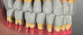
After the eruption of baby teeth, their roots have an uncovered apex for several years, which is why they are called unformed.
Symptoms of pulpitis in children
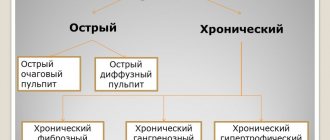
Symptoms and treatment of pulpitis in children vary depending on the form and severity of the disease. Pulpitis has two forms - acute and chronic. The first is relatively less common, but the symptoms in this case are more pronounced. There are two stages of the inflammatory process:
- Serous. The pulp becomes inflamed, and the tooth canals are filled with serous fluid. The child experiences severe pain in the tooth, which intensifies at night or with exertion—chewing on the side of the causative tooth. Often there is only one episode of pain. Within a few hours, the inflammation moves into the next stage.
- Purulent. Purulent contents begin to form in the root canals of the tooth. The severity of this stage depends on several parameters: the state of the child’s immune system, the activity of microorganisms, the state of the root system of the tooth, and the age of the young patient. The pain can be tolerable if the child’s immunity functions normally, microorganisms multiply slowly, and pus escapes through the carious cavity of the tooth. However, more often there is quite pronounced pain, prolonged attacks, and the unpleasant sensations radiate to other teeth. They intensify with mechanical, temperature effects on the causative tooth; the child’s appetite worsens or he refuses food altogether. In rare cases, there is an increase in body temperature and enlarged lymph nodes.
It is worth noting that chronic pulpitis can be asymptomatic, and there are often cases of the development of an inflammatory process in a previously filled tooth. The fact is that it can be quite difficult to properly treat the carious cavity of a child’s tooth before filling it - this is due to the emotional state of the patient and the forced haste of the dentist. This leads to the fact that the filling is installed in a cavity that is not sterile or is not sufficiently dried from saliva - pathogenic microorganisms continue to multiply, penetrating deep into the tooth and causing pulpitis.
With chronic pulpitis, pain can only occur when food debris gets into the carious cavity. The gangrenous form of the disease is characterized by the appearance of pain some time after eating hot food, a feeling of fullness and heaviness in the tooth, and bad breath.
Pulpitis
Treatment of patients with pulpitis
Treatment of children with pulpitis should be carried out taking into account the fact that foci of chronic infection of odontogenic origin can adversely affect the course of general diseases. In this regard, there is a need to reconsider the issue of indications for the sanitation of temporary teeth with chronic forms of pulpitis.
The question of the advisability of treating a child with chronic pulpitis of a primary tooth should be decided after an X-ray examination and comparison of the clinical and radiological manifestations of the disease.
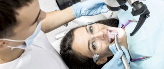
When visiting the dentist, the biggest fear is the drill. Dental interventions are more likely than others to be associated with pain and other unpleasant sensations. Therefore, the problem of premedication is especially relevant in pediatric dental practice. Psychological and pharmacotherapeutic effects on restless children with heightened emotional reactions relieve excessive stress.
For premedication, minor tranquilizers are prescribed - sibazone and mebicar in an age-specific dosage 30-40 minutes before the start of treatment. For young children, it is preferable to use sibazon, and to achieve a stronger tranquilizing effect, a combination of sibazon with mebikar.
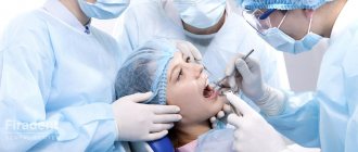
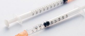
For pulp anesthesia, various methods of anesthesia are used: infiltration, conduction, application, intraligamentary, reflex analgesia, electrical anesthesia, as well as anesthesia: mask, intubation, intravenous.
When treated with vital pulp extirpation, intrapulpal anesthesia is sometimes used using a needle-free injector.
Traditional methods of anesthesia - conduction and infiltration - cause a negative reaction in children in the form of fear of the syringe and needle. In this situation, intraligamentary anesthesia is the most appropriate, which has been increasingly used in dental practice in recent years. There are only isolated studies on the use of this method of pain relief in children. In pediatric dental practice, domestic ointments are used for topical anesthesia: 5% pyromecaine and 5% pyromecaine with methyluracil on collagen.
General anesthesia is performed for children who cannot tolerate the anesthetic, with an unbalanced psycho-emotional state, accompanied by fear, fainting, increased gag reflex, as well as children with epilepsy, cerebral palsy, mental retardation, etc. In pediatric dental practice, mask anesthesia with a mixture of fluorothane, nitrous oxide and oxygen is more often used for general anesthesia. By inhibiting salivation, fluorotane has a great narcotic effect (50 times more active than nitrous oxide), ensuring rapid entry into a state of anesthesia and restoration of consciousness. The use of this method provides highly effective anesthesia of the pulp.
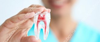
Pulp tissue has a significant ability for reparative and plastic processes. The main task in the treatment of children with pulpitis is to eliminate odontogenic infection and, if possible, preserve the viability of the pulp, especially its root part, as well as the prevention of periodontal diseases.
During treatment, it is very important to provide conditions for the completion of the formation of teeth, temporary and permanent, and the physiological resorption of temporary teeth. Conservative and surgical treatment methods are used. Conservative therapy is aimed at preserving the entire pulp. Surgical treatment consists of partial (pulpotomy or amputation) or complete (pulpectomy or extirpation) removal of the entire pulp. The choice of treatment method is determined by the nature of the inflammatory process, the state of the child’s health, the group affiliation of the tooth, the location of the carious cavity, the degree of root formation or its resorption (for temporary teeth).
- Biological method
Preserving the vitality of the pulp of temporary teeth promotes their function until physiological change, prevents the development of complications in the periodontium and jaw bone, and in permanent teeth ensures the complete completion of formation.
Complete preservation of the pulp in children is possible with acute partial pulpitis, including traumatic origin, and chronic fibrous pulpitis. Some authors indicate the possibility of using a biological method for acute general pulpitis that is not accompanied by a reaction of the surrounding soft tissues, but in most cases in children this recommendation is clinically untenable.
The use of a biological method in the sanitation of teeth with pulpitis helps reduce repeated visits from patients by 10 times and saves the dentist’s work.
A contraindication for this method is active resorption of the roots of primary teeth. This method showed the best results in children at the stage of compensation of the carious process. In children with a decompensated form of caries and low levels of nonspecific resistance of the body, conservative treatment of temporary teeth with pulpitis turned out to be ineffective.
Most doctors are supporters of the radical method of treating patients with pulpitis, since in widespread practice the biological method gives a large number of complications. This is due to errors in diagnosis, lack of clinical tests that accurately determine the form of pulpitis and the extent of the inflammatory process. The discrepancy between clinical and pathological diagnoses is 70-90%.
To preserve the vitality of the pulp, it is indirectly coated with the drug, if the pulp is not opened, and directly, during which a medicinal paste is applied to the exposed pulp. Drugs selected for conservative treatment must meet the following requirements: have a pronounced antibacterial and anti-inflammatory effect, stimulate pulp regeneration and not cause irritation, in addition, they must have no allergic component and microbial resistance to them.
Most clinicians both in our country and abroad, when using the biological method, give preference to drugs based on calcium hydroxide. Therapeutic materials containing calcium hydroxide have antimicrobial activity (due to a pronounced alkaline reaction), stimulate the plastic function of the pulp, resulting in the formation of replacement dentin - a “dentin bridge”. The best results are observed in the case of using “Calmecin” in the treatment of temporary molars in children aged 4 to 7 years, when the roots are in a state of physiological rest, and in permanent immature teeth, especially in the treatment of chronic fibrous pulpitis.
In recent years, the number of negative results of treatment with a biological method using drugs based on calcium hydroxide has increased: the possibility of calcification of the coronal and root pulp cannot be excluded; the destruction of all microorganisms in softened dentin is not ensured.
However, at present, preparations based on calcium hydroxide remain popular. In pediatric dentistry, they use Calmecin, Dycal (Germany), Sterimax, Life (USA), etc. Depending on the indications, biological treatment for pulpitis is carried out either in one or two visits.
When treating in two visits, for the first time drugs with antimicrobial and anti-inflammatory effects are used: antibiotics, glucocorticoids, enzymes. However, neither antibiotics, nor glucocorticoids, nor other antibacterial agents should be left under a permanent filling, so as not to cause inhibition of the dentin-forming function of the pulp and selective suppression of microorganisms.
When treating teeth with immature roots using the biological method, glycosaminoglycan preparations are used - honsurid, luronite. Medicinal pastes containing collagen, bone meal with heparin, and lysozyme with vitamin A are also offered. Zinc-eugenol paste continues to be used, although N.M. Chuprynina believes that this paste rarely gives a positive result.
- Vital amputation method
A viable root pulp serves as a reliable barrier to the penetration of microorganisms into periapical tissues and prevents the development of odontogenic foci of inflammation. Therefore, the vital amputation method is aimed at preserving the vital activity of the root pulp. The apical part of the root pulp, periodontium and growth zone represent a single biological whole. The root pulp is well supplied with blood, the tissue of the growth zone contains a large number of cellular elements with high protective and shape-forming abilities. The root pulp is built like deep-fibrous connective tissue with a small number of cellular elements and is capable of metaplasia and the construction of dentin-, cement- and osteo-like tissue. These features of the root pulp contribute to its resistance, especially in the apical part, to adverse influences.
The indications for using the vital amputation method are the same as the biological method: acute partial and chronic fibrous pulpitis. Incomplete root formation and just begun resorption of the roots of primary teeth serve as direct indications for the use of the vital amputation method. When the crown of a permanent immature tooth is broken with the pulp exposed over a significant extent, vital amputation of the pulp is indicated if no more than 2-3 days have passed since the injury. With significant resorption of the roots of primary teeth, the reactivity of the pulp is reduced, and the method of vital amputation is contraindicated. Opening of the tooth cavity and removal of the coronal pulp is carried out after infiltration or conduction anesthesia. For children with unbalanced psycho-emotional perception, local anesthesia can be combined with psychotherapeutic and medicinal preparation with small tranquilizers in an age-appropriate dosage.
Having opened the tooth cavity, the pulp is removed from the crown and canal mouths and deep amputation is performed to 1/2 or 1/3 of the length of the root, depending on the form of pulpitis, and in case of pulp injury, on the period that has passed since the injury. During pulp removal, it is necessary to avoid the formation of a laceration. To stop bleeding, hemostatic agents are applied to the wound surface for 10-20 minutes. The wound surface of the root pulp is covered with the same medicinal pastes that are used in the conservative method of treating patients with pulpitis. The paste is applied carefully, without pressure. After vital amputation, in teeth with unformed roots, the root continues to grow in length, and a dentinal bridge is formed in the area of the wound surface.
In case of significant infection of the pulp in single-rooted permanent teeth, a method of preserving the apical part of the root pulp and the growth zone is indicated. To do this, the maximum possible removal of the pulp with a boron is carried out under anesthesia, and a mixture of phenol and formaldehyde (in a ratio of 2:1) is applied to the stump for the purpose of mummification and disinfection. Treatment is completed by applying formaldehyde paste to the stump. The paste is prepared extempore: 1 drop of formalin, 1 drop of glycerin, a crystal of thymol and zinc oxide. This creates a layer of mummified pulp, which is separated from the viable apical part and its growth zone. The effectiveness of treatment is monitored after 3-12 months and until the end of root formation. If it is determined that root formation has stopped, treatment is carried out as for chronic periodontitis, i.e. complete removal of the pulp.
- Vital extirpation method
The method can be performed in temporary and permanent teeth with complete root formation, if the root canals are passable. If these conditions are met, the method is applicable for all forms of pulpitis and is carried out in the same way as in adults.
When performing this method, it is necessary to ensure good anesthesia. However, in pediatric dentistry, the vital extirpation method is not widely used, since it is time-consuming and labor-intensive, and children are not always able to withstand long-term multi-stage treatment.
- Devital amputation method
Most often in pediatric dental practice, this method is used for acute general and chronic fibrous pulpitis in temporary molars, as well as in permanent immature molars. Devital amputation is not indicated for chronic gangrenous and aggravated chronic pulpitis. If the tooth cavity is not opened after removing the softened dentin, then it is advisable to open it with a spherical bur No. 1, having previously applied application anesthesia.
Arsenic paste is used as a devitalizing agent, which has a necrotizing effect on the pulp. The use of arsenic paste is due to its ability to quickly diffuse into the tissue. If the arsenic paste remains in the tooth cavity for more than 24-48 hours, arsenic anhydride reaches the periodontium and causes foci of destruction in it.
Necrotization of the pulp with arsenic paste still remains the main method of treating children with pulpitis, since this method makes it possible to spare the child’s psyche as much as possible and to carry out the treatment painlessly on the second visit. In this case, there is no need for local anesthesia, which children are so afraid of. Arsenic paste is used in the same doses as in adults. During the second visit, the coronal pulp is removed, carefully opening the tooth cavity and taking into account the topography of the root canal orifices. A tampon with a resorcinol-formalin mixture (liquid) is left in the tooth cavity, which has the ability to diffuse along the dentinal tubules, and after mummification of the pulp, fill the space between the reduced root pulp and the canal walls. On the third visit, in conditions of a dry mouth, a temporary bandage and tampon are removed, and resorcinol-formalin paste is applied to the bottom of the tooth cavity, which, due to diffusion, completes the mummification of the pulp.
Mummifying substances do not disrupt the process of root formation and resorption of the roots of temporary teeth.
With a low-pain pulp, during the period of active resorption of the roots of temporary teeth, phenol-formalin can be used as a necrotizing agent (the tampon is left for 4-5 days). Since arsenic paste is highly toxic, it is proposed to use pastes containing paraformaldehyde to necrotize the pulp. It causes vasodilation in the pulp, followed by stasis and necrosis, but does not lead to pathological changes in the periodontium, even after prolonged exposure. Treatment is carried out using the amputation method in three visits. Paraformaldehyde paste in a volume equal to the head of a spherical bur No. 3 is applied for 5-6 days. There are ready-made para-formaldehyde pastes: “Sinarsen”, “Fasipulpin”, “Devipulp”, etc.
However, when prepared for future use, they quickly lose their activity. This is due to the fact that paraformaldehyde depolymerizes in air under the influence of water and temperature.
If acute pulpitis in children is accompanied by a pronounced inflammatory reaction of the periodontium and surrounding soft tissues, lymphadenitis, then arsenic paste should not be applied on the first visit. You should carefully open the tooth cavity, create an outflow of exudate and prescribe anti-inflammatory treatment: orally - acetylsalicylic acid (the dose depends on age) after meals; sulfonamide drugs, calcium gluconate, drinking plenty of fluids. The application of arsenic paste is carried out after the inflammatory phenomena have subsided.
- Devital extirpation method
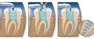
The method is indicated for all types of pulpitis in single-rooted temporary and permanent teeth and permanent formed molars with well-passable canals. The stages of treatment for devital extirpation are the same as for adults.
The method of complete pulp removal is the most reliable in terms of eliminating odontogenic infection and preventing periodontitis, if the pulp is completely removed and the canals are sealed throughout. However, the tops of the roots of temporary teeth are often curved due to the permanent tooth germ located underneath them, and it is not always possible to completely fill them. In this case, resorcinol-formalin paste, which has a mummifying effect, should be used for filling.
For well-passable canals, non-irritating oil-based pastes (eugenol, sea buckthorn oil, etc.) are used for filling.
Errors in treatment and diagnosis are associated with insufficient history taking, incorrect assessment of the signs and extent of pulp inflammation, and underestimation of pain symptoms. They can also arise when there is insufficient justification of the indications and contraindications for the treatment of teeth with biological pulpitis and the method of vital amputation of the coronal pulp, underestimation of the unique course of acute general pulpitis and the reaction of the surrounding soft tissues in young children. A lot of troubles are associated with arsenic paste. If the temporary dressing is not applied tightly, then the arsenic paste that leaks into the surrounding tissue can cause necrosis of the mucous membrane of the gums, cheeks, and tongue. With prolonged contact with tissue, necrosis and sequestration of part of the alveoli are possible. Due to an overdose or prolonged stay of arsenic mouth in a carious cavity, acute arsenic periodontitis develops. In children, the diffusion of arsenic paste into the periodontium occurs faster than in adults, due to the anatomical features of temporary teeth. Acute periodontitis that occurs has a long course and is difficult to treat. For treatment, use the arsenic antidote unithiol, as well as a solution of iodinol and potassium iodide.
A common mistake in treating patients with pulpitis in primary molars is perforation of the bottom of the tooth cavity, when the anatomical features of the structure of hard tissues and pulp of temporary teeth are not taken into account.
In recent years, many criticisms have been leveled at the popular method of treating temporary and permanent immature teeth with pulpitis—devital pulp amputation. Indeed, often due to a diagnostic error in chronic gangrenous pulpitis with significant pulp necrosis, treatment of temporary molars is carried out using the method of devital amputation. Often, the opening of the cavity of a temporary molar is not performed completely, but “somehow.” In order to reduce visits, treatment is carried out not in three, but in two visits. As a result of these medical errors, the necrotic pulp in the root canals does not have time to sufficiently mummify under the influence of impregnation agents, and chronic granulating periodontitis gradually, painlessly develops.
Forecast
Untimely and improper treatment of children with acute and chronic pulpitis can lead to a rapid transition of the odontogenic inflammatory process to the next stage: periodontitis, purulent periostitis, acute osteomyelitis.
When should you visit a dentist?
The sensitivity of the neurovascular bundle of a tooth in a child is lower than in an adult, so the symptoms of its inflammation may be erased or absent altogether. It is important to contact a pediatric dentist when the first symptoms of caries appear - a dark spot on the tooth, or the child complains of pain. You should hurry up with your visit if:
- severe pain;
- increased pain when eating hot or cold food/drinks;
- bad breath;
- elevated body temperature;
- inflammation of the gums around the tooth;
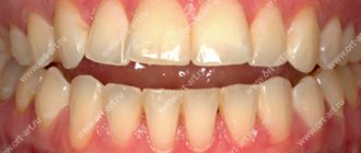
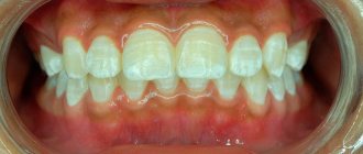
- darkening of the enamel.
Pulpitis of a baby tooth: is it necessary to treat?
Treatment of pulpitis in children should be carried out in any case and as soon as possible. It is unacceptable to wait for a baby tooth to fall out, stopping only the symptoms. Failure to provide assistance to a child can result in serious complications - periostitis (in common parlance - gumboil), periodontitis - inflammation of the periodontal tissues. In addition, the infectious process in the neurovascular bundle of a baby tooth can spread to a permanent tooth when it erupts.
If a long time must pass before the eruption of a permanent tooth, premature destruction of a baby tooth due to pulpitis can lead to malocclusion - displacement of healthy teeth in a row, change in their position, which will require complex orthodontic treatment in the future.
If a baby tooth with pulpitis is soon to be replaced by a permanent one, the doctor may resort to extraction. In any case, this disease should not be ignored.
Principles of treatment of teeth with formed roots
The tactics for treating chronic periodontitis are identical to the approach for treating temporary teeth. If the following conditions are met, it is possible to cope with the disease in one visit:
- there is no gangrenous decay with a putrid odor in the canal;
- there are no granulations grown into the canal;
- there was no exacerbation of such diseases in the anamnesis;
- it is possible to qualitatively process the canal in one visit and dry it;
- the child is somatically healthy and does not take antibiotics, cytostatics, corticosteroids or other immunosuppressive drugs.
In this case, during one visit, the root canal is treated instrumentally and medicinally, obturation is performed with a permanent root filling, and then the crown part is restored. It is much more common to practice treatment in several stages.
On the first visit, the following manipulations are performed:
- necrotomy, formation of a carious cavity;
- opening the canal and removing putride masses from it using a pulp extractor or several - with a very wide canal;
- removal of granulations grown into the canal with injection anesthesia or short-term treatment of granulations with special preparations, for example camphorophenol (very carefully so as not to burn the mucous membrane);
- instrumental treatment of the canal, washing and drying;
- administration of a medication with an antiseptic effect on the turunda or in the form of a paste;
- isolation with an airtight bandage for temporary obturation of the carious cavity.
The medicine is left on average for 1-7 days, after which permanent obturation is performed - subject to the conditions of readiness of the root canal. Obturation is carried out with a sealer with gutta-percha, after which X-ray control is necessary, and only then restoration of the crown.
Treatment methods for pulpitis
The tactics of a pediatric dentist depend on the condition of the child’s tooth and dental system, the severity of his condition, and whether there are concomitant diseases of the oral cavity. If the pulpitis is very severe and there is a risk of a threat to its health, the tooth must be removed. However, in the vast majority of cases, it is possible to keep the tooth permanent until its natural replacement, since removal can lead to disruption of the bite or the position of the remaining healthy teeth in the row.
Traditional treatment of dental pulpitis in a child consists of devital amputation of the pulp and is carried out in several visits. The stages of treatment of pulpitis in children are presented as follows:
- The doctor opens the tooth cavity, removes carious tissue, and applies a paste to devitalize the pulp. It is designed to “kill” the neurovascular bundle and eliminate pain. If the paste contains arsenic, it is used for a period of 1-2 days; if an arsenic-free product is used, it is used for at least 7 days.v
- The second visit involves placing a special mixture into the canals for tissue mummification - a resorcinol-formalin mixture is used.
- The third visit is to fill the diseased tooth.
This method is practiced much less frequently today, since there is a possibility that the infectious process will persist in unsealed tooth canals - mummification of the pulp involves its “drying out” and the formation of voids in which pathogenic bacteria continue to multiply.
The modern approach to the treatment of pulpitis of primary teeth in children is extirpation - complete removal of the inflamed pulp. This can be done with or without prior killing or devitalization of the pulp. In many cases, devitalization is more preferable, since it relieves the child of any unpleasant sensations during the removal of the neurovascular bundle.
After the tooth canals are freed from pulp and carefully processed, the doctor fills them with an anti-inflammatory paste - it tends to dissolve along with the roots during the natural change of teeth to permanent ones. For this purpose, zinc eugenol paste is often used. This method of treatment is comparatively more effective, and if the canals are treated with the utmost care, the infection will not reactivate. Other modern methods of treating pulpitis in children include vital amputation. It consists of preserving the viability of part of the pulp - the upper part of the nerve is removed under local anesthesia, and a medicine with an antibacterial and anti-inflammatory effect is applied to the remaining root pulp. The drug closes the lower part of the neurovascular bundle without affecting its viability, and the tooth is subsequently filled.
Clinical researches
“Results of clinical use of the Asepta series of products: already at the first control examination (after 1-2 days) of using the Asepta line of products, there is a decrease in complaints of discomfort, but bleeding persists when brushing teeth. On the 7th day, complaints of gum bleeding persisted in 4 patients. Upon examination, a decrease in hyperemia and swelling of the gums was noted, but bleeding persisted upon probing. After the final application of the gel with propolis, normalization of clinical manifestations was revealed, which is manifested by the absence of bleeding during brushing and probing.”
Sources:
- The effectiveness of the use of Asept “adhesive balm” and Asept “gel with propolis” in the treatment of chronic generalized periodontitis and gingivitis in the acute stage (Municipal Dental Clinic No. 4, Bryansk, Kaminskaya T. M. Head of the therapeutic department Kaminskaya Tatyana Mikhailovna MUZ City Dental Clinic No. 4, Bryansk
- Study of the clinical effectiveness of treatment and prophylactic agents of the Asepta line in the treatment of inflammatory periodontal diseases (A.I. Grudyanov, I.Yu. Aleksandrovskaya, V.Yu. Korzunina) A.I. GRUDYANOV, Doctor of Medical Sciences, Prof., Head of Department I.Yu. ALEXANDROVSKAYA, Ph.D. V.Yu. KORZUNINA, asp. Department of Periodontology, Central Research Institute of Dentistry and Maxillofacial Surgery, Rosmedtekhnologii, Moscow
- The role of anti-inflammatory rinse in the treatment of periodontal diseases (L.Yu. Orekhova, A.A. Leontyev, S.B. Ulitovsky) L.Yu. OREKHOVA, Doctor of Medical Sciences, Prof., Head of Department; A.A. LEONTIEV, dentist; S.B. ULITOVSKY, Doctor of Medical Sciences, Prof. Department of Therapeutic Dentistry of St. Petersburg State Medical University named after. acad. I. P. Pavlova
- Report on the determination/confirmation of the preventive properties of personal oral hygiene products “ASEPTA PLUS” Remineralization doctor-researcher A.A. Leontyev, head Department of Preventive Dentistry, Doctor of Medical Sciences, Professor S.B. Ulitovsky First St. Petersburg State Medical University named after. acad. I.P. Pavlova, Department of Preventive Dentistry
- Clinical studies of antisensitive toothpaste “Asepta Sensitive” (A.A. Leontyev, O.V. Kalinina, S.B. Ulitovsky) A.A. LEONTIEV, dentist O.V. KALININA, dentist S.B. ULITOVSKY, Doctor of Medical Sciences, Prof. Department of Therapeutic Dentistry, St. Petersburg State Medical University named after. acad. I.P. Pavlova
Treatment of pulpitis of teeth with unformed roots
The root system of a baby tooth is formed over a long period of time after eruption, so there are often situations when caries begins to form on a tooth whose roots have not yet closed the apex. This leads to some treatment difficulties:
- short roots and wide channels;
- the upper part of the root is the “growth” zone, injury to which is an obstacle to root formation;
- the likelihood of infection of the permanent tooth germ;
Treatment of pulpitis of teeth with immature roots in children requires special care. The doctor must carefully monitor that the filling material is not carried beyond the expansion of the root apex. It is worth noting that complete removal of the pulp and treatment of the canals is impossible - the optimal solution here is vital and devital extirpation. Pulp amputation may also be used. The biological method, which consists in preserving a viable pulp and relieving inflammation, is also practiced quite often. Its essence is to prepare the tooth, apply a medicinal paste with calcium hydroxide, after which the tooth is filled with temporary material. A few days later, if there are no complications and the unpleasant symptoms disappear, the doctor installs a permanent filling.
Diagnosis of acute pulpitis
Various methods are used to diagnose acute pulpitis.
Basic research methods.
During the survey, it is important to establish the so-called “pulpitis” nature of the pain.
During the examination, a deep carious cavity is discovered (with an infectious cause of pulpitis).
Probing its bottom is sharply painful at one point or over the entire surface. Possible perforation into the tooth cavity.
The reaction to percussion is still painless in case of partial pulpitis, slightly painful in the case of general pulpitis, painful in case of purulent pulpitis.
Palpation of the transitional fold is painless.
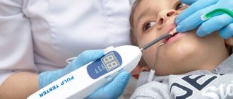
Additional research methods include studying the reaction to temperature stimuli, electrical excitability, and radiography. Temperature stimuli provoke an intense, prolonged pain attack.
Electroodontodiagnosis indicators: 20-25 µA for acute focal pulpitis (the value may be normal in the area of another tubercle, on the side of the not yet inflamed pulp). Acute diffuse pulpitis corresponds to values of up to 30-40 μA or more.
Radiography is effective for determining the location of a hard-to-reach carious cavity (contact surface) in relation to the tooth cavity. It also helps to identify periodontal pockets and inflammation in the periapical tissues. This is useful in the differential diagnosis of acute forms of pulpitis.
How to prepare a child for treatment?
Treatment of pulpitis of primary teeth in children is a rather complex task, so it’s great if the child is already familiar with the dentist’s chair. The first visit should be preventative - to familiarize yourself with the office environment, the doctor, and the instruments.
Also, the task of parents is to psychologically prepare the child. Required:
- talk to your child about doctors in a positive way - tell them that the dentist treats teeth and makes sure they don’t hurt;
- play dentist with toys and other family members;
- avoid mentioning pain or unclear, scary terms;
- do not deceive the child - explain that dental treatment may not be very pleasant, but it will not last long and the tooth will no longer hurt;
- remain calm, act gently but confidently if the child resists treatment;
- choose a suitable time for visiting - it is better in the morning, when the baby is alert and active;
- take a toy with you to calm the child;
- give the doctor the opportunity to establish contact with the small patient - do not rush him;
- Do not scare the child under any circumstances, do not threaten, do not blackmail.
If the situation cannot be controlled completely, you will have to reschedule the appointment for another day. However, the disease should not be ignored - treatment of pulpitis of baby teeth in children must be carried out one way or another. In special cases, treatment under general anesthesia may be considered, but this should be discussed with your doctor.
Features of treatment of different forms of periodontitis in children
Acute apical periodontitis
It is also called apical because the source of inflammation is located near the apex of the root. The acute form of the disease is accompanied by excruciating constant pain. Possible increase in temperature, symptoms of general intoxication of the body.
We recommend treating the acute form of inflammation as early as possible, before it becomes chronic.
The doctor creates an outflow of infectious fluid through the root canal, prescribes applications of anti-inflammatory ointments, bed rest and plenty of fluids.
Chronic periodontitis
The chronic form often develops for years without pronounced symptoms. It can be determined by x-ray. For treatment, a swab moistened with a 10% formaldehyde solution is inserted into the dental cavity. For multi-rooted molars, a resorcinol-formalin mixture is used, which penetrates well into all tubules.
Granulomatous periodontitis
With granulomatous periodontitis in children, “bumps” filled with granulations (dead epithelial cells) form in the oral cavity. Treatment includes 3 visits to the doctor. A month after therapy, a control x-ray is prescribed.
Possible complications of treatment of pulpitis in children
Child anxiety is the main difficulty faced by pediatric dentists. Due to the emotional state of the patient, the specialist has to rush or fails to perform the manipulation carefully, which may result in the following complications:
- the paste is not applied to the nerve - this leads to the fact that the pain does not subside, and the procedure must be repeated again;
- burn of the gums by the paste due to its close location to the tooth;
- bleeding when processing unformed roots;
- perforation of the root of a baby tooth;
- breakage of instruments in the root canal.
You can avoid possible complications or minimize the likelihood of problems arising by normalizing the child’s psychological state and contacting an experienced, qualified doctor.
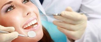
Conservative methods
At the slightest opportunity to save the tooth, gentle conservative treatment is used. Endodontic treatment involves repeated root canal treatment.
First visit
- Anesthesia.
- Cleaning out the carious cavity with a drill, removing softened dentin (bone dental tissue).
- Expansion of the root canal mouth using a special hand instrument.
- Removal of the “dead” pulp – the neurovascular bundle inside the cavity.
- Expansion of the canal and its mechanical cleaning.
- Rinsing the cavity with an antiseptic solution - sodium hypochlorite or chlorhexidine bigluconate.
- Opened apical (root) openings for the outflow of exudate.
- Filling the canal with an anti-inflammatory drug: for permanent teeth - a paste based on calcium hydroxide, for temporary teeth - an oil-based paste.
The doctor leaves the tooth in this state for some time - from 2 to 10 days. In the intermediate period, rinsing the mouth with antiseptic solutions (Chlorhexidine, Miramistin) is prescribed, and less often, taking antibiotics.
Treatment of a tooth with dead pulp
Second visit
- Mechanical cleaning of the canal, removal of medication.
- Washing with an antiseptic.
- Installation of a permanent filling - hermetically closing the cavity with gutta-percha, hydroxyapol or other material.
Physiotherapy
Physiotherapeutic methods are used as an aid in the fight against inflammation. These are inexpensive and painless procedures, children tolerate them well:
- electrophoresis of antiseptics - enhancing the antiseptic effect using pulsed current;
- phonophoresis – introduction of an antiseptic under the influence of ultrasound;
- laser therapy – the laser beam sterilizes the root canals and has a direct bactericidal effect.
Laser therapy
Prevention of pulpitis in children
Treatment of pulpitis in permanent teeth in children, as well as their “predecessors” - baby teeth - is a rather complex procedure that may require several visits to the doctor. Preventing a disease is easier than treating it - all you need to do is pay attention to hygiene procedures and teach your child how to brush their teeth correctly from a very early age.
It is also important to monitor the child’s diet - strengthen the enamel with solid foods (carrots, apples, etc.), provide a balanced diet to saturate bone tissue with minerals. It is better to limit sweets and give only water at night. Prevention of caries is the basis for preventing its complications, including pulpitis.
Features of diagnosis and treatment of caries of permanent teeth in adolescents.
To maintain the health of your teeth for a long time, you need to start carefully monitoring their health as soon as they erupt.
To do this, just follow a few simple rules.
- Provide quality hygiene, including professional dental hygiene for the child.
- Try to regularly attend preventive dental examinations at your clinic every 6 months, because... The development of caries in children in mixed dentition occurs very quickly.
- As a preventive measure at this age, it is recommended to do fissure sealing - this is “sealing” the recesses on the chewing surfaces of the lateral teeth
special filling material containing fluoride. This procedure mechanically closes the “weak” spots of the tooth and, at the same time, strengthens its enamel, releasing fluoride into the tissue.
- Teenagers often develop whitish spots on their teeth. It is necessary to find out what type of lesion such a defect belongs to - carious or not. This can be done by undergoing caries diagnostics with the Diagnocam apparatus in our clinic - this type of examination is digital and does not expose the child to radiation, therefore it is suitable for regular use. If a carious lesion is detected at an early stage, it can be cured without resorting to preparation using the modern ICON infiltration method
- If a child has crooked teeth and crowded teeth prevent them from being properly cleaned, then it is necessary to consult a pediatric orthodontist and begin orthodontic treatment.
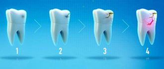
- Treat caries in a timely manner in the early stages of the disease - this will allow you to save permanent teeth as much as possible from preparation, because the earlier caries is detected, the less affected tooth tissue the dentist will have to remove.
This approach to the child’s dental health, if not eliminates it, will significantly reduce the risk of serious carious lesions. If time was lost at the first symptoms of the disease, then a complication of caries – pulpitis – will have to be treated.