Caries is a slowly developing pathology in which the enamel and hard tissues of the tooth are destroyed. The disease can occur in different forms, but each of them goes through four stages. Each of them has its own symptoms. In the article we will consider the features of the development of caries and methods of its treatment.
In this article
- Causes of caries development
- First stage of caries
- Diagnosis of the first stage of caries
- Second stage of caries
- Diagnosis of the 2nd stage of caries development
- Stage 3 caries
- Diagnosis of stage 3 caries
- 4th stage of caries development
- Diagnosis of caries at stage 4
- Treatment of caries
- Prevention
Caries is a dental disease accompanied by demineralization of enamel and slow destruction of hard dental tissues - dentin. The main cause of the disease is the spread of cariogenic microorganisms in the oral cavity. Caries occurs in acute or chronic form, developing on incisors and molars, on milk and molars. Occurs in children and adults. Any type of this pathology goes through 4 stages in its development. On each of them it can be cured. However, the likelihood of complications increases if treatment begins not at the first, but at subsequent stages. Let's look at the degrees of caries, their specific symptoms and treatment methods.
Causes of caries development
In addition to the main cause of caries, the impact of bacteria on enamel and dentin, there are endogenous and exogenous factors in the development of this pathology. Among the first:
- poor oral hygiene;
- weak immunity;
- systemic infectious diseases;
- bad habits: smoking, alcoholism;
- tartar and plaque.
Exogenous factors include causes that do not depend on a person:
- bad ecology;
- poor water quality;
- heredity;
- unformed tooth enamel.
Each person's susceptibility to caries is individual. For some people it never occurs even once in their lives, for others it almost never goes away. In any case, dentists recommend coming for an examination every 6 months in order to detect symptoms of pathology in time. They are different at each stage of caries development. Let's consider them separately.
Pregnancy and baby's teeth
Pregnancy is a time for a huge miracle to happen. It's amazing that in just nine months a real baby grows out of a tiny cell; the embryo, which looks like a flat disc in the fourth week from conception, by seven weeks turns into a little man (with a tail!), by seventeen it grows to the size of a pear (and the tail is a thing of the past), and by thirty-eight weeks it is completely ready for life outside the mother womb.
In this article we will not talk in detail about all the magical transformations of the fetus; we will touch upon only one - but very important - topic.
Teeth: where do they come from, how do they develop, can an expectant mother take care of their health in advance?
First stage of caries
The initial stage of caries development is called the white or chalky spot stage. At this degree, a mild clinical picture is noted. The disease is asymptomatic, its signs can only be detected during dental diagnostics. The person himself does not notice any changes.
This is the danger of the first stage of caries: the pathological process begins to destroy individual areas of tooth enamel. Demineralization occurs, as a result of which hard tissue may be exposed. Areas of enamel affected by bacteria appear as white spots. It becomes loose, matte and looks different from the healthy shiny surface of the tooth. As the pathology progresses, the spot increases in size and acquires a darker shade - yellow or brown.
As a rule, caries begins due to poor oral hygiene. Cariogenic microbes begin to multiply, and their secretion products corrode tooth enamel. Often, areas of demineralization are observed in places that are difficult to reach for cleaning - in gum pockets, interdental spaces and on wisdom teeth.
Stages of tooth formation in children
Few expectant parents know that the foundation for their child’s baby teeth begins to form as early as the 7th week of pregnancy, and from the 5th month the formation of permanent teeth begins. The future dental health of the baby depends entirely on the habits, lifestyle and nutrition of the mother.
At the age of 6-8 months, babies' first teeth erupt - the two lower incisors. Then, at the age of 8-9 months, the top two appear. The timing of teething is quite individual and depends, among other things, on genetic factors. The normal range is considered to be the eruption of the first teeth at 5-9 months. The third, fourth and fifth teeth gradually erupt. The formation of a temporary bite is completed by the age of 2.5-3 years, when the child has 20 milk teeth - 5 teeth on each side on the upper and lower jaws.
In this case, the process of maturation of the enamel of primary teeth occurs for another 2 years after the tooth erupts in the oral cavity, i.e. The enamel on the central incisors finally matures by about 3 years, and on the lateral teeth, accordingly, even later. In its anatomical structure, a child’s baby tooth differs from a permanent tooth: its walls are much thinner. For example, on the lateral surface the thickness of the hard tissues of a baby tooth (enamel, dentin) is only 1 mm. Such a tooth, compared to a permanent one, is much more vulnerable to various types of problems - first of all, the development of caries and its complications. And if the tooth is still forming, the enamel is in the maturing stage, then it can be destroyed very quickly, especially against the background of a sharp decrease in the child’s immunity: during and after a viral infection, after an illness with antibiotic treatment.
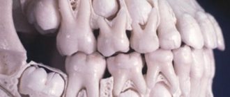
At the age of 6-7 years, the physiological change of temporary teeth to permanent teeth begins. The order of their appearance is the same as during the eruption of primary teeth. The first to appear are the lower and upper central incisors, and at the same time the so-called sixth teeth appear: first the lower, then the upper. The sixth teeth do not change, but grow. The pattern of appearance of all teeth is similar: first, the tubercles become noticeable above the gum, then the chewing surface. At the moment when a tooth erupts, its root is approximately half formed, it is short and has a wide canal opening. During its formation, the root grows in length and its walls thicken.
The last milk tooth changes by the age of 11-12 years. At the same time, for two years after eruption, the enamel matures, and the formation of the root of a permanent tooth lasts even longer: from 2 to 4 years. Thus, only in adolescence can we say that the situation has stabilized.
It is important to know!
From the moment the first teeth erupt at the age of about 6 months and up to 3 years, the child develops a temporary bite. The anatomical structure of a baby tooth differs from a permanent tooth; its walls are much thinner, which means the risk of caries is higher. Therefore, we recommend having preventive examinations with a doctor once every 3-4 months. It is not easy for parents to spot the beginning of caries in a hard-to-reach place, and the process that has begun can develop too quickly and lead to undesirable consequences. At the same age, we recommend visiting an orthodontist for the first time in order to, if necessary, correct the formation of the bite in a timely manner. At the age of 6-7 years, the process of physiological replacement of temporary teeth with permanent teeth begins, which is completed by 11-12 years. At the same time, the maturation of enamel and the formation of the root of permanent teeth continues for another 2-4 years after the appearance of the tooth. Thus, teeth are finally formed and take their place in the oral cavity only by the age of 16. Until this point, it is important to pay increased attention to hygiene and prevention.
Diagnosis of the first stage of caries
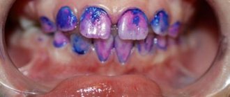
The dentist may notice a white spot during an external examination of the mouth. Doctors also use the method of exposing teeth to organic dyes. Typically, methylene blue or red indicator, tropeolin or carmine are used. It is applied to tooth enamel, left for several minutes and washed off. Colored areas indicate demineralization. To establish an accurate picture, the teeth are cleaned of plaque, and the enamel is treated with hydrogen peroxide.
It is easy to identify caries at the initial stage, but the following factors can complicate the diagnosis:
- inaccessibility of the localization of the pathological focus;
- similarity of the clinical picture with endemic fluorosis.
Fluorosis is accompanied by an accumulation of white spots, and caries is characterized by the formation of single foci of demineralization.
Second stage of caries
The 2nd stage of caries development is called the superficial stage, which is characterized by damage to hard tissues. The carious lesion is located in the enamel coating, but it begins to collapse, exposing the dentin. With this degree of pathology, discomfort is noticeable when eating sweet and sour foods, and, less often, hot, cold and salty foods. Some people complain of pain when brushing their teeth. The pain disappears immediately after the stimulus is removed.
The second stage begins due to the lack of treatment of caries at the white spot stage. At first, the demineralization process is reversible; the doctor prescribes special formulations containing phosphorus, calcium, magnesium and other minerals. For superficial caries, such therapy usually does not help.
Periodontal and root formation
This stage of development of temporary teeth precedes their eruption and continues 1-.5-2.5 years after the completion of this process. The crowns are usually already fully formed by this time. The place of root origin is Hertwig's epithelial sheath, consisting of external and internal cells of the enamel organ.
The process of development of the root system and periodontium includes the following stages:
- ingrowth of epithelial cells into the underlying mesenchyme;
- separation and formation of a channel of a certain shape;
- formation of root dentin with the participation of odontoblasts;
- development of cement on the surface of dental dentin;
- formation of dense periodontal tissues by fibroblasts;
- fixation of the root with collagen fibers to the alveolar bone;
- formation of secondary cement on the basis of primary cement after eruption.
4th stage of caries development
The last stage of caries development is called deep. It occurs as a result of progression of the previous stage or due to the chipping of a previously installed filling. Excessive consumption of sweets and poor hygiene can speed up the process of tissue destruction.
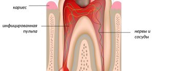
Deep caries is considered an advanced form of the disease. At this stage, the carious cavity extends to all hard tissues and almost reaches the pulp. In addition, the patient's crown is destroyed. The tooth may break into several pieces. This stage is often complicated by pulpitis or periodontitis. My mouth constantly smells bad. Painful sensations are observed not only during eating and palpation, but also at rest. If a pathological process develops under the filling, it becomes loose and falls out.
Delay in the eruption of primary teeth
A delay in the process of teething in infants and children under 1 year of age is called retention. This often happens with canines, but can also affect molars and incisors.
A delay of 1-2 months remains within normal limits. If this period is exceeded, this is a clear signal about the occurrence of pathology, which, in turn, may be a consequence of diseases suffered by the mother during pregnancy.
Also, so-called redundant (or supernumerary) teeth can become an obstacle to timely teething. At the same time, teething too early can be an indicator of disturbances in the endocrine system of the child’s body.
If you notice a significant discrepancy in the timing of the eruption of your child’s baby teeth, make an appointment with your child to the dentist and pediatrician.
Treatment of caries
The choice of treatment method depends on the stage of development, form of progression and type of caries. With the first degree of pathology, it is enough to conduct several sessions to remineralize tooth enamel. The procedures are done in the dentist's office. He also prescribes special gels and pastes that he can use at home. Sometimes he is prescribed medications that help restore the balance of minerals in the body.
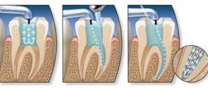
Treatment of subsequent stages is carried out by filling. It is carried out according to the following algorithm:
- the dentist gives an anesthetic injection;
- cleanses the tooth cavity of necrotic tissues;
- disinfects the surface;
- applies a filling;
- grinds and polishes it.
Treatment of caries can be complicated if it occurs on the front teeth. It is not so easy to fill them, especially if the enamel and dentin are severely damaged. In addition, a regular filling will be noticeable. To eliminate a cosmetic defect, you have to install veneers or select a composite material by color and apply it in several layers.
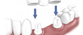
In case of extensive damage and the impossibility of restoring the tooth, it is removed. An implant can be installed instead. Functionally and externally, it will not differ from other teeth, but not everyone can afford such a procedure.
Prevention
To prevent caries, you should follow a number of simple rules:
- visit the dentist once every six months;
- Brush your teeth twice a day;
- use dental floss and mouthwash;
- eat less sweets;
- Perform ultrasonic cleaning periodically;
- stop smoking;
- limit your consumption of coffee and tea;
- Change your toothbrush every 2-3 months.
To further cleanse the oral cavity, you can use irrigators - devices that clean the teeth and interdental spaces using a pulsating stream of water. However, such devices should not become an alternative to a toothbrush.
If you notice symptoms of tooth decay or other dental disease, consult your doctor immediately.
Resorption and preparation for shift
At the age of 5-6 years, the process of completely replacing the temporary bite begins. This period begins immediately after the resorption of milky roots and the beginning of the growth of permanent rudiments. Temporary teeth begin to undergo resorption and ejection from the alveoli. Odontoclasts, which carry out demineralization and intracellular destruction, as well as pulp tissues that secrete osteoclast-like cells, which are responsible for the destruction of dentin and predentin from the internal side, actively participate in the process. Removal of the crown of temporary teeth, as a rule, occurs under the influence of chewing pressure. The root remaining in the hole undergoes a natural process of destruction and resorption. The location of the crown is quickly epithelialized due to granulation tissue.
The process of loss of temporary teeth occurs symmetrically on both jaws and ends with the appearance of permanent teeth. The individual nature of its course is determined genetically.


