Various types of fractures lead in frequency among injuries to permanent teeth; the central incisors of the upper jaw are most often damaged. As a percentage, fractures account for about 67% of all types of injuries to permanent teeth; for baby teeth this figure is much lower - about 7%.
The success of treating dental fractures directly depends on the speed at which the patient seeks dental care; in some cases, this must be done in the first hours after the injury occurs.
Causes of fractures
Tooth fractures occur when:
- a strong blow to the tooth during a game, a fall, an accident, or doing any work;
- contact of solid food with a tooth with a carious cavity or with a previously treated tooth with large fillings;
- presence of bad habits (crunching nuts in shells, opening beer bottles with teeth);
- abnormal bite;
- hitting the antagonist tooth with dental forceps when removing a tooth on the opposite jaw.
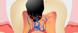
A tooth fracture can occur in any part of the tooth; In direction, fractures are longitudinal, oblique and transverse.
Crown fractures, methods of their treatment
Incomplete crown fracture
An incomplete crown fracture (fracture) is a tooth crack that appears as a result of acute (dislocation, bruise) and chronic (abnormal bite, bad habits) traumatic injuries, while the integrity of the tooth is not compromised. The cracks look like lines that run randomly across the enamel; they can appear on teeth located next to a tooth that has undergone more severe traumatic effects.
During a normal inspection, cracks may not be visible; they are detected using transillumination. Transillumination is an examination method that involves shining a cold light beam through the injured tooth. Transillumination lighting is used not only to detect cracks, but also to identify contact caries, subgingival dental plaque, and pulpitis.
When a patient with an incomplete crown fracture seeks help, an electroodontodiagnosis (EDD) is performed to monitor the condition of the pulp after injury.
Typically, cracks do not penetrate deeper than the enamel-dentin border of the tooth, and therefore do not cause complaints in patients, except for minor sensitivity to temperature and chemical stimuli, which sometimes occurs when eating. Over time, the enamel at the site of damage darkens, caries develops, and the tooth may split along the crack.
Clinical manifestations of tooth fracture
A tooth fracture is accompanied by acute intense pain; other symptoms include:
- difficulty opening the mouth;
- difficulty closing the jaws;
- pathological loosening of a damaged tooth;
- bleeding gums.
Painful sensations also occur when exposed to temperature stimuli. In this case, the nature of the pain depends on the location of the fracture and the degree of its mobility. When examined by a dentist, swelling of the tissues adjacent to the tooth and hemorrhages in the mucous membranes are also observed.
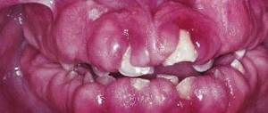
A crown fracture is visible visually and is often accompanied by exposure of the pulp.
In case of a root fracture, tooth mobility and pink discoloration of the enamel are observed.
Treatment
Small cracks are treated with remineralization therapy - the enamel is coated with products containing calcium and fluoride. Veneers are also used for treatment - thin ceramic or composite plates that cover the surface of the tooth. The technique is used on the front teeth; for cracks in the chewing teeth, artificial crowns are installed.
According to their location, cracks can be vertical, horizontal, or inclined; deep vertical ones can eventually open access to the pulp, which leads to its death. In this case, after cleaning and filling the canals, a crown is installed on the tooth. If there is a vertical crack running through the entire root, the tooth must be removed and replaced with a denture.
Enamel fracture (chip)
When the enamel is fractured, its surface layer usually breaks off. Patients do not complain of pain, they are concerned about cosmetic defects. Chipping most often occurs at the corner of the tooth; the sharp edges of the chip injure the mucous membranes of the oral cavity.
X-ray examination does not reveal any changes in the dental tissue; transillumination examination can reveal enamel cracks at the edges of the chip. When a chip is combined with a tooth bruise, pulp death is possible.
Treatment for an enamel fracture involves grinding off the sharp edges and applying fluoride varnish to the fracture plane to protect the tooth from caries. Chips are restored using artistic restoration - extension is carried out with composite materials. If the pulp dies, it must be removed followed by filling the root canals.
Classification of dental injuries
The ICD-10 (International Classification of Diseases) classification of dental injuries includes more than ten types of dental injuries. To summarize, all mechanical damage is divided into:
- for injuries to the tooth itself or fractures;
- tooth displacement or dislocation;
- damage to soft tissues and jaws.
It is also worth noting a bruise - minimal damage to a tooth when it looks intact and visually no damage is visible - no chips, no cracks. But due to the impact, the blood supply to the tooth pulp may be disrupted, and subsequently the tooth may darken. Tooth bruises belong to the first class of injuries according to ICD-10. It hurts to touch, bite and chew food.
First aid for a bruise is to give pain relief if necessary, and consult a dentist as soon as possible.
Crown fracture without opening the pulp chamber
This fracture affects not only the enamel, but also the dentin; the pulp is not exposed. Patients complain of pain when eating, caused by thermal, chemical and mechanical irritants. The closer the pulp is to the fracture plane, the more severe the pain.
A dental examination reveals:
- defect of part of the crown limited to dentin;
- pain on percussion;
- pain when probing the exposed dentin surface.
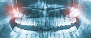
X-ray diagnostics are performed to exclude a root fracture, EDI and transillumination examination.
Treatment
To treat this type of fracture in baby teeth, a protective bandage is applied to the exposed dentin, covered with glass ionomer cement on top. The tooth is left in this condition until it is replaced with a permanent one. If the integrity of the bandage is damaged, the bandage is changed; X-ray monitoring of root formation is periodically carried out if it has not been formed.
Subsequently, thermal diagnostics and EDI are carried out to check the viability of the pulp: one, three and six months after the injury, then every six months until the root is formed.
When treating permanent teeth, sharp edges are ground off, teeth are restored with filling materials and crowns. If the non-viability of the pulp is detected due to a strong blow, it is removed.
Forecast and prevention of dental injuries
Whether the tooth will be saved during treatment will depend on the type of damage itself, the time, and how timely the person consulted a doctor. Often, the state of a person’s general health and age are added to such criteria.
Today, modern methods of dentistry and orthodontics are so progressive that they help preserve their natural teeth, restoring them from damage even in the most complex cases of injury.
If the tooth still cannot be saved, then the doctor will suggest replacing it with an artificial option that has high functional and aesthetic characteristics.
Prevention of dental injuries will depend on the behavior and caution of the person himself, both at home and at work. When engaging in dangerous sports, a person should always use a protective helmet.
Treatment
When a permanent tooth with an unformed root is damaged, therapeutic actions should be aimed at preserving the vitality of the pulp to ensure further root formation. To do this, direct pulpotherapy is performed.
The method of direct pulpotherapy consists of applying a therapeutic pad based on calcium hydroxide to the pulp, which is covered with an insulating pad on top and then the tooth is restored. The method is effective if no more than two hours have passed since the tooth injury, and the surface of the opening of the tooth cavity is no more than 1 mm in diameter.
If it is impossible to use the method of direct pulpotherapy, the method of partial pulpotomy is used - removal of the surface layers of the inflamed pulp to preserve the viability of the rest of it. After partial pulpotomy, the wound surface of the tooth is covered with therapeutic and insulating pads, and the condition of the pulp and developing roots is subsequently monitored.
If the pulp dies as a result of injury, the apexification method is used to stimulate the formation of the root apex - cleaning the root canal and filling it with calcium hydroxide paste, which is removed after the root apex is closed. Calcium hydroxide also prevents resorption (resorption) of the roots of pulpless, injured teeth.
If the injury occurs on permanent teeth with fully formed roots, the pulp is completely removed, then the tooth is restored.
Complete fracture of the crown at the level of the neck
Examination of the patient reveals the absence of a crown, severe pain and bleeding in the area of the injured tooth. An x-ray is performed to rule out a root fracture.
Treatment consists of removing the pulp, followed by filling the root canal and restoring the tooth using core inlays. If a permanent tooth has been injured and its root has not yet formed, apexification is performed; After the root has formed, the root canal is filled with permanent materials and the crown is restored.
If a complete fracture of the crown occurs on a temporary tooth, it is depulped or the tooth is completely removed. Before the permanent one erupts, the missing baby tooth is replaced with a temporary removable denture to prevent the development of anomalies in the child’s dental system.
Corono-root fracture
This type of fracture is rare and cannot always be determined by visual examination. The injury occurs after a vertical blow to the tooth; the fracture gap passes through the crown and root simultaneously.
Patients experience mobility of part of the tooth and pain when chewing food. The fracture is confirmed by radiography.
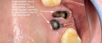
The treatment method is chosen depending on the degree of damage to the tooth: endodontic treatment is carried out, followed by the installation of a root pin and tooth restoration, or the tooth is removed and prosthetics are performed.
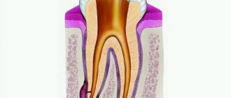
Root fractures
The frequency of fractures of the roots of primary teeth is 0.5%, permanent - 2% of the total number of tooth fractures. A fracture can occur in the apical, middle or coronal third; more often, its transverse direction is diagnosed, less often - oblique. Sometimes two or more fracture lines are found, which indicates a comminuted fracture.
A root fracture is usually combined with tooth dislocation; patients complain of tooth mobility, aching pain in the tooth that occurs spontaneously and intensifies when biting and closing the jaws.
TOOTH DISlocation
Tooth dislocation is a dental injury with damage to the supporting apparatus. To put it more simply, the tooth is displaced and the periodontal ligaments are damaged. A tooth dislocation can be:
- Incomplete tooth dislocation;
- Impacted dislocation (intrusive);
- Complete tooth dislocation.
Tooth dislocation can be caused by various factors, for example:
- Hit;
- Injury;
- Dislocation of a nearby tooth due to aggressive tooth extraction, or due to incorrect selection and application of forceps for tooth extraction;
- Biting on hard food;
- Bad habits (opening bottle caps with teeth; biting hard wire, etc.).
Symptoms of tooth dislocation
Symptoms of incomplete tooth luxation
Symptoms of incomplete tooth dislocation are pain when biting on a tooth, pain when chewing food, possibly bleeding of the causative tooth, and its displacement.
Symptoms of impacted tooth dislocation
Symptoms of an impacted (intrusive) tooth dislocation will be shortening or complete disappearance of the visible part of the tooth crown, the tooth is motionless, percussion is slightly painful. It must be remembered that with impacted dislocation of the central incisors on the upper jaw, the teeth can penetrate into the nasal cavity.
- Symptoms of complete tooth dislocation
The patient can bring the tooth in his palm.
Treatment of tooth luxation
Treatment of incomplete tooth luxation
Treatment for incomplete tooth luxation involves repositioning the tooth and splinting it. Reposition must be performed no later than 2 hours from the moment of injury, otherwise the risk of developing pulp necrosis increases.
Treatment of impacted (intrusive) tooth dislocation
Treatment of impacted (intrusive) tooth dislocation consists of orthodontic reposition for 3-4 weeks.
Treatment of complete tooth dislocation
Treatment for complete tooth avulsion consists of replantation, splinting and endodontic treatment.
Only those that have been outside the mouth for no more than two hours are suitable for replantation of natural teeth. Drying of tooth cells leads to early resorption. But if such a nuisance does occur, patients should place the tooth either in a glass of milk, or hold it behind the cheek or under the tongue. And it’s better not to lose teeth at all C:
The article was written by N. Shidlovskaya specifically for the OHI-S.COM website. Please, when copying material, do not forget to provide a link to the current page.
Diagnostics
Examination of the patient first reveals a tooth dislocation, a root fracture is confirmed later by X-ray examination; It is recommended to perform a 3D computed tomography, since a regular x-ray may not detect a fracture. In addition to determining the area and direction of the fracture, the study makes it possible to determine the condition of the walls of the alveoli, periodontium, and the position of displaced fragments.
The dentist is also able to determine a root fracture using the following technique: with his right hand he slightly moves the crown of the tooth back and forth, while the finger of his left hand located on the vestibular surface (oriented towards the vestibule of the mouth) of the alveolar part of the jaw will feel the movement of part of the broken root associated with crown This method is not suitable if the fracture occurred in the apical part of the root, so an x-ray examination is required.