Dentists use various classifications of caries to determine the extent of its development. One of the oldest such systems is the scale created by the American scientist Greene Black. With its help, doctors assess the condition of teeth with carious lesions and determine the method of treatment. Let's take a closer look at the features of this classification.
In this article
- What is caries: main characteristics of the disease
- Black caries classes
- Other types of caries
- Diagnostics
Dentistry techniques are constantly being improved, but the basis for modern scientific developments are theories developed in the 19th and 20th centuries. For example, many dentists in their practice are guided by Black’s classification, which helps determine the degree of development of caries and methods of its treatment.
In 1986, American practicing dentist Green Vardiman Black developed a scale that allows you to assess the condition of a patient’s teeth with carious lesions. With the help of his classification, it became easier for doctors to make a diagnosis. Previously, dentistry was not considered as a science. After Black's scientific research, the idea of it changed, and it began to develop faster.
The American scientist created not only a classification of caries, but also the first foot-operated drilling machine. In addition, he developed a filling formula that is still used today. However, the most famous of all the developments of the dentist is the scale of carious lesions based on symptoms and the extent of the disease. Let's tell you how Black's classification differs.
Classification of caries lesions according to the process
In this direction, there are 3 types of dynamics of the course of this disease: fast, slow and stabilized.
Also, this pathogenic process can be considered by the extent of its localization: caries manifests itself on one tooth, on several elements, or is systemic in nature and affects most of the different teeth in the upper and lower rows.
What is subcompensated caries and what are its features?
Subcompensated caries occurs in approximately 25% of children with dental caries. This form is characterized by an average rate of spread of the pathological process. Characteristic features are dull enamel and superficial lesions without deep tissue involvement. Most often, subcompensated caries does not cause discomfort or pain, so it can only be detected during an examination by a dentist.
People with this form of caries are recommended to have scheduled visits to the dentist two to three times a year, as well as oral sanitation and preventive measures.
Classification of caries according to the sequence of occurrence
As in the previous gradation, experts distinguish 3 types of carious lesions.
The first category includes caries that appeared on a tooth for the first time.
The second is a re-injury to a previously filled tooth. In the vast majority of cases, this disease spreads around or under the filling.
The third category includes the so-called recurrent caries lesion. It occurs due to insufficient treatment of the area or a poorly installed filling.
Secondary caries is all new carious lesions that develop near a filling in a previously treated tooth. Secondary caries has all the histological characteristics of a carious lesion.
The reason for its occurrence is a violation of the marginal seal between the filling and the hard tissues of the tooth; microorganisms from the oral cavity penetrate into the resulting gap and optimal conditions are created for the formation of a carious defect along the edge of the filling in the enamel or dentin.
Recurrent caries is the resumption or progression of the pathological process if the carious lesion was not completely removed during previous treatment. Recurrence of caries is often detected under the filling during an X-ray examination or along the edge of the filling.
There are a large number of caries classification systems, almost all of them are repeated. Therefore, for an accurate diagnosis, it is very important for a specialist to correctly determine the depth of the cavity, the nature of the course and the main reason why the carious pathology formed.
The effectiveness of treatment and the absence of relapse processes in the future will depend on the reliability of the diagnosis.
Clinic and pathogenesis of initial dental caries
Clinic and classification of caries
The clinical signs of dental caries have been well studied.
However, there are different opinions on the question of whether dental caries can be classified as a disease of the body. I. G. Lukomsky, E. E. Platonov, A. E. Sharpenak and others insisted on a positive interpretation of this issue. A. I. Rybakov shares the same opinion. At the same time, many foreign authors consider caries to be only a pathological process, which lacks two or three more important signs to be characterized as a disease of the body. In our opinion, there is a significant gap between the modern clinical definition of caries and previously existing ideas about it. Previously, the diagnosis of “dental caries” (caries) included the concept of destruction of hard tooth tissues. Currently, the diagnosis of “caries” is made not only when tooth tissue is destroyed, but also at the stain stage, which is a sign of demineralization without the formation of a visible cavity.
There are justified attempts by individual authors to supplement the existing clinical classifications of dental caries with modern data from biochemical and morphological studies. They are dictated by the need to fill the existing gap between clinical and experimental-theoretical research.
I. G. Lukomsky (1949) proposed a classification of caries, which has not lost its significance to this day. The author distinguished two main clinical manifestations of caries - a carious spot and a carious defect. Dentin caries is usually a continuation of the carious process that began in the enamel.
Zonal classification of caries according to I. G. Lukomsky
- 1. Carious stain: a) chalk - acute caries;
- b) pigmented - a chronic process.
A carious spot is the first stage of a “spotted enamel lesion.” Superficial, medium and deep caries - the stage of the defect. Medium caries is the stage at which the process extends beyond the enamel-dentin border up to the prepulp dentin, and deep caries is a lesion of the prepulp dentin.
Darling (1956) drew attention to the fact that some white spots may be a consequence of hypomineralization and do not develop into a defect stage, essentially not being caries in the generally accepted view. Even earlier, Gottlieb (1944) believed that caries does not begin until there is yellow pigmentation in the area of future development of pathology. In this regard, the zonal (clinical and topographical) classification of caries by I.G. Lukomsky, much less other classifications of caries that do not include the spot stage, are not able to summarize modern data on caries.
G.N. Pakhomov (1968) proposed to distinguish between several types of carious spots. He pointed out that between the white and pigmented spots there are a number of transitional forms, different both in the color of the affected surface (from white and light gray to brown and black) and in the pathomorphological picture. The author indicated the following types of carious spots:
- 1) white spots of any size;
- 2) brown carious spots occupying less than 1/4 of the proximal surface of the tooth;
- 3) brown carious spots occupying 1/4 of the proximal surface;
- 4) brown carious spots occupying 1/3 or more of the proximal surface;
- 5) black carious spots occupying 1/4 or less of the proximal surface of the tooth.
G.N. Pakhomov believed that in the case of multiple carious spots on the teeth of one person, the color of these spots is approximately the same. The classification of carious spots, developed by G. N. Pakhomov, is an important addition to existing classifications and significantly expands modern ideas about the forms of initial caries, however, clinically it is complex, since it is difficult to accurately determine the size of the spot (especially in the area of close contact of the chewing group of teeth ).
Based on morphological characteristics, the classification is also not exhaustive.
I. G. Lukomsky indicated the favorite places of caries (fissures and folds of enamel, approximal surfaces of the tooth crown and cervical areas) and, depending on this, distinguished carious lesions on smooth surfaces, fissure caries, approximal and cervical caries.
Blackwell (1971) additionally pointed out that caries can develop in the area of the necks of teeth, at the junction of enamel and dentin, with gingival retraction associated with periodontal pathology or human age.
When most teeth were simultaneously affected, caries was called systemic (I. G. Lukomsky, 1948).
In the clinic, suspended caries is often observed. Examining the teeth of London schoolchildren at 2-year intervals, Mellanby and Coumoutos (1946) noted that in 11-21% of children caries tended to stabilize. Usually, suspended caries is observed in children more often on the occlusal surface of molars, which are more susceptible to abrasion during chewing (Stones, 1966). I. G. Lukomsky called suspended caries self-healing, which is possible if the caries does not cross the enamel-dentin boundary.
According to the international classification of diseases, caries is divided into enamel caries, dentin caries and cement caries.
Based on modern data on the pathogenesis and clinical picture of dental caries, caries can be defined as a chronic pathological process of hard dental tissues.
Classification of dental caries
- I. Clinical forms 1. Spot stage (carious demineralization): a) progressive (white or light yellow spots);
- b) intermittent (brown spots);
- c) suspended (dark brown spots).
- A. Enamel caries (superficial).
- 1) fissure caries.
- 1) fast-flowing caries;
- 1) single lesions;
The proposed classification of caries does not cover all possible forms and types of manifestation of caries, but is a working scheme on the basis of which it is possible to systematize the numerous manifestations of this pathological process.
It should be borne in mind that demineralization, or a white spot, is also a clinical sign of a number of other nosological forms of pathological processes in the enamel (necrosis, hypoplasia, fluorosis, etc.). On the other hand, caries, which begins in deep fissures and pits in dentin (if there is no enamel) or cement (if gum atrophy has occurred), is not characterized by the stage of a white carious spot.
In this regard, white and pigmented spots of enamel should only be conditionally considered the initial manifestation of caries.
Initial forms of caries, isolated cases of demineralization and disintegration of hard dental tissues are not accompanied by disorders of the dental system and changes in the body. Some clinical signs of caries in the early stages (demineralization) have not been sufficiently studied. On the contrary, the signs of caries in the stage of disintegration of hard tissues (cavity formation) are well known.
Our main attention will be paid to the early stages of the carious process.
Topographic classification of caries distribution
In many countries, this classification is most widely used.
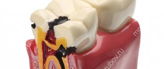
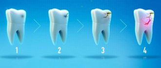
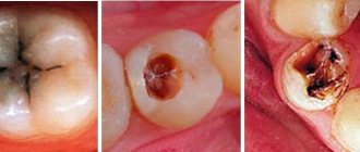
It takes into account the depth of the lesion, which is very convenient for the practice of the dentist. There are 4 stages of development of this disease:
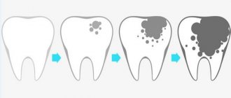
- The appearance of a carious spot. The source of demineralization of the dental element. The process of this harmful phenomenon can last either slowly or quickly, depending on the individual characteristics of the patient’s body.
- Superficial caries is characterized by local damage to the enamel on the tooth.
- Moderate caries manifests itself in damage to the surface layer of dentin.
- Deep caries clings to the peripulpal dentin and affects the tooth right down to the nerve endings.
How to understand that it is caries?
Caries in the spot stage can be mistaken for hypoplasia or fluorosis; superficial caries has similar features to wedge-shaped defects and dentin erosion. It is important not to confuse caries with other diseases. To distinguish caries from fluorosis and hypoplasia, the affected area is stained with a special dye. Dyes are activated only during caries. The period of development of the pathology is also taken into account: hypoplasia and fluorosis develop in infancy - before teething, and caries - after eruption on the “weak” areas of the tooth from negative external influences.
Differences between chronic caries and acute
Let's take a closer look at the features of the chronic and acute forms of this disease.
The acute form of caries is characterized by the rapid development of destructive changes in the hard tissues of the tooth, the rapid transition of uncomplicated caries to deep caries.
The affected tissues are soft, slightly pigmented (light yellow, grayish-white), moist, and can be easily removed with an excavator.
Chronic caries is characterized as a slow process (several years).
The spread of the carious process (cavity) is mainly in the planar direction. The altered tissues are hard, pigmented, brown or dark brown in color.
Dr. Greene Vardiman Black
Dr. Greene Vardimar Black is a well-known personality who is at the forefront of the development of dental science in the United States of America. He was born in 1836 in the city of Winchester.
At the age of 17, the young man became interested in medicine and worked for several years as an assistant to dentist D.S. Spira, while simultaneously gaining theoretical knowledge on this topic.
After completing his education, Greene Vardimar Black opened his own dental office in Jacksonville. In addition to providing services to the public, Dr. Black never stopped studying science and improving himself.
In 1870, a specialist invented a mechanical drill equipped with a foot drive. The gold amalgam composition developed by Dr. Black is also used in modern dentistry.
In addition, the specialist brought the terminological base to the standard, and also developed a classification of carious cavities and cutting dental instruments.
Dr. Black compiled several books that described methods for preparing the tooth surface, touched on the features of therapeutic dentistry, and also described some pathologies. In addition, Mr. Black taught dental science at the College of Chicago and also served as dean of the Northwestern University School of Dentistry.
Type of caries according to degree of activity
There are 3 types of caries in this category: compensation, subcompensation and decompensation.
Compensatory caries is characterized by a slow or non-progressive process.
Damage to the surface of the teeth is insignificant and does not cause any discomfort in the patient.
With regular and systematic hygiene procedures, as well as special preventive measures, it is possible to stop the development of the disease at its initial stages.
Subcompensatory caries is characterized by an average rate of progression, at which it can go unnoticed and not cause concern to the patient at all.
Decompensation caries is expressed by intensive development and dynamics of progression, accompanied by such acute pain that it affects both the patient’s ability to work and everyday life.
Because of this, the disease is often called acute caries. It requires immediate treatment procedures, since otherwise the process may spread to third-party teeth with the subsequent addition of pulpitis and periodontitis.
Three forms of dental caries
Vinogradova’s classification is based on the degree of activity of dental caries:
- The compensated course of caries is characterized by the fact that the pathological process develops slowly, carious cavities are hidden under dense dentin, they are rare, and the disease progresses relatively slowly.
- The subcompensated form of the disease develops slightly faster than the compensated form. It is characterized by the appearance of dark areas on the enamel and horizontal spread without penetrating deep into the tooth.
- Decompensated caries is often multiple and spreads very quickly. With such caries, all teeth located near the cheeks are often affected.
The decompensated form is considered the most severe, in most cases accompanied by complications in the form of pulpitis or periodontitis.
Let's talk about all forms of caries in more detail.
Clinical principles for preparing carious areas
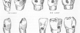
To carry out all the necessary therapeutic manipulations, many specialists rely in their work on Black’s classification of caries.
For any of the above types of tooth damage from caries, it is necessary to carry out full preparation and filling.
The durability of your tooth (or several) depends on the quality of these manipulations.
Experienced dentists, when removing soft carious dentin, can leave its deep pigmented elements to avoid damage to the tooth pulp. After carrying out this work, there should be no affected tissue left on the walls of the cavity.
At all stages of preparation and filling, the dentist sets the main goal - to destroy the carious areas of the affected tooth, disinfect the remaining parts and apply a hermetically sealed structural material that can restore the structure of the tooth and help it fully perform its functions in the future.
Manifestations of caries in children
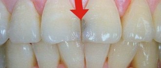
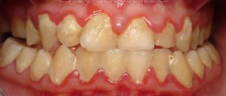
The appearance of light or dark spots on tooth enamel
The first sign of caries in children is white spots on the enamel, resulting from calcium loss. At this stage, treatment may be minimal; often the doctor prefers minimally invasive methods, for example the ICON technique. The dentist applies a special preparation to the affected tooth tissue, thereby blocking the proliferation of bacteria and the development of the infectious process - caries is “preserved.”
If the white areas darken, dentin, the deep tissue of the tooth, is involved in the pathological process.
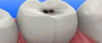
Cavity formation
In a child, the caries process develops very quickly, and the next stage is the formation of cavities. Most often they are visible upon examination even with the naked eye, and the child complains of pain while eating.
Reaction to food temperature
With the further development of the carious process, the affected tooth begins to react to hot, cold and sweet foods and drinks.
Pain
Pain occurs while eating - when food gets on a carious tooth. If the infectious process has spread to the pulp, the child will complain of pain constantly. Often unpleasant sensations occur during night sleep.
5.Bad breath
Food particles accumulate in carious cavities, bacteria cause rotting, so parents often complain about bad breath.
Periodontitis with decompensated caries
In addition to pulpitis, with decompensated caries another serious complication often develops - inflammation of the tissues surrounding the tooth root. This disease is called periodontitis.
The periodontium is a ligamentous apparatus that holds the tooth in the jaw and provides shock absorption. If an inflammatory process develops in the periodontal tissues, this can lead to loosening and tooth loss. Against the background of periodontitis, ulcers can form, the removal of which will require surgical opening of the gums.
Periodontal inflammation can be acute or chronic. In the first case, inflammation develops rapidly, accompanied by darkening of the tooth, acute pain, swelling of the affected tissues, and inflammation of the cheek. Chronic periodontitis is characterized by more smoothed symptoms, pain syndrome may be absent. Treatment for the chronic form is long and difficult and can take several months.
Periodontitis greatly weakens the immune system and creates a source of chronic infection in the body.