Kochneva I.S., Sarukhanov G.M.
Every woman is unique, amazing and beautiful in her own way. Everyone, looking in the mirror, wants to please others and herself, and everyone, looking in the mirror in the morning, asks the question: “My light, mirror, tell me. Am I the sweetest in the world..." And every time she wants to hear in response: “Yes, you are beautiful - no doubt about it...”. And, of course, every woman dreams of always being young and attractive. Rejuvenation in general and facial rejuvenation in particular becomes the goal. In pursuit of youth, most women resort to operations such as a facelift or blepharoplasty, but at the same time they forget about rejuvenating such an important area as the lips and perioral area, which require no less close attention than the rest of the face.
But, before we delve into the description of methods and modern approaches to oral rejuvenation, in our opinion, it is necessary to determine its main anatomical components.
Rice. 1. Anatomy of the mouth and perioral area
- cutaneous part of the upper lip;
- column of the philtrum (two vertical ridges above the upper lip);
- philtrum groove;
- cupid's bow;
- white lip roll - a thin strip of light skin surrounding the outside of the red border of the lips;
- tubercle;
- commissure – the junction of the lips at the corners of the mouth;
- vermillion (red border of lips).
Rice. 2. Muscles surrounding the oral area
The orbicularis oris muscle is formed by muscle bundles located in the thickness of the lips. It begins in the area of the corners of the mouth, where its fibers are woven into the skin at the commissure and surround the lips in the form of a sphincter. The function of this muscle is to close the mouth and pull the lips forward.
The muscle that depresses the angle of the mouth or DAO (Musсulus Depressor Anguli Oris) originates and is attached to the bony edge of the lower jaw and ends in the skin of the commissure of the lips.
The subcutaneous muscle of the neck (M. platizma) covers the entire anterior surface of the neck with a uniform thin layer. M. platizma starts from the clavicle area, is woven into the superficial tissues of the cheek area and at the level of the corners of the mouth (its lateral bundles Lateral Platisma - LP). M. platizma can act as a strong lower lip depressor, meaning it can forcefully lower the lower lip. In some people with well-developed platysma muscle of the neck, this part of the platysma (LP) can be clearly visible during active movements during chewing, swallowing, and while talking.
It is important to distinguish between the action of DAO, which pulls the corner of the mouth downwards (Fig. 3a) (vertical traction), and the action of LP, the fibers of which pull the corner of the mouth posteriorly and downwards (posterior oblique traction) (Fig. 3a). Their action is complemented by contraction of the muscle depressor labii inferioris “DLI”. The motor innervation of the DAO comes from the marginal branch of the mandibular nerve marginalis mandibulae nervus MMN), which projects to the region of the marionette fold (Fig. 2).
Rice. 3. a — action of the DAO muscle; b — action of the lateral part of the platysma (LP).
Oral cavity: anatomy in ontogenesis
During human embryonic development, the oral cavity begins to develop as early as the 12th day. Visually, it is a protrusion of the ectoderm, which is located between the cardiac protrusion and the medulla. During this period, it is called the fossa, or oral cavity.
The tongue develops at 4-5 weeks of ontogenesis. Together with the chewing muscles, it is the result of a modification of the gill arches. Further development of the oral cavity, the anatomy of which becomes much more complex, allows the fetus to taste the amniotic fluid. This is the environment in which he finds himself. At week 7, taste buds appear on the tongue. By the beginning of the second month of embryonic development, the formation of the palate is completed.
Features of the mucous membrane
The anatomy of the oral cavity (the photo demonstrates its structure) is represented by the following components: lips, tongue, cheeks, teeth, gums, salivary gland ducts, palate and tonsils.
An important role in ensuring its functions is played by the mucous membrane, formed by multilayered squamous epithelial tissue. Below it are the basement membrane and the submucosal layer. A characteristic feature of the oral epithelium is its high ability to regenerate, which is carried out due to its germ layer, as well as resistance to the negative effects of infections and environmental irritants.
The mucous membrane itself is formed by connective tissue cells. It is where the nerve endings, capillary and lymphatic vessels are located. The mucous membrane itself has specialized cellular structures that perform essential functions. These include macrophages, mast cells and plasma cells. They provide phagocytosis of foreign particles, regulation of blood vessel permeability, and synthesis of immunoglobulins.
There are different types of receptors in the oral mucosa. These include pain, tactile and temperature. But the mucous membrane does not perceive taste. This function is performed by the muscular organ of the oral cavity - the tongue.
As a result, we can say that the mucous membrane of the human oral cavity provides protective, sensitive and plastic functions.
“FACE RECURVE” CONCEPT IN ORAL REJUVENATION
In 2006, famous French plastic surgeons Claude LeLouarn and Jacqees Buis developed their original concept of “Face Recurve”. The development of this revolutionary concept was based on new ideas about the mechanisms, principles, and stages of facial aging, on the basis of which the approach to rejuvenation of the face and the perioral area in particular was radically changed. According to Claude LeLouarn and Jacqees Buis, the formation of clinical signs of an aged face takes place in 2 stages (stages): stage 1 – “cutaneous” (superficial) and stage 2 – “structural” (deep) (Fig. 25).
Rice. 25. Stages of facial aging (Claude LeLouarn).
Stage 1 - “skin aging” includes changes that occur over time on the outer “shell” of the human body, that is, on the skin. This manifests itself in the loss of elasticity, firmness of the skin, its thinning, enlarged pores, the appearance of age spots or, conversely, areas of depigmentation, fine wrinkles, etc.
Stage 2 - “structural aging” affects deeper layers: adipose tissue (lipoatrophy), muscular-fascial system, bone tissue (resorption). Clinically, this manifests itself in the form of flattening of the middle zone of the face, deepening of the nasolacrimal (dark circles under the eyes) and nasolabial furrows, folds of grief, the appearance of bags under the eyes (fat “hernias”), jowls, etc. And further progression of this process leads to sagging facial tissues (ptosis).
According to the “Face Recurve” concept, facial aging develops mainly under the influence of constant contraction of certain facial muscles. These muscles initially have a curved shape and in young people are located in the deep fat layer. Over time, facial muscles straighten and shorten. (Figure 26). These changes in the shape of the facial muscles of Claude LeLouarn and Jacqees Buis were confirmed by x-ray studies.
Rice. 26. a) the muscle of a young person (has a curved shape), b) the muscle shortens and straightens with age (Claude LeLouarn).
Under the action of muscle contraction, the underlying fat is pushed out more superficially. Thus, grooves are formed (underneath them lies a “tight”, straightened muscle), surrounded by excess fat along the periphery. This is how circles under the eyes, uneven cheekbones, “sorrow” folds and sagging cheeks, as well as vertical folds on the neck are formed (Fig. 27, 28).
Rice. 27. a) facial muscles in a relaxed state b) facial muscles during contraction. The green arrow indicates the direction of muscle contraction, the blue arrows indicate the direction of pushing deep fat outward from the muscle fibers (Claude LeLouarn).
Rice. 28. a) the orbicularis oculi muscle and the zygomatic muscles in a relaxed state; b) the same muscles contract (the fat underlying them is pushed outward); c) blue arrows - the direction of pushing out fat during contraction of muscle fibers: fatty “hernias” of the lower eyelids bulge, the nasolacrimal groove worsens, and uneven contours of the cheekbones intensify (Claude LeLouarn).
According to the authors, during the aging process of facial skin, gravity is only of secondary importance in those areas that are most susceptible to repeated contractions of facial muscles. Some bundles of these muscles are peculiar age markers (signs) and are responsible for the formation, for example, marionette folds (similarly called “folds of grief”), medial fibers of the neck muscle (m.platizma), nasolabial fold, etc. (Fig. 29).
Rice. 29. Age “markers” - indicated in yellow (explanation in the text) (Claude LeLouarn).
On a young face, the facial muscles are elongated, curved and relaxed at rest (Fig. 30a). Only when these muscles are tense can signs of future markers of aging be seen (Fig. 30c). During the aging process, age markers (deep nasolabial fold, folds of grief, medullary cords of the neck, etc.) are clearly defined even in the absence of facial expressions (Fig. 30b).
Rice. 30. a) 20 years old facial oval at rest, b) 60 years old facial oval at rest, c) 20 years old facial oval with tension of facial muscles.
Based on their findings, Claude LeLouarn and Jacqees Buis developed the “Face Recurve” technology, which involves timely impact on certain groups of facial muscles, as well as correction of deep fat layers. In addition to a new approach to facial rejuvenation, the authors proposed a whole range of new cosmetic and surgical procedures aimed at preventing and correcting age-related changes in the face. French surgeons were the first to propose a certain algorithm of actions depending on the age and condition of the patient’s face. According to the concept of facial aging, this algorithm is divided into several stages, each of which corresponds to a specific set of preventive and therapeutic measures aimed at rejuvenation.
(Inquisitive readers can familiarize themselves with the full version of the concept of Claude LeLouarn and co-authors on the corresponding author’s websites).
In the opinion of the authors of this article, the theory of aging and, accordingly, the principle of facial rejuvenation “Face Recurve” is most applicable in rejuvenating the mouth and perioral area. Judge for yourself.
Depending on age, Claude LeLouarn and Jacqees Buis propose to divide “facial aging” into several stages and, according to this “division,” carry out preventive and therapeutic measures for rejuvenation.
Language
The anatomy of the human oral cavity also provides the formation of taste sensations. They occur when various chemicals interact with specialized receptors. Agree, the perception of taste is purely individual. But scientists distinguish its main varieties. These include sour, bitter, sweet and salty.
Taste receptors are called chemoreceptors. They are located in taste buds, each of which connects to the mouth opening at a time. Despite the general plan of the structure, they are all specialized. So, at the tip of the tongue there are receptors that perceive sweet, at the edges - sour, at the root - bitter. The larger area is capable of perceiving salty taste. It is located at the tip and along the edges. The tongue is also involved in the formation of sounds, wetting, mixing and swallowing food.
Anatomy of the mouth and teeth
Mechanical processing of food is carried out using teeth. Normally, there are 32 of them. In the sockets of each jaw there are 4 incisors, 2 canines, 4 small and 6 large molars. All of them are specialized. So, with the help of incisors and fangs, food is bitten off, and with the help of molars, it is already crushed to a mushy state.
Based on the characteristics of the external structure of a tooth, there are distinguished root, neck and crown. The latter is its visible part and is located above the gum. The tissue that covers the crown is called enamel. It is considered the hardest in the human body. The neck is formed by a less durable substance - cement. The connective tissue that fills the tooth cavity is the pulp. It contains nerve fibers, lymphatic and blood vessels. Therefore, it is through the pulp that nutrition and growth of teeth occur.
How do these oral structures form? The formation of teeth occurs during the embryonic period. But they appear 6 months after the birth of the child. There are 20 of them in total. They are milky and are replaced by permanent ones up to 10 years. The last teeth to grow are wisdom teeth, which appear by age 25. For humans, they are an atavism, since they have lost their meaning in the course of evolution.
Treatment
Therapy of the disease involves strengthening the body’s immune forces and influencing the affected area, removing signs of pathology. If stomatitis develops in children, the pediatrician will tell you how to treat it.
The choice of medications depends on:
- affected areas;
- type of pathogen;
- severity of the disease;
- health status and age of the patient.
The disease can be effectively treated at home at an early stage.
To eliminate the symptoms of the disease, you can use the following drugs in tablet form:
- Imudon (for children over 3 years old);
- Hexoral Tabs (after 4 years);
- Hexalize (from 6 years old).
It is useful to rinse your mouth with antiseptic solutions, including:
- Stomatophyte;
- Chlorhexidine;
- Miramistin;
- Chlorophyllipt;
- Iodinol.
You can use stomatitis ointment in your mouth. Kalgel, Sinka, Oxolinic ointment, Actovegin gel are suitable.
For aphthous stomatitis, antibiotics and antihistamines may be prescribed. With any type of disease, it is important to maintain the body's defenses. For this purpose, immunity modulators and vitamin complexes with minerals are used.
For bacterial stomatitis, children and adults can use Vinilin. It is also called "Shostakovsky's balm." It is a strong antiseptic that kills bacteria in the mouth.
It has wound healing and antimicrobial effects. Vinilin has no side effects and rarely causes allergic reactions. It is necessary to dip a cotton swab in the balm and lubricate the affected area with it. The procedure is carried out 2-3 times a day. After it you cannot eat or drink for 30 minutes.
It is important to avoid getting medications into the body of children and adults. Before using a pharmaceutical drug, you must familiarize yourself with the precautions and make sure that it is indeed suitable.
Receptors
Scientists say that there are about 2000 taste buds in the oral cavity. In response to the arrival of food, they become irritated. The signals that are generated in this case are sent along nerve fibers through the intermediate to a specialized section of the cerebral cortex. This is where the sense of taste is formed.
It is truly individual for all people. Taste is determined by the threshold of sensitivity. It is not the same for different chemicals. This indicator is highest for bitter, low for sour. But people perceive salty and sweet in the same way.
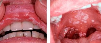
SIGNS OF AN AGING MOUTH
Rice. 5. Photo of the mouth of an elderly person – a) with closed lips; b) the mouth is slightly open.
- The upper lip becomes disproportionately long and completely covers the upper incisors, the red border turns inward and visually decreases.
- The lower teeth are exposed (Fig. 5, b) when the lips are opened.
- Gradual drooping of the corners of the lips under the action of the muscle of the same name (DAO) and the appearance of furrows in the corners of the mouth (marionette lines).
- A fine network of vertical wrinkles appears around the mouth (“purse-string wrinkles”).
- Lips lose their original volume and become thinner.
- The clear outline or the so-called white roller disappears.
- The filtrum becomes lengthened and flattened.
All these signs are associated with loss of elasticity and thinning of the skin, atrophy of the subcutaneous fatty tissue, decreased elasticity of the fibers of the orbicularis oris muscle and the depressor anguli oris muscle, partial resorption of the bones of the facial skull, alveolar processes of the jaws, and malocclusion due to loss of teeth.
Based on the above signs, the principles of mouth rejuvenation are determined.
- It is necessary to raise the mouth as much as possible (oral lifting) in order to cover the lower incisors (midface lift, impact on the mentalis muscle - mentopexy, resection of the muscle that depresses the lower lip, DAO resection, lifting the oral mucosa).
- Smooth out purse-string wrinkles and even out the skin texture around the mouth (mesotherapy, biorevitalization, chemical peeling, laser resurfacing, botulinum toxin injections, contour plastic surgery or lipofilling).
- Emphasize the artistic contour of the lips - white rollers (use of gels based on hyaluronic acid, lipofilling of the lip contour, thread lifting - GORE-TEX or AlloDerm collagen thread, installation of Permalip silicone lip implants, etc.)
- Restore the volume and elasticity of the lips lost with age (it is very important not to overdo it). It is necessary to ensure that the dynamics of the mouth do not change and that the individuality of each smile remains. There should be no distortion of the smile area (for example, the use of fillers or large amounts of fat). Bring to balance the ratio of the upper and lower lips in volume to 1:1.6.
- Avoid external incisions on the lips, as the resulting scars may subsequently be noticeable and will give the lips an unnatural appearance.
Now let's look at each of the points in more detail.
Chemical food processing
The anatomy of the oral cavity and pharynx is such that they are also a kind of reservoir for the primary breakdown of food. The food itself, its image or even the smell stimulate the secretion of saliva. This happens with the help of glands, the ducts of which open into the oral cavity. With the help of saliva, complex carbohydrates are broken down into simple ones, microorganisms are neutralized, and the food bolus is moisturized and enveloped. Then, with the help of the tongue, it is pushed towards the pharynx, moving into the esophagus and stomach.
SIGNS OF A YOUNG MOUTH THAT THE SURGEON CONSIDERS WHEN LIP REJUVENATION
Rice. 4. a - schematic representation of a young mouth; b - photo of a young mouth; c - photo of a young mouth smiling.
- Slightly exposed upper teeth.
- The lower teeth are completely covered by the lower lip.
- The corners of the mouth are slightly raised.
- The skin of the upper and lower lips is smooth without vertical wrinkles.
- The lips are quite full.
- A clear white lip roll is a thin strip of light skin surrounding the outer red border of the lips or the contour of the lips.
- The grooves of the philtrum are clearly visible - two vertical ridges above the upper lip.
- The shape of the edges of the lips with artistically expressed anatomical curves.
With a harmonious smile, the upper teeth are exposed by 2/3. The lower lip only slightly exposes the lower teeth.
Composition of saliva
According to its physical properties, saliva is a colorless liquid with a mucous consistency. More than 98% of its content is water. The breakdown of complex sugars is ensured by salivary enzymes - maltase, amylase and lysozyme. The latter substance also performs a protective function, neutralizing pathogens and healing oral wounds.
Saliva also contains mucus called mucin. It provides moisture and envelopment to food. Thus, both mechanical and chemical processing of food is carried out by the oral cavity. The anatomy of this section of the digestive system is completely interconnected with the functions performed.
IMPROVING THE RELIEF AND STRUCTURE OF THE SKIN AROUND THE MOUTH
In order to improve the condition of the skin around the mouth and remove fine wrinkles, it is necessary to use various cosmetic techniques. There are various ways to improve the condition of the skin around the lips, and only a competent cosmetologist will be able to choose exactly the technique that is most effective in each specific case.
- Skin biorevitalization and facial mesotherapy will help to qualitatively improve and rejuvenate the condition of the skin around the mouth. Both of these techniques have a deep and long-lasting moisturizing effect. As a result, the skin is smoothed, filled with moisture, becomes velvety and acquires a healthy color. Both methods are aimed at rejuvenating the skin: increasing turgor and elasticity, smoothing out fine wrinkles, as well as moisturizing. Biorevitalization involves the use of low concentration hyaluronic acid and gives an instant, but less lasting effect of saturation and rejuvenation. The result of mesotherapy cocktails containing vitamins, microelements, collagen, elastin, coenzymes, etc. manifests itself more slowly, but produces more lasting changes, also preventing skin aging.
- The rejuvenating effect of peelings and Botulinum toxin injections is associated with smoothing skin texture and smoothing wrinkles. Any peelings (physical (laser or mechanical polishing) and chemical (glycolic, yellow and many others) significantly affect the condition of the skin around the mouth: increase blood supply, stimulate cell renewal, thereby smoothing out the relief, as a result of which small wrinkles are smoothed out and become almost invisible deep, and new purse-string wrinkles will not appear for a long time. After peeling, the skin will look even, smooth, young. Botulinum toxin injections, blocking nerve endings and excluding some groups of facial muscles from active facial expressions, will also help smooth out purse-string wrinkles above the upper lip and raise the corners of your mouth.
- Contouring of lips and deep wrinkles. To obtain a more pronounced rejuvenation effect, it is necessary to fill the lips, which over time lose their youthful plumpness. This can be done using hyaluronic acid fillers or lipofilling. (These techniques will be described in more detail below).
How does salivation occur?
The process of salivation occurs reflexively. To “launch” it, it is necessary to irritate the receptors of the oral mucosa. As a result, nerve impulses arise, which are subsequently sent to the salivation center of the medulla oblongata. This process is unconditional reflex.
But if we just imagine a sour lemon or a fragrant pie, then saliva will immediately begin to secrete into the oral cavity. Such stimuli are conditioned.
So, the oral cavity, the anatomy of which was discussed in our article, performs the following functions:
- determining the quality and taste of food;
- mechanical and chemical processing of food;
- protecting the body from pathogens and low-quality products;
- formation of a food bolus;
- breaking down complex carbohydrates into simple ones.