Development of teeth in the embryonic period and after birth
At the 6-7th week of embryogenesis, the first signs of teeth appear. At the time of birth, the crowns of the central incisors are formed in the child, the crowns of the lateral incisors are not fully formed, half of the crown of the canines is present, as well as the chewing surfaces of the remaining teeth. Many surfaces have incomplete mineralization.
In each jaw of a newborn there are 18 dental follicles, including 8 permanent and 10 temporary. They all have different degrees of mineralization. By about five years of age, the growth of the rudiments of permanent teeth is activated and the process of physiological resorption of the roots of baby teeth begins. Against the background of these changes, a change in bite begins - temporary to permanent.
Methodology
There are several ways to take an X-ray of a child's jaw or lower skull.
Intraoral radiography
Intraoral x-rays are carried out using special dental devices, which help take targeted images of one or more adjacent teeth (one to four). Such an examination allows us to assess the condition of the hard tissues of primary and permanent teeth, periodontal and periodontal tissues, jaw bones for the diagnosis of retention and follicular cysts, oncological tumors, anomalies in the location and number of erupted teeth and rudiments, etc.
When performing a study, a blank of x-ray film is placed in a special paper envelope, which is inserted into the oral cavity and placed in the area of the desired tooth. The X-ray machine is brought to the face from the outside and placed as close as possible to the tooth/teeth being examined.
In pediatric dentistry, intraoral radiography is often prescribed to diagnose pathologies of the palatal suture. Although, it must be said that if we are talking about a very young patient (2-3 years), it is quite difficult to carry out such an x-ray, because it can be difficult to explain to a child why it is needed and that it will not hurt.
Extraoral methods
The most common method of performing extraoral x-rays is a panoramic photograph, or orthopantomogram. In such photographs, you can see in a direct projection the entire upper and lower dentition, as well as the structure of the patient’s jaw. Orthopantomogram images allow you to make a detailed interpretation and description of the condition of teeth and gums, to recognize even hidden pathologies or those that have just begun to develop (for example, periodontal disease, caries).
You can see what a panoramic x-ray of a child’s jaw looks like in a photo on the Internet.
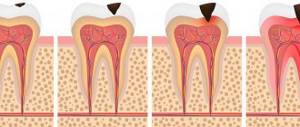
Features of the shadow picture of temporary teeth
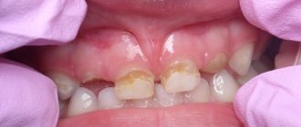
On x-rays, the roots of primary teeth are shorter and the bifurcation angle of molars is greater than that of permanent teeth. The root canals are wider, and the dental cavity is larger in volume. The size of the periodontal gap in a child may vary, and this is considered a normal variant.
The dental follicle appears on an x-ray as a rounded clearing and has a clear rim of compaction. In its cavity there is a rudiment, which changes at different stages. First, points are visible along the cutting edge, which gradually form a single contour of the crown.
Deviations from the norm
The most common causes of deviations from the norm are developmental anomalies, congenital pathologies and birth injuries. Often injuries occur if the fetal head does not match the size of the mother's birth canal. However, almost all consequences can be corrected by the first year of life.
During this period, it is necessary to follow a number of recommendations related to caring for the baby. Often, the irregular shape of the head (occipital part) is a consequence of lying on the back for a long time. A flat neck sometimes indicates a lack of vitamin D, which can cause rickets in young children. When the fontanel is still open, it is not recommended to direct a stream of water onto the baby’s head.
Congenital diseases are much more difficult to cure. Particularly dangerous is dropsy of the brain or hydrocephalus. It can be detected during pregnancy and treatment can begin immediately.
Sometimes pathological deformation of the head in a child occurs:
- sloping back of the head (plagiocephaly);
- curvature of the occipital and frontal area (scaphocephaly);
- elongated shape (acrocephaly).
Asymmetry and curvature of the skull often lead to neurological pathologies, as well as retardation in physical and mental development.
What unformed root tips look like on an x-ray
In unformed tips, the length of the root is almost normal, the walls are parallel to each other. The wide root canal ends in a funnel-shaped extension. In the area of the apex, the periodontal fissure merges with the growth zone, which incompetent specialists sometimes identify as a pathology.
Throughout the entire length of the root and at the apex, a compact plate of the socket wall is differentiated. This picture is typical for the upper central and lateral incisors in 8-year-old children, for the lower central incisors in 6-year-old children, as well as for the lower lateral incisors and first lower molars in 7-8 year old children.
Features of the structure of a child’s skull
Immediately after birth, a child’s skull has a certain appearance and structure that is very different from the skull of an adult. The baby's skull has soft bones. They have a movable structure, since during childbirth this feature greatly facilitates the passage of the newborn through the birth canal.
The shape of the skull largely depends on the method of birth. The skull of small children born by Caesarean section has an even shape. During natural childbirth, the baby's skull is slightly elongated.
One of the structural features of a newborn baby’s skull is the disproportion between the head and body. The baby's head looks disproportionately large. Its circumference is a couple of centimeters larger than the chest. This is an age-related feature and an indicator of the child’s health.
The bones of the skull are greatly separated. The space between the bones is filled by a certain layer, which consists of connective tissue and non-ossified cartilaginous tissue. In this case, the brain region significantly predominates over the facial region. If in an adult the ratio of the volume of the facial region to the brain is 1:2, then in a newborn child this figure is 1:8.
A child's skull has another important distinctive feature - the presence of fontanelles. Fontanas are usually called certain non-ossified areas of the skull, which are located in places where future sutures form.
Experts note the complete absence of sutures in newborn children, as well as the weak development of diploe - the spongy substance of the bones of the cranial vault, and a weakly defined relief on the outer and inner sides of the skull.
The air sinuses in newborn children are underdeveloped. Muscle tubercles, lines, and ridges also remain poorly expressed, since their development and normal functioning require muscles, and at this moment they are not yet working at full strength.
The jaws also remain undeveloped, since the chewing function is not formed. There are no alveolar processes, the lower jaw consists of two unfused halves. For this reason, the face of a newborn baby almost does not protrude forward, like that of an adult.
Formation of fontanelles
During the initial stages of embryonic development, the upper part of the skull has a membranous formation that covers the brain. After the cartilage stage, at approximately 2-3 months of intrauterine development, bone nuclei begin to form. Subsequently, they fuse with each other, forming bony plates or the base of the bones of the roof of the skull.
By the time of birth, between the already formed bones there are areas of narrow stripes and rather wide spaces - fontanelles. With the help of these areas of the membranous skull, which have the ability to protrude and recess, a significant displacement of the bones of the skull occurs. It is this feature that ensures that the baby’s head passes through the mother’s narrow birth canal during the birth process.
The skull of a small child has several fontanelles:
- anterior or greater fontanelle;
- small;
- wedge-shaped;
- mastoid.
The anterior or greater fontanel is located in the area of the junction of the parietal and frontal bones. It has a rhombus shape. The process of ossification of the large fontanel is completed by 2 years.
The small or posterior fontanelle is located between the parietal and occipital bones. Its ossification is completed in the third month after birth.
The wedge-shaped fontanel is paired. It is located on the sides of the skull in the anterior section between several bones: the sphenoid, frontal, parietal and temporal. Ossification of this fontanel occurs immediately after birth.
The mastoid fontanel is also paired. The process of ossification occurs together with the sphenoid fontanel. Location: behind the sphenoid, at the junction of the temporal, parietal and occipital bones.
It is noteworthy that wedge-shaped and mastoid fontanels are more often observed in premature infants. Babies born at term often lack the occipital region.
Closing times depend on the following factors:
- hereditary predisposition to rapid or prolonged closure of fontanelles;
- nutritional features;
- dates of birth;
- genetic abnormalities of intrauterine development;
- metabolic processes in the baby’s body;
- excess or deficiency of vitamin D;
- pathologies of the digestive tract, as a result of which mother’s milk is poorly absorbed.
The speed at which the fontanelles close depends on the intensity of the child’s growth.
The anterior and posterior fontanelles are often palpated by the midwife during childbirth. When the baby's head enters the birth canal, doctors use the location of these fontanelles to determine the placement of the head in the birth canal. The normal position is when the fontanel is in front. The change often indicates an unwanted rotation of the fetus in the uterus.
NEW LEVEL
Micrognathia, or jaw hypoplasia, is commonly referred to as an unusually short lower jaw. However, there is no standard or precise definition of this condition. The fact is that when comparing the proportions of the head of an adult and a newborn baby, we will see a very large difference in the ratio of the sizes of the lower jaw. In the last weeks of pregnancy, the fetus is in cramped conditions, its head is usually tilted, its chin is pressed to the chest. This position temporarily inhibits the growth of the chin, which is compensated by the relatively large size of the tongue compared to the tongue of an adult. But the most important thing is that the lower jaw of a newborn at birth is, as it were, at the start of the rapid changes that will occur to it in the next days and weeks with active motor load in the form of breast sucking. That is, the size of a newborn’s chin in most cases is not a fait accompli, but a point of development . With a high probability, a healthy child who receives breastfeeding on demand will sooner or later catch up or surpass his peers. Just like after a while all healthy babies begin to crawl and sit up, some a little earlier, others a little later.
We are talking about the characteristics of healthy children, but for children with genetic diseases, which are characterized by underdevelopment of the lower jaw, breastfeeding has no less therapeutic effect. That is, a breastfeeding consultant in this situation does not so much tell the mother how to adapt to the baby’s characteristics, but rather acts as a coach who helps level out these characteristics.
In clinical practice, the difference in jaw size is considered significant if the lower lip and lower jaw do not reach the upper gum. Sucking the breast on demand provides a good load on the facial muscles, and in breastfeeding children, the size of the jaw, as a rule, normalizes with age. For example, one study proved that in children 5-11 years old who were breastfed for up to 6 months, micrognathia was much less common than in their peers from the control group. (Lus, Garib, Auroca 2006)
Babies with a short jaw often have difficulty sucking at the breast. But not because of insufficient emphasis on the chin, as one might think, but because of the incorrect position of the tongue, which limits muscle movement and creates additional tension. In children with a short jaw, the tongue size is usually completely normal. In order for such a language to fit into a too limited space, the child is forced to resort to “tricks”. Most often, the baby acquires the habit of resting the tip of the tongue on the lower gum, while the root of the tongue is in constant tension, and a characteristic “bridge” appears.
Another option is to curl the tip of the tongue upward. In both cases, constant tension and unnatural position of the muscles lead to motor difficulties and form compensatory movements that complicate the sucking process. The most common is pushing out the nipple when latching on the breast . Instead of pulling the nipple deep into the mouth, the tongue makes the usual pushing movements. Such incoordination of tongue movements can be dealt with quite quickly using a silicone pad. Moreover, nursing mothers who are faced with this manner of latching on their babies usually complain of severe pain in the nipples and cracks. If the mother doesn’t like the idea of using a pad as a training device or the baby refuses to feed in it, finger feeding and tongue smoothing can have a similar effect.
An excellent effect is achieved by the so-called “thrown back” positions at the chest, which allow for a more asymmetrical grip.
If nipple pain continues despite training and special feeding techniques, you can encourage mom to replace several feedings a day with pumping to give her nipples a rest and healing. Time is the best assistant in such a situation. Most babies with a short lower jaw master the art of sucking at the breast by 12 weeks and stop causing pain to their mothers when feeding.
Photo source: https://newborns.stanford.edu/PhotoGallery/Micrognathia1.html and the author's own photo.
Tatyana Kondrashova, “New Level”.
Go to other articles for consultants of the “New Level” project