It is believed that at the age of 6-7 years the first milk teeth (lower central incisors) should fall out, and at the same time the first permanent molars grow. But what if the pattern breaks down? Can the upper incisors fall out first or, for example, first the lateral and then the central incisors? What could this mean, and should parents pay special attention to this?
What if my teeth fell out too early? Or did the molars start growing before the incisors fell out? Is it normal for molars to start growing at 4-5 years of age? With what it can be connected? When should a situation alert parents?
Intramaxillary development
The first stage of formation of temporary teeth begins at 6-8 weeks of embryogenesis and lasts until the middle of the first year of a child’s life. It includes the following processes:
- the formation of a dental plate from multilayered squamous epithelium located in the oral fossa of the embryo with the further formation of a ridge and the growth of tooth buds in the form of flask-shaped buds;
- the formation of epithelial (enamel) organs due to the ingrowth of mesenchyme into the kidneys with the subsequent formation of dental papillae and sacs;
- histogenesis of dental tissues (formation of dentin, formation and mineralization of enamel, development of pulp from the mesenchyme of the dental papilla with the formation of nerve fibers and blood vessels).
Each newborn has inside both jaws the rudiments of all 20 temporary and 16 permanent teeth, which have varying degrees of development and mineralization.
What are the anatomical features of “children’s” molars?
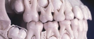
- The first upper molars are smaller in size than the second ones. The first has two widely spaced roots, which sometimes merge to the apex. The rudiment of the first “adult” premolar is formed between the roots. Most often, the first upper molars have four root canals, although there are also three or two (in 19 and 5% of cases, respectively).
- The first lower molars differ from others by having an elongated prismatic crown. On their chewing surface there are four tubercles: lingual, higher, and buccal. The first “children’s” molar has 2 roots, with the medial one being longer and wider than the distal one. Often the first lower molars have three canals.
- The structure of the crown of the second upper molars of the dentition resembles the first upper permanent tooth. Four tubercles are clearly visible on the surface, and sometimes a fifth one is noticeable. The buccal surface is almost square with slightly convex sides. An enamel ridge is often visible on the palatal side. Such teeth have three roots, in 85% of cases there are 4 root canals, much less often - 3.
- The second lower molars already have five cusps with less deep grooves than those of “adult” teeth. Both roots are flattened and curved at the top. In 85% of cases, teeth have three canals, although there are exceptions.
With the eruption of permanent, “adult” molars, the change of teeth begins, which lasts from 5-7 to 12-14 years. It is the first molars, which do not have primary analogues, that hold the bite, ensuring the correct placement of the remaining permanent teeth in the arch.
Teething
This stage is characterized by the active movement of temporary teeth from their places of origin inside the jaw until the crown completely exits into the oral cavity. This process is accompanied by active changes in the surrounding tissues, increased development of the root part, restructuring of the alveolar bone structure, as well as the formation and strengthening of the periodontium.
The first temporary teeth, which are usually the lower central incisors, erupt at the age of 5-6 months. Behind them appear the upper antagonists, other incisors and first primary molars. The last to erupt are the lower and upper canines. Typically the process lasts from 4-6 months to 2-2.5 years. With late eruption, the timing of the appearance of temporary teeth shifts from 8-10 months. up to 3.5 years.
Periodontal and root formation
This stage of development of temporary teeth precedes their eruption and continues 1-.5-2.5 years after the completion of this process. The crowns are usually already fully formed by this time. The place of root origin is Hertwig's epithelial sheath, consisting of external and internal cells of the enamel organ.
The process of development of the root system and periodontium includes the following stages:
- ingrowth of epithelial cells into the underlying mesenchyme;
- separation and formation of a channel of a certain shape;
- formation of root dentin with the participation of odontoblasts;
- development of cement on the surface of dental dentin;
- formation of dense periodontal tissues by fibroblasts;
- fixation of the root with collagen fibers to the alveolar bone;
- formation of secondary cement on the basis of primary cement after eruption.
Milk bite
The dynamics of jaw development should be considered in connection with the formation of dental follicles, their eruption and loss. This can most easily be seen using the example of the ontogenesis of the lower jaw (Fig. 34).
The rudiments of baby teeth are formed already in the first weeks of intrauterine development. Mineral salts begin to be deposited into the organic matrix from the 5th month, so that during the period of birth, the crowns of primary teeth have various stages of calcification (see Fig. 35, 36). The eruption of primary teeth generally occurs from 6 to 30 months. in the following order: I, II, III, IV, V. Usually the lower teeth erupt before the upper ones, both on both sides of the jaw. Root development ends approximately 2 years after eruption, and 4 years after this, their resorption begins.
With normal development of the dental system at the age of 6-8 months. The process of teething begins. There are two periods in primary occlusion, namely from 6 months. up to 3 years and from 3.1 to 6 years. The first period is characterized by close standing of the teeth, the absence of their wear, orthognathic relationship and the location of the upper and lower dentitions in the same frontal (tuberal) plane. In other words, the closure of the dentition of the primary occlusion on the distal surfaces most often ends without overlap, i.e. without a step (see Fig. 37, 43). This is explained by the fact that the lower second primary molar, which has three buccal cusps, is as much wider than the second upper primary molar as the lower central primary incisor is narrower than the upper one.
Rice. 35. Timing of mineralization of teeth in the antenatal period (Vinogradova T.F.).
In order to monitor the development of a child, the clinician must know the age-related signs of the correct formation of the dental system and in particular the bite, the formation of which occurs in parallel with the growth and development of the masticatory apparatus. Before teeth appear, the height of the lower third of the face corresponds to the height of the alveolar processes of both jaws and the height of the body of the lower jaw and is supported by the not yet fully formed articular disc. When the incisors erupt, three points of support appear: two in the joints and the third on the front teeth. Before the eruption of molars, the lateral sections of the alveolar processes are in contact and the height of the lower third of the child’s face does not correspond to either the upper or middle third.
By 6 months two central incisors erupt on the lower jaw, which moves slightly anteriorly, but the sucking function is preserved and decreases at approximately 8-9 months, with the appearance of the upper incisors. By 12 months all incisors must erupt, the chewing function begins to form and the activity of growth of the lower jaw in width in the frontal area sharply decreases, tissue ossification occurs at the junction of the two halves of the lower jaw and the type of swallowing changes, in which the tip of the tongue begins to rest on the upper front teeth.
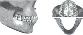
The first physiological increase in the interalveolar height (synonym in old textbooks is the height of the bite), and at the same time the lower third of the face, begins with the appearance of the first primary molars, which maintain the height and ensure the transition from sucking to the initial period of chewing function. Then further eruption of primary teeth (canines, second molars) occurs. The signs of normality are considered to be: average periods of eruption, pairing and sequence of eruption. Milk teeth differ in size, shape and color. Their shape is more convex, sharply demarcated from the root, and the severity of the neck is clearly felt by the probe. The crowns of primary teeth have a more bluish tint, a pronounced sign of curvature and are smaller in size than the permanent ones of the same name. The upper row of teeth is slightly larger than the lower one, and both of them have the shape of a semicircle (Fig. 38).
Rice. 37. Primary occlusion of a 3-year-old child, the distal surfaces of the second molars are in the same plane - indicated by a vertical line (explanation in the text).
Rice. 38. Primary occlusion: a - shape of the dentition of the upper and lower jaw, 6-3 years old, c - lateral view of the occlusion.
The shape of the dentition of the lower jaw changes depending on the shape of its body. Age-related changes in the shape of the body of the lower jaw are characterized by three periods: up to 5-6 months. During intrauterine life, the shape of the jaw is triangular, then up to 2 years the jaw arch has a semicircular shape and after 3-4 years the body of the lower jaw takes on the shape of a parabola (Chaikovskaya I.P.). The shape of the lower dental arch apparently goes through the same stages. It should be noted that the molars and anterior teeth are placed with their occlusal surfaces in the same plane, i.e. do not form either a sagittal or transversal curve. On the issue of the relationship between the dentition, i.e. bite, there is confusion in various manuals, namely, some talk about orthognathic (scissor or psalidodont), others - about direct (pincer, labiodont). These disagreements are explained by the fact that different authors characterize different periods of primary occlusion, i.e. in the first period, closure occurs according to the type of orthognathic, and in the second - direct bite. In a child aged 1.5 years, already chewing food, characteristic features of the joint are noted. With the advent of chewing teeth, the function of all elements of the joint becomes more complex and more perfect. Since until this time the interalveolar height was maintained on the disc, and now on the molars, the head from the depth of the fossa moves closer to the anterior wall. The disc becomes more formed, namely its posterior section thickens and fills the articular fossa, having a convex shape. The middle section of the disc, adjacent to two convex surfaces (articular tubercle and head), has a biconcave shape. This part of the disk is the most loaded. The articular tubercle increases and the articular cone atrophies. The layer of cartilage covering the mandibular head and articular fossa also decreases due to atrophy, since the risk of injury to the tissues lying in the articular fossa disappears. The anterior inclination of the articular head increases, and it, together with the already formed disc, moves anteriorly.
In accordance with further teething and the formation of speech, there is a gradual reverse development of special anatomical and physiological adaptations of the oral cavity. The basal part of the jaws thickens, the relief and architecture change, the alveolar process develops even more, the curvature of the mandibular canal begins, the branch grows and the angle decreases. It is mainly the posterior parts of the lower jaw that grow in length due to constant irritation and pressure from the molars located in this area. If you measure the distance from the socket of the second primary molar to the perpendicular drawn from the angle of the lower jaw, then in a newborn it will be 10 mm, and by 2 years it will already be 20 mm. The anterior sections of the jaw increase little, which can be seen by measuring the distance between the mental protuberance and the mental foramen, which remains almost unchanged. Growth in thickness also predominates in the lateral sections, where external and internal oblique lines gradually form. Approximately similar changes, but less pronounced, are observed in the upper jaw, which grows mainly in the areas of connection with the bones of the base of the skull. The maxillary sinus has a great influence on this process, the development of which mainly determines the growth of the upper jaw in height. In a newborn, the maxillary sinus is just emerging, and its development contributes to the eruption of all milk teeth and permanent molars (see Fig. 39). The canines and molars lie just under the orbit, and as the maxillary sinus develops, the tooth sockets move away from the bottom of the orbit. Between the sockets and the orbital area, spongy bone tissue appears, which subsequently resolves and turns into a cavity (Zucerkandl). The maxillary sinus increases mainly in the lateral direction (Walker F.I.) and by 2 years its volume is 1.5 cm3, and by 6 years - 2.5 cm3. Thus, due to greater growth in the distal sections, the upper and lower jaws move forward and downward in relation to the base of the skull (Varas E.Ya.). The completion of the eruption of all primary teeth is normally characterized by a number of signs: 10 teeth on each jaw, the upper anterior teeth slightly overlap the lower ones, there is fissure-tubercle contact of the molars, the anterior teeth may not have three and diastemas (one option), and may be located with primary (physiological) three between the lateral incisors and canines, and on the lower jaw also between the canines and first molars (three primates).
Along with the development of the dental system, the function of biting and chewing food develops, and therefore, features of the shape and function of the masticatory muscles appear in comparison with a newborn. In particular, the cross-sectional area, magnitude and direction of the resulting force of the synergistic muscles change. In other words, the masticatory muscles adapt and change depending on the function performed by the oral cavity before teething, during primary and permanent dentition. Knowledge of the functional dependence of the muscles on the activity of the dentition is very important for the orthodontist, because in case of dysfunction of the dentition (abnormal bite or deformation), it is necessary to look for one of the causes of the disease in the structure and function of the muscles.
Rice. 39. Development of the maxillary sinus according to Torrigiani (/), horizontal section of the skull through the maxillary cavity (diagram): a - newborn, 6 - adult; ch.p. - maxillary sinus.
Rice. 40. Primary dentition with the formation of secondary trema and diastemas.
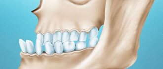
The second period of primary occlusion is characterized by significant wear of teeth, the appearance of teeth and diastemas, especially in the frontal area (Fig. 40). The presence of the latter indicates the preparation of the alveolar processes for the eruption of permanent teeth. The difference in the width of all front teeth of the primary and permanent dentition is 5 mm in the upper jaw and 3.8 mm in the lower jaw. Simultaneously with the appearance of diastemas and three behind the second primary molar, a free platform is created for the eruption of the first permanent molar. The morphological elements of the temporomandibular joint also correspond to this period, namely the absence of inclination of the articular head, the initial stage of development of the articular tubercle. All this explains the fact that in the primary occlusion there are no sagittal and transverse curves of the dentition.
Stabilization
This period is characterized by a complete stop of all processes of tissue formation and development. The crown and root reach the required shape, size and level of strength that allows them to perform their basic functions.
The period of stabilization of primary teeth lasts on average 2.5-3 years. At this time, it is important to ensure optimal chewing load on the bite, which will ensure the normal development of facial and other muscles, as well as periodontal tissues and jaw bones. If a child has caries or other diseases, it is during this period that treatment of temporary teeth will be most effective in terms of preserving them, preventing the spread of infection and ensuring normal conditions for changing the bite.
Physiological features of the development of baby teeth
The front, or frontal, teeth are located very tightly to each other and only when the child reaches the age of four, diastemas become visible between them - physiological gaps that increase after a year or two against the background of jaw growth. If there are no diastemas at the age of six, this indicates insufficient jaw growth, leads to crowding and requires correction of the bite in children. Physiological abrasion of surfaces, which contributes to the timely development of the masticatory apparatus, begins at three years of age.
Resorption and preparation for shift
At the age of 5-6 years, the process of completely replacing the temporary bite begins. This period begins immediately after the resorption of milky roots and the beginning of the growth of permanent rudiments. Temporary teeth begin to undergo resorption and ejection from the alveoli. Odontoclasts, which carry out demineralization and intracellular destruction, as well as pulp tissues that secrete osteoclast-like cells, which are responsible for the destruction of dentin and predentin from the internal side, actively participate in the process. Removal of the crown of temporary teeth, as a rule, occurs under the influence of chewing pressure. The root remaining in the hole undergoes a natural process of destruction and resorption. The location of the crown is quickly epithelialized due to granulation tissue.
The process of loss of temporary teeth occurs symmetrically on both jaws and ends with the appearance of permanent teeth. The individual nature of its course is determined genetically.