Causes of dental abnormalities
The main reasons that result in the development of dental anomalies are factors that have a direct impact on the jaw during its formation. Anomalies of hard dental tissues are a consequence of a lack of vitamin A in the body, which takes an active part in synthesis.
Disruption of the body's endocrine system, manifested by secondary insufficiency of the peripheral endocrine glands, is the cause of disruption of the mineral metabolism of teeth, which leads to their eruption at an early age and rapid destruction. Also, dysfunction of the parathyroid glands can, on the contrary, slow down the timing of teething, cause hypoplasia of hard dental tissues and lead to complete or partial absence of teeth.
Diseases suffered by a child in the period 1-3 years from birth, which were accompanied by various kinds of complications, affect the process of jaw development, which leads to the formation of an irregular (abnormal) shape of the dental arch.
Often, the process of abnormal development of the structure of the jaw and the shape of the teeth begins in childhood, and it is provoked by such bad child habits as thumb sucking, lip and tongue biting, prolonged pacifier sucking and prolonged bottle feeding - all this also affects the process of tooth formation . In some cases, when primary teeth fall out early, a child may develop some type of anomaly. In adulthood, teething anomalies are observed during the growth of wisdom teeth, when the position and number of teeth on the arch do not allow them to grow freely.
Another reason for the development of dental anomalies is genetic predisposition, and the pathological process can begin both on temporary and permanent teeth.
Fluorosis
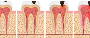
Fluorosis manifests itself in the form of whitish, later yellow, brownish spots, stripes, as well as enamel erosion. If the disease is neglected, it can lead to serious tooth destruction and even damage to bone tissue. The listed symptoms are provoked by long-term and constant exposure to large doses of fluoride on the body. A person can receive the substance at home (for example, with drinking water used in the area where he lives) or at work (if it involves constant contact with fluoride) due to safety violations. The doctor accurately determines fluorosis by characteristic external signs. Treatment begins with stopping the patient's contact with fluoride (replacing drinking water, other work). Affected teeth are remineralized and restored. To return them to their normal appearance, whitening and installation of crowns are possible.
Classification of dental anomalies
The basis for the classification of dental anomalies is the external and internal signs of the tooth structure, indicating their unnatural formation and development.
- Anomalies of individual teeth:
- congenital complete or fragmented absence of teeth;
- the number of teeth exceeding the natural number is supernumerary.
- Anomaly in the size and shape of teeth:
- disproportionately large teeth;
- vestigial incisors;
- upper incisors with atrophy of the chewing surface;
- anterior molars with a shortened surface and enamel hypoplasia;
- teeth with chalk stains on the enamel.
- Anomalies in the structure of hard dental tissues:
- excessive or insufficient formation of hard dental tissues;
- early or late growth of primary teeth;
- unnatural position of the tooth germ;
- different widths of the dentition (lower and upper);
- oblique or crossbite;
- discrepancy between the lateral teeth on both sides;
- overlap of the lower incisors with the teeth of the upper row.
- Anomalies of the dentition:
- dysfunction of dentition formation;
- unnatural position of single teeth;
- growth of teeth in the cheek and lip area;
- growth of teeth in the palate and tongue;
- medial and distal bite;
- non-occlusion of teeth with antagonists;
- location of the tooth above the occlusal surface;
- tooth growth across the jaw arch;
- the location of adjacent teeth is out of place;
- large or small distance between teeth;
- non-eruption of the maxillary incisors.
- Anomalies in the shape of the dentition:
- narrow dentition;
- saddle-shaped dentition;
- V-shaped dentition;
- dentition in the shape of a quadrangle;
- asymmetrical dentition.
- Bite abnormalities:
- sagittal anomalies (progenia and prognathia);
- transversal (open, true, traumatic and combined bite).
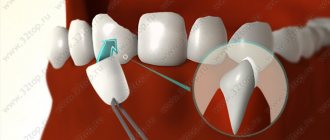
Wedge-shaped defect
A wedge-shaped defect is a special defect in the shape of the outer side of the tooth in the neck area. It is called so because of its V-shape, the inner surface is smooth. Occurs due to poor hygiene, excessive mechanical stress due to malocclusion or, for example, rough brushing with a hard brush. It provokes a moderate increase in enamel sensitivity, and in advanced cases, tooth fracture. Seriously ruins the appearance of your smile. It is easily diagnosed upon examination and treated by remineralization and filling of the enamel, installation of crowns, and, in the future, correction of the bite.
Anomalies in the number of teeth
One of the most common pathologies is a deviation from the standard number of teeth in one row. The main symptom of deviation is single or multiple absence of teeth due to their failure to erupt. Also, a smaller number may be due to the combination of several teeth into one during the period of their development and eruption. Most often, such an anomaly occurs on the front teeth and incisors located in the lateral part. This localization of the process causes microdentia - a disease characterized by tooth reduction or absence in the pathological zone on one or the other side of the upper jaw. In dentistry, the anomaly of missing teeth is called adentia. It can be partial or complete. Additionally, a tooth may develop in the jaw but not erupt, which is called impaction. When teeth are retained, their formation is almost complete, but the roots of the tooth are absent. The absence of teeth is due to a violation of the formation process, when the tooth germ has not formed. Most often, the cause of an abnormal number of teeth is a person’s genetic predisposition. The above anomalies are treated with prosthetics, sometimes in combination with orthodontic treatment.
Along with an incomplete set of teeth, supernumerary teeth also occur - when the number of teeth in a row significantly exceeds their normal number. The localization of such an anomaly most often occurs in the area of the front teeth. The shape of teeth located in an unnatural place is spike-shaped, but in some cases they can be similar to nearby teeth. The main reason for the increased number of teeth is dysfunction of the development of the dental epithelial plate. The treatment technique for such dental anomalies is selected individually for each case and depends on factors such as the location of the superset of teeth, the influence of the teeth on the permanent teeth. If the permanent teeth on the jaw are displaced, then treatment consists of removing the abnormal teeth and subsequent orthodontics. In cases where the grown abnormal tooth does not disturb the position of permanent teeth, its removal is not performed - the shape of the tooth crown is changed using prosthetics.
Pathogenesis
Dental anomalies are a group of congenital or acquired disorders of the location of molars and the entire dentition that affect the functioning of the jaw. According to research results, pathology occurs in 50% of children and 30% of adults.
Each tooth has its own special characteristics, such as appearance and size. They are determined by its location and the period of formation. Deviations from normal sizes, shapes, shades or structure are considered an anomaly and require treatment, as they negatively affect the functioning of the jaw.
Most often, the gastrointestinal tract is affected, since the unnatural position of the teeth makes it difficult for the patient to chew food thoroughly.
Treatment of pathology is carried out in stages, often requiring surgical intervention. If the position of the canine and incisor is incorrect, orthodontic or orthopedic methods are used to correct the bite.
Anomaly in tooth size
Excessively enlarged teeth have a huge base compared to the base of healthy teeth. Often, such a pathological deviation is diagnosed in older patients, when a permanent dentition has already formed. An exception to clinical cases is the diagnosis of overly large teeth in older children and teenagers, when they still have a temporary bite. Most often, the anomaly of increased tooth size affects the incisors, sometimes other teeth in the row.
The reasons for the development of the pathological process in modern medicine are known and well studied. The main one is dysfunction of the body's endocrine system, as a result of which several tooth buds merge into one, which leads to gigantism. The increased size negatively affects the process of teething located in close proximity, since it can slow it down.
In rare cases, the location of giant teeth may be outside the dentition. In addition to functional disorders, this pathology can cause mental distress in the patient due to the unaesthetic appearance of the teeth.
As a therapeutic therapy, dentists perform the removal of a pathological tooth; it is possible to remove it along with adjacent teeth. Removal is performed in conjunction with prosthetics, which helps eliminate large gaps between teeth. If it is possible to remove an abnormal tooth without damaging nearby growing teeth and correct their position, then after removal there is no need for prosthetics and filling voids.
Teeth that are abnormally small are often an abnormality of the permanent bite, affecting the incisors of the upper, lower, or both jaws. Unlike giant teeth, small teeth have small crowns.
Experts note the fact that today the true cause of the development of anomalies of small teeth is unknown. One of the etiological factors in the development of pathology is the genetic predisposition of the patient. It is also known that most often in patients with an anomaly of small teeth, parents suffered from such anomalies as small teeth in one, and large jaws in the other.
A characteristic feature of the anomaly is the location of the teeth at a great distance from each other, which leads to a violation of the aesthetic appearance and psychological disorders of the patient. Treatment includes prosthetics and the installation of crowns on pathological teeth.
Symptoms
Compared to the non-standard position of primary teeth, anomalies of molars are less common.
All about impacted teeth
Signs of malalignment of teeth are:
- Distal displacement. Characterized by varying degrees of deviation of one or more molars back in relation to the row.
- Mesial displacement . The incisor or canine moves forward in relation to the dentition.
- Vestibular location . Most often, the fangs are affected, which move towards the vestibule of the oral cavity.
- Vestibular placement of anterior teeth. In this case, there is a movement of the incisors forward, towards the inner surface of the lip. The degree of pathology is established during examination of the alveolar process.
- Oral location . A distinction is made between lingual placement, when the row deviates towards the tongue. Pathology occurs in many patients when replacing baby teeth with molars. Usually the process affects incisors and premolars in the absence of free space in the row. There is also a palatal arrangement, characterized by movement towards the upper jaw. Common causes include misdirection during teething. Occurs during the formation of a permanent dentition.
- Palatal position . Deviation of one or more anterior molars towards the palate of the upper row. The pathological process affects the upper incisors. The defect is formed when the alveolar process is not fully formed. Diagnosed by examination, in relation to neighboring ones.
- Supraposition. The molar is located above the occlusal curve. Develops as a result of incomplete eruption or insufficient development of the jaw. Occurs more often in children.
- Infraposition. The tooth is located below the occlusal line.
- Tortoanomaly. It is characterized by varying degrees of rotation of the molar along the vertical axis. Occurs most often with supernumerary conditions.
- Transposition. A change in the location of the teeth when the canine is located in place of the premolar.
In many cases, deviations occur together with improper development of the jaw, resulting in occlusion. Clinical manifestations directly depend on the type of anomaly and are visible to the naked eye. The degree is established using instrumental diagnostic methods.
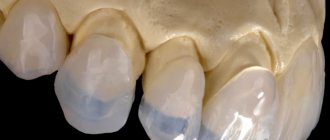
Anomalies in tooth shape
Tooth shape anomalies include various deviations that differ significantly from the natural appearance of the tooth. So one of the common forms of this deviation are teeth, which in their shape resemble spikes, or rather, have a wide base at the gum edge and taper wedge-shaped towards the end of the tooth. This anomaly is typical for incisors located in the center and on the sides. In some cases, it may be located on the lateral teeth of both jaws. The exact reason for the formation of such teeth is unknown, but dentists suggest that it is due to the disrupted development of tooth buds. Elimination of pathology is performed by prosthetics or removal of abnormal teeth. When teeth are removed, dentures are installed in their place, which can be either removable or permanent.
In general, any deviation from the natural shape of a tooth is considered an anomaly. Because of this, any pathological process associated with the shape of the tooth is classified as malformed teeth. Most often they are located in the front of the upper jaw. The nature of the appearance of such teeth has not been studied, the exact reason for their formation is unknown; doctors identify dysfunction of the development of the jaw and tooth buds as a likely factor that provoked this process. Anomalies are treated, as in the previous case, by prosthetics, removal, or a combination of the two methods presented.
Treatment
The goal of treatment is to normalize the size, appearance and location of the molars. For this purpose, various designs are used, which can be removable or non-removable. The treatment regimen depends on the type of disorder.
When the tooth deviates backwards, mesial movement of the tooth is indicated, but only if there is free space in the row. Treatment is carried out individually, depending on the degree of molar deviation.
When establishing a lateral type of deviation, the removal of the supernumerary tooth is required. If the presence of microdentia is diagnosed, the pathology is eliminated by installing dentures. The procedure is performed on adults and children aged 14-15 years, since by this age the jaw is fully formed.
Everything you need to know about edentia
The Engel arc is used for vestibular placement. Stationary or sliding is used, depending on the degree of violation. It is also possible to install a positioner.
When a deviation towards the tongue is established, movement of one or more molars is required. In this case, the doctor must determine the availability of space. The procedure is performed using orthodontic instruments.
Deviation towards the upper palate requires the installation of special plates. In the case when the incisors are turned towards the palate, a Bynin, Schwartz or Reichenbach-Brückl plate is used. Wearing a positioner with a setup system is also shown.
A lingual or twisted position resulting from macrodentia requires surgical removal. The dentist carefully examines the location of the midline. You can remove a cutter located in the center or on the side. When no line displacement is observed, an incorrectly located canine or incisor must be removed. But if the line deviates, the tooth located on the other side is removed.
Description and 4 effective methods of treating an underdeveloped lower jaw
If a tortoanomaly has been diagnosed, metal structures or crowns with special fasteners are installed that create versatile forces. The function of support points is performed by the hooks on the basic devices.
When inserting rings used to restore the normal position of molars, a force is directed away from the incisor or canine that was malformed. Positioners are also used to restore rows.
Anomalies in the structure of hard dental tissues
This pathology is characterized by deposits on the surface of the tooth. With hypoplasia and hyperplasia, an accumulation of chalky spots appears on the teeth, and all teeth in a row are immediately affected, occlusion is disrupted, and unnatural bumps appear on the chewing surfaces.
When a patient has an anomaly such as amelogenesis imperfecta, the surface of his teeth becomes covered with pigment spots that have a characteristic brown or yellow color.
With dentinogenesis imperfecta, dysfunction of dentin formation is observed. In this case, the teeth acquire a yellow-brown color and are characterized by rapid wear.
Edentia
Adentia is a phenomenon characterized by the absence of one, several or all teeth in a patient. The reasons can be different - from disturbances in the formation of the dental system to the loss of normal grown organs. Broken teeth are not only unsightly - they also do not cope well with chewing, speech, and sometimes lead to deformation of the facial skeleton and destruction of the remaining teeth. Adentia must be diagnosed and treated in time! To make a diagnosis, a doctor needs only a visual examination and palpation, and less often, an x-ray. The defect is corrected with an individually selected prosthetic program.
Diagnosis of dental abnormalities
In order to establish the correct final diagnosis, the patient must undergo a preventive examination by specialized specialists: pediatrician, ENT specialist, endocrinologist, geneticist. Due to the fact that the etiology of various dental anomalies has not been fully studied, any disturbance can provoke a pathological process.
After a complete examination, the dentist will be able to establish an accurate diagnosis and prescribe the necessary treatment, which will involve surgeons, orthodontists, periodontists and therapists.
For differential diagnosis, laboratory tests and the results of radiography and tomography are necessary. The specialist will perform a thorough examination of the face and oral cavity, collect all anatomical data, on the basis of which the deviation index will be determined.
Casts of the jaw and face will help establish a complete picture of the disease.
Granuloma
Granuloma is a localized inflammation of the root of a tooth caused by prolonged exposure to infection. It manifests itself as pain and swelling of the gums, the development of gumboil, abscess, cyst, damage to the jaw bone, sepsis, and infection of nearby organs. It is almost never diagnosed during examination; it is suspected based on clinical signs and confirmed by x-ray or radiovisiography. It can be treated both without surgery - by filling the granuloma cavity with a filling compound and administering antibiotics - and surgically. In the latter case, the tooth can be removed, or the fluid can be drained and only the affected areas of the root removed.
Forecast and prevention of dental anomalies
In the conditions of modern medicine and advanced equipment, dentists and surgeons can (if not completely, then maximally) eliminate the manifestations of dental anomalies. If you contact a specialist in a timely manner in order to establish an accurate diagnosis and determine a treatment plan, especially if the deviation is identified in early childhood, the prognosis for recovery will be favorable. Doctors will be able not only to restore the functional abilities of the jaw, but also to eliminate aesthetic defects.
The main preventive measure to prevent the development of any type of dental anomalies is a careful attitude towards pregnancy. It is during the period of intrauterine formation that pathological processes can arise in a person. It is important to monitor your health in the first days after the birth of your baby. Early breastfeeding and a balanced diet will help avoid many dental anomalies. You should undergo a full preventive examination by specialists, including a dentist, at least once every six months.