2024
The bite is the location of the jaw rows and individual teeth relative to each other.
Not only the external attractiveness of a person’s smile, but also the functionality of the entire oral cavity depends on the correctness of their ratio.
Permanent bite is the final stage of formation of the human dental system, which is completed by the age of 24-25.
After this, correcting existing anomalies in the structure of the jaw rows is much more difficult, and correction requires a lot of time.
Stages of development
The beginning of the formation of a permanent bite is observed in children at the age of six, when the replacement of milk teeth with molars occurs.
By the age of 12, this process is usually completed and active growth and development of the erupted elements of the jaw rows begins.
Experts distinguish three stages in the formation of a permanent bite:
- Development period. Between the ages of 13 and 15 years, adolescents experience active growth of the alveolar processes of the jaw. At this time, eruption of the second molars is observed. As a rule, this stage is completed by the age of 18.
- Pre-formation period. From 18 to 24-25 years of age, people experience growth and development of dental arches, and the jaws reach their final size.
At this stage, the eruption of the third molars, the so-called wisdom teeth, occurs. However, experts note that these fragments may appear in humans much later or may not erupt at all if their rudiments have not been formed, or there is not enough space in the jaw row.This stage is also characterized by mesial movement of the elements of the jaw row, which contributes to the optimal distribution of the chewing load.
- The period of final formation. By this time, a person often has all 32 units cut through.
The processes of growth and development of the jaw rows slow down and stop, but displacement in the mesial direction can continue throughout life, which is explained by the gradual erasure of their contact surfaces.
In the process of forming a permanent bite, the proportions of the face also change, in particular its height, depending on the eruption of chewing units.
The growth and development of the jaw rows entail a displacement of the facial bones relative to each other, which ensures that the correct proportions of the face are maintained.
Intramaxillary development
The first stage of formation of temporary teeth begins at 6-8 weeks of embryogenesis and lasts until the middle of the first year of a child’s life. It includes the following processes:
- the formation of a dental plate from multilayered squamous epithelium located in the oral fossa of the embryo with the further formation of a ridge and the growth of tooth buds in the form of flask-shaped buds;
- the formation of epithelial (enamel) organs due to the ingrowth of mesenchyme into the kidneys with the subsequent formation of dental papillae and sacs;
- histogenesis of dental tissues (formation of dentin, formation and mineralization of enamel, development of pulp from the mesenchyme of the dental papilla with the formation of nerve fibers and blood vessels).
Each newborn has inside both jaws the rudiments of all 20 temporary and 16 permanent teeth, which have varying degrees of development and mineralization.
Functions and position of teeth
The total number of teeth located in a person's mouth can range from 28 to 32, depending on whether the third molars have been cut.
Depending on the structure, all elements of the jaw line are divided into several groups, each of which performs certain functions in the process of eating food:
- Incisors – eight central teeth, which are located four in the upper and lower jaw rows. These are elements with one root and a sharp cutting part, the purpose of which is to bite off food.
- The canines are located in the jaw row behind the incisors. There are 2 fangs on each jaw. These elements have a special pointed edge that helps them participate in the digestion process by tearing food particles away from a larger piece.
- Premolars are also called small molars. These elements are located 4 in each jaw row and are necessary for grinding and grinding food. This is achieved by the prismatic shape and the presence of two tubercles on the chewing surface.
- Molars are the largest elements of the jaw row.
Their number in the oral cavity reaches 12 units - 6 in each jaw row. The molars are rectangular in shape and have three strong roots. Their chewing surface is equipped with four tubercles, which allows them to thoroughly crush and grind food before swallowing it.
Wisdom teeth are the third molars in the jaw row, which erupt later than the other elements.
They play a small role in the digestive process; their main purpose is to protect the remaining molars from loosening as a result of high chewing load.
At what age does a mixed dentition occur and the features of its formation.
Read here about senile progeny and methods of its treatment.
At this address https://orto-info.ru/zubocheliustnye-anomalii/okklyuzii/profilaktiki.html we will talk about devices used to prevent malocclusions.
Row shape
Physiological or correct permanent dentition is determined by the following factors:
- there are no large gaps between the frontal incisors;
- the median facial line passes exactly between the frontal incisors of the upper and lower rows;
- the overlap of the coronal part of the lower units with the upper elements of the jaw during closure does not exceed 1/3 of their length;
- when the jaws are brought together, there is no space between their elements, and the location of the palatal cusps of the lateral teeth of the upper jaw corresponds to the fissures of the lower molars;
- when connecting the jaws, each element of the row, with the exception of the central incisors of the lower line and the upper third molars, has two antagonists;
- the upper dental arch has the shape of a semi-ellipse, and the lower one - a parabola;
- the length of the upper jaw row is slightly greater than the size of the lower one.
The optimal bite from an orthodontic point of view is an orthognathic bite. It ensures a complete process of biting and chewing food.
Dentists also include the following forms of occlusion as physiological forms of occlusion:
- Straight. Its peculiarity is the contact of the cutting surfaces of the teeth in the anterior sector when connecting the jaws.
- Bioprognathic involves a slight inclination of the teeth of both jaws in the vestibular direction.
- Opisthognathic - a slight inclination of the elements of the jaw rows to the inside.
- Progenic involves a slight advancement of the elements of the bottom row forward.
Teething
This stage is characterized by the active movement of temporary teeth from their places of origin inside the jaw until the crown completely exits into the oral cavity. This process is accompanied by active changes in the surrounding tissues, increased development of the root part, restructuring of the alveolar bone structure, as well as the formation and strengthening of the periodontium.
The first temporary teeth, which are usually the lower central incisors, erupt at the age of 5-6 months. Behind them appear the upper antagonists, other incisors and first primary molars. The last to erupt are the lower and upper canines. Typically the process lasts from 4-6 months to 2-2.5 years. With late eruption, the timing of the appearance of temporary teeth shifts from 8-10 months. up to 3.5 years.
Types of deviations
In some cases, at the first stage of the formation of a permanent dentition, some malfunctions occur, as a result of which further growth and placement of teeth occurs with disturbances.
Orthodontists distinguish the following forms of pathological occlusion:
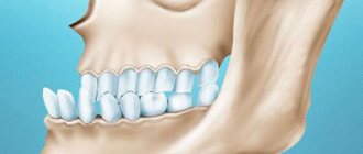
- Distal. Significant underdevelopment of the lower jaw row or excessive development of the upper one leads to a strong protrusion of the central incisors when the jaws are closed. Pathology threatens the early development of caries, periodontal disease, and impaired swallowing function.
- Mesial. The anomaly is characterized by excessive protrusion of the lower jaw and overlap of the upper elements by the lower teeth.
In addition to disturbances in the patient’s appearance, expressed in an excessively protruding chin and concave profile, the pathology causes functional disorders. These include periodontal diseases, joint pain, and discomfort during chewing. - Open. With this form of occlusion, the patient experiences non-closure of the jaw rows in the frontal or lateral areas.
Symptoms of the anomaly are elongation of the face and severe impairment of diction. The pathology can be either congenital or acquired as a result of mechanical trauma to the jaw. - Cross. The anomaly involves a discrepancy in the size of the jaw in any part of it.
As a result of this phenomenon, a displacement of one of the dentition occurs, which is why a person experiences an intersection of the upper and lower units. The pathology makes it impossible to fully chew food, entails impaired diction, changes in the appearance of the face, as well as numerous problems with the gums. - Deep . When the jaws are connected, the upper teeth overlap the lower teeth by 2/3 or completely, as a result of which the mucous membrane of the gum tissue is often injured. A person experiences difficulty biting and chewing food and pronouncing certain sounds.
Orthodontics
BRIEF INFORMATION ON EMBRYOLOGY AND FUNCTIONAL ANATOMY OF THE MASTICAL APPARATUS
The embryonic facial skeleton is formed from seven processes of the first branchial arch.
Three processes are paired - lateral nasal, maxillary, mandibular and one unpaired - frontal. The upper jaw develops from the upper section of the first gill arch, and the lower jaw develops from the lower section.
The differentiation of the oral and nasal cavities in the embryo begins after the 4th week. Before this, both cavities are fused and have a five-pointed shape characteristic of all vertebrates.
JAW DEVELOPMENT
The development of the upper jaw occurs as follows: in the second month of uterine life, the pterygoid processes branch inward from the rudiments of the jaws, and the frontal process descends between both maxillary processes. In the third month, the opposite processes of the upper jaw grow together and form the upper jaw. At this stage of embryo development, the separation of the oral cavity from the nasal cavity begins: the nasal septum and premaxillary bone are formed from the frontal process, and the process of the upper jaw provides the anlage for connecting the upper jaw with the ethmoid bone and the palatine bone with the pterygoid. The palatine processes fuse by the end of the 8th week, and their fusion begins at the lip. At the 9th week there is already a vault of the hard palate, and at the 12th week there is a vault of the soft palate.
The fusion of the palatine processes with the nasal septum occurs in the same period. The maxillary sinus is formed in the form of a groove on the side of the lacrimal canal at the 3-4th month of intrauterine life. The alveolar process develops as teeth develop and erupt and roots form, which is associated with the formation of root sockets and intersocket septa.
The development of the lower jaw parallels the development of the upper jaw.
Ossification of the skeleton of the masticatory apparatus begins early from the so-called ossification points. The lateral processes of the upper jaw ossify in the second month, and both halves of the palatine processes ossify by the end of the second month from the bone core lying between the ascending and horizontal parts. Ossification of the premaxilla occurs later than ossification of the maxilla. The premaxillary bone is connected to the upper jaw after the palatal plates fuse and the oral cavity is formed, approximately by the tenth week.
The basis for the lower jaw is Meckel's cartilage, bounded on the sides by connective tissue, which, when calcified, forms the dermal or integumentary bone, and Meckel's cartilage is absorbed and partially goes to other formations. Both parts of the lower jaw fuse during the first year of extrauterine life.
In a newborn, the skull is much larger than the facial skull, the chin is almost not pronounced, and the angle of the lower jaw is 140-150° (Fig. 1).
Other pathologies
The formation of a pathological permanent bite can occur due to a large number of reasons.
Dentists identify the main factors influencing the condition of teeth during their formation:
- disturbances during the formation of the rudiments of milk or permanent teeth due to a lack of calcium in the body of a pregnant woman or child;
- heredity;
- pathologies of intrauterine development or chronic diseases;
- presence of bad habits , for example, prolonged sucking of a pacifier or fingers, biting nails, resting the chin on the palms of the hands;
- mechanical injury to the jaw.
Experts in the field of orthodontics note that the formation of an abnormal permanent bite causes not only disturbances in the aesthetics of the appearance of the oral cavity, but also numerous functional disorders .
The most common of them are the following diseases:
- disturbances in the functioning of the digestive system as a result of inadequate chewing of food;
- inflammation of the gum tissue due to injury;
- pathological abrasion of the enamel due to the incorrect relationship of the opposite elements of the jaw rows;
- loosening of teeth , characteristic in cases of uneven distribution of chewing load;
- breathing and diction problems.
In the absence of timely treatment, acquired diseases will progress, leading to new severe consequences for the body.
What is Costen's syndrome and the symptoms accompanying the pathology.
From this publication you will learn how the pacifier affects your child’s bite.
Here https://orto-info.ru/zubocheliustnye-anomalii/chelyustey/krivaya-ne-prigovor.html all the most important things about methods for correcting a crooked jaw.
Numerous scientific works in dentistry, otorhinolaryngology, traumatology and plastic surgery are devoted to the study of the structural features and topography of the facial part of the head, gender characteristics and age dynamics.
The anatomical structure of the bones of a given area largely determines its functionality. In particular, the features of the maxillofacial area of the head determine the functional ability of the digestive and respiratory systems, the phonetic apparatus, and also have great aesthetic significance.
At the same time, the development of bone tissue of the alveolar process is of interest to specialists from the field of practical dentistry, including those involved in the issues of dental implantation. Understanding this process largely determines the outcome of treatment.
It is necessary to take into account the pronounced dependence of the structure of the maxillofacial region on a number of physiological and pathological factors, primarily on the age-related restructuring of the bone tissue of the jaws, in particular their alveolar areas.
Numerous sources of scientific literature contain a large amount of information about the course of these processes, but over the past decades there has been a pronounced tendency towards changes in their duration and timing compared to the data of fundamental research 40-50 years ago.
Obviously, the reasons for such deviations are various factors, the influence of which on the body has intensified over the past few decades, both in connection with technological progress and the lifestyle of modern man. First of all, the condition of the oral cavity, jaws and teeth is strongly influenced by improper and irrational nutrition, low level of oral hygiene, as well as living in environmentally polluted areas.
Factors that influence the course of the formation and development of both the body as a whole and the maxillofacial region in particular include the phenomenon of acceleration, which is also confirmed by numerous scientific sources.
The research results convincingly prove that today, much earlier than indicated in the sources of the last century, the processes of eruption and replacement of teeth occur, which means that the period of their intramaxillary development is significantly reduced. As a result, the degree of mineralization of their hard tissues at the time of tooth eruption into the oral cavity is insufficient, and this, in turn, leads to rapid destruction of crowns and premature tooth loss.
Sources of professional dental literature indicate that another cause of adentia is the high incidence of periodontal diseases. Domestic and foreign authors confirm that pathological processes affecting the periodontium extend to the bone tissue of the alveolar processes of the jaw, the mucous membrane of the gums and the root of the tooth, leading to the loss of practically healthy teeth and, as a result, the restructuring of the alveolar areas.
It is necessary to take into account the complex anatomical structure of the upper jaw and the presence of the maxillary sinus, characterized by individual differences, the relative position of the sinus, the roots of the maxillary dentition and the bone tissue of the alveolar process of the upper jaw in different age periods. An in-depth study of these anatomical formations is important to ensure high-quality restoration of the structure and functionality of the dentofacial apparatus in pathological conditions of various etiologies, as close as possible to physiological ones.
Patterns of formation of skull bones
The processes of formation and development of the musculoskeletal system, in particular the skeletal system, have always attracted the attention of researchers. Domestic and foreign authors continue to thoroughly study the features of development and age-related restructuring of the skull, especially its maxillofacial part.
Numerous scientific studies are devoted to the study of the characteristics of the formation of the skull during the intrauterine period of ontogenesis. The author of the fundamental work “Fundamentals of Medical Craniology” V.S. Speransky described 3 stages of the intrauterine period of skull development - membranous, cartilaginous and bone.
At the first stage, the components of the membranous skull are mesenchymal cords, the placement of which is associated with the growth of the embryo’s head. Starting from the fourth week of intrauterine development, the formation of a cartilaginous skull occurs, the development of which originates from the occipital bone and spreads in the ventral direction. Ossification of the cartilaginous skull begins in the 3rd month of intrauterine development, but in some bones of membranous origin, ossification centers appear earlier.
The process of ossification of the skull has been well studied, but the results obtained often differ significantly from each other (according to 28 authors, there are from 115 to 120 ossification centers in the skull, some of which lie in the cartilage, some in the mesenchymal base).
However, the vast majority of researchers argue that the first points of ossification in the areas of jaw formation are formed in the 7th week of embryogenesis, and the boundaries between individual bones of the skull at the sites of future sutures are visualized starting from the fourth month of intrauterine development.
It is known that most skull bones have several centers of ossification, and therefore at the time of birth of a child the number of skull bones is much greater than in adulthood, and the fusion of individual parts in such bones continues for a certain time after birth.
The characteristic appearance of the embryo is determined by the gill arches, which are formed during 4-5 weeks of intrauterine development. The formation of the jaws begins in the cartilage zone of the first branchial arch, which consists of a dorsal part (maxillary protrusion located under the orbit) and a ventral part (mandibular protrusion), which contains Meckel's cartilage and forms a kind of rod. Around the latter, the formation of the lower jaw occurs.
Numerous studies by morphologists, anthropologists, craniologists, as well as representatives of practical medicine, whose area of professional interest is the area of the head, as well as organs and systems associated with it functionally or topographically, have been devoted to the study of the patterns of development of individual bones of the skull.
V.S. Speransky claims that the formation of the skull is characterized by an uneven course in different time periods. In particular, until the end of the intrauterine period of development, the skull is 70-80% represented by formed bone tissue. In this case, the bones of the base of the skull are separated by cartilaginous layers, between the bones of the vault there are remnants of membranous tissue that forms the theme, expanding at the intersection of the sutures, and the bones of the facial skull are combined using syndesmoses.
The development of the head, the bony basis of which is the skull, after the birth of a child is characterized not only by an increase in size, but also by a change in the shape and spatial relationships of its component parts. In particular, this concerns the relationship between the brain and facial areas of the skull.
Each of them is characterized by a different type of growth and depends on the structural and functional characteristics of a given area. The bones of the cranium are structurally connected to the brain and its membranes, as well as to the sensory organs. The bones of the facial skull, primarily the jaw, are associated with teeth of both generations and depend on the course and duration of the processes of their formation, formation, growth, development, change and loss, as well as on the degree of pneumatization of the air-bearing bones.
The upper jaw, frontal and sphenoid bones contain air sinuses, and the ethmoid bone contains a labyrinth. All this determines certain features of the growth and formation of bones in the maxillofacial region, which today attract the attention of scientists and practicing doctors.
Growth and remodeling of skeletal bones in general, and the skull in particular, occur as a result of the synchronous flow of two interrelated processes - opposition and resorption of bone tissue. Thus, the restructuring of bone structures is especially important for the upper jaw, since simultaneously with changes in its size and external shape, processes of formation and restructuring of the maxillary sinus, anlage, development, eruption and replacement of teeth occur in the thickness of the bone body.
Another significant feature characteristic of the head skeleton is that at different age periods the intensity and direction of growth of the skull changes. This fact must be taken into account in pediatrics, especially in the practice of neurosurgery, otorhinolaryngology and maxillofacial surgery.
Craniometric studies show that the size of the head changes especially rapidly during the first 3-4 years of a child’s life. Their intensity decreases somewhat at the age of 5-6 years and intensifies again after 7 years.
At the same time, different directions of growth prevail at different periods of time, and the longitudinal and transverse dimensions of the skull alternately change. The periods of the most intensive growth of the skull in length are 2-3 and 7-8 years, and the most active growth of bones in width continues during the 1st year of a child’s life.
The results of craniometric studies tell us that the formation of the alveolar areas of the jaws and the associated vertical growth of the anterior part of the face is completed after reaching 10 years of age.
The directions of skull growth in individuals of different sexes during adolescence are different. In boys, during this period, longitudinal head growth prevails, and in girls, the size of the skull increases synchronously in the vertical and horizontal directions. Scientists agree that the increase in the size of the skull in width continues in girls up to 15, and in boys up to 16 years; in height - up to 16 years for girls, and up to 18 for boys.
However, with the end of the main stage of growth and formation of the skull, its restructuring does not end. Under the influence of local factors that determine the individual characteristics of each part of the face (the intensity of the load on the masticatory muscles, the prevalence of nasal or mouth breathing, bad habits), remodeling processes continue throughout life.
This fact must be taken into account not only during surgical interventions in this area, but when planning orthodontic or orthopedic measures.
Craniometric studies undertaken in various areas of the facial skull over various time periods indicate that in the fetal period the size of the jaws increases by 3-4 times, and the most active growth is observed in width. After birth and until the formation of the permanent occlusion is completed, increased growth of the maxillofacial region occurs, as a result of which the size of the jaws in three mutually perpendicular planes increases by 7-9 times.
Development of bones of the maxillofacial region and alveolar process
Since with age the growth rate in individual areas has different dynamics, the proportional ratios of the width, height and length of the jaws change significantly throughout life. Consequently, in each age period, regardless of the general gradual slowdown in the rate of bone growth, the jaws have areas of increased and relatively slow growth.
In areas of increased growth, a quantitative increase in bone structures occurs, and in areas of slow growth, their qualitative restructuring occurs. In this regard, growth zones should be understood not as permanent, clearly defined areas, but as places where, in a given period of time, opposition processes prevail over resorption processes.
In adults, the formed upper jaw (UM) is a paired bone of a complex structure, consisting of a body and four processes - alveolar, palatine, zygomatic and frontal. The thickness of the bone body contains the air-bearing maxillary sinus (MSS), and the cells of the alveolar process contain fixed tooth roots that form the maxillary dentition.
Research shows that the alveolar process is built of spongy bone tissue, covered with a compact layer, which is better expressed on the palatal side. This structure of the bone tissue of the HF in general and the alveolar process in particular is largely determined by the load acting on the bone during chewing.
It is due to the uneven load, which is distributed and spread in a certain way by the bone beams, that in certain areas of the processes and body of the upper jaw its spongy structure is compacted, forming buttresses and thus changing the external relief of the bone.
The presence and severity of buttresses is associated both with the presence or absence of teeth (after their loss, not only the area of the alveolar process of the jaw is resorbed, but also bone tissue, a buttress is formed at the level of a given dentofacial segment and along the line of pressure distribution), and with the condition of periodontal tissues, pathology which changes the distribution of chewing load on the jaw.
Some studies are devoted to the structural features of the maxillofacial region and structures of the oral cavity, depending on the constitutional structure of the skull. The results of anthropometric studies indicate that dolichocephals always have a high palate and high alveolar processes of the upper jaw, while brachycephals always have a low palate and low but massive alveolar processes.
In adulthood, after the completion of the basic formation of the permanent occlusion and growth of the skull, a number of factors are identified that influence the condition of the bone tissue and the functional ability of the jaws. These include the general condition of the body, the presence or absence of chronic diseases and metabolic disorders, as well as the method of nutrition, which determines the load on the teeth, periodontium and jaw bone tissue.
However, of course, the main factor determining the characteristics of jaw restructuring in adulthood is the preservation or loss of teeth.
Among the causes of acquired adentia, trauma is common, as well as tooth loss due to periodontal diseases, complications of carious lesions and pathological processes in the bone tissue of the jaws (periostitis, osteomyelitis).
Restructuring of the bone tissue of the alveolar process of the upper jaw and the alveolar part of the lower jaw after tooth loss consists not only of a decrease in bone mass, a decrease in the height of the alveolar process at the level of one or several dentofacial segments. According to research results, with edentia, the quality of bone tissue also changes - the volume decreases due to a significant thinning of the bone layers, which is also confirmed by a decrease in their density.
According to a number of authors, whose professional interests concern the state of bone tissue and its age-related dynamics, changes in the density of skeletal bones, as well as its individual parts, are due to several systemic and local reasons.
General reasons include, first of all, age-related metabolic changes occurring in the body - changes leading to a decrease in the content of mineral elements (calcium) in the bones, as well as organic compounds. This mainly implies the development of osteopenia and osteoporosis.
Most scientists consider the main local reason for the decrease in the quality of bone tissue of the maxillofacial region and alveolar process to be the redistribution of the load during chewing - a sharp decrease and uneven distribution of pressure on bone tissue in edentulous areas with various types of tooth loss.
After tooth loss, atrophy of the alveolar processes occurs differently in dolichocephals and brachycephals. When the size of the alveolar process is small, bone atrophy under the influence of compression pressure increases, which causes a change in the width of the arch. The processes of bone tissue resorption have different intensities in different zones of the cellular areas, but they are most pronounced on the vestibular side.
When teeth are lost, atrophy of the alveolar process is not only an aesthetic problem. It leads to a change in the proportions of the face and may in the future become the root cause of the restructuring of a number of anatomical formations, including the displacement of the neurovascular bundles passing through the incisive and palatal canals, individual facial muscles, as well as all masticatory muscles, asymmetry of the jaws and dentition, and, as the consequence is dysfunction and even destruction of the temporomandibular joint.
Another important factor to consider when examining dental patients is the different visualization of the alveolar process in different areas of the jaw. In particular, in the central part of the jaw at the level of the incisive and canine dentoalveolar segments, the height and shape of the alveolar process are well visualized, and changes in their morphological parameters can be detected in a timely manner.
But in the lateral areas of the alveolar process, primarily at the level of segments of large molars, manifestations of bone resorption will be more difficult to establish, especially taking into account the shape, size and topography of the maxillary sinus.
Therefore, often an objective determination of the condition of the bone tissue of the alveolar process of the upper jaw in the lateral areas, both in the presence of teeth and in edentia, is impossible during a standard dental examination and requires additional examinations.
When planning surgical and orthopedic measures aimed at restoring the aesthetic appearance and functional ability of the maxillofacial area, it is necessary to take into account not only the condition of the bone tissue in general and the degree of atrophy of the alveolar process of the upper jaw in a certain area in particular, but also a number of others, no less important factors. These are the patient’s gender and age, the presence of systemic and chronic diseases, diet and quality of nutrition.
It is also necessary to take into account local factors that affect the condition of the bone tissue of the jaws, in particular the volume of previous dental interventions and the dental status in general, including the shape of the palate and the type of bite.
It should also be remembered that the presence of the maxillary sinus in the thickness of the body of the upper jaw of an adult person contributes to significant relief, but at the same time, weakening of the bone. Most often, the lowest point of the sinus is located in the projection of the first large molar, although both the shape and size of the sinus are characterized by significant variability, as well as their symmetry and relationship with the roots of the maxillary teeth.
Given the presence of a large number of scientific publications on the peculiarities of the formation and development of the maxillary sinus, its relationship with the rudiments (in the process of formation) and roots of teeth of the milk and permanent generation of teeth, the main emphasis in the study of this area is on its pathology. About 15-30% of patients in otorhinolaryngological and dental clinics suffer from sinusitis of various etiologies.
In the scientific literature we find the results of studies devoted to the study of the shape, size and degree of pneumatization of the sinuses, their topography and features of the relationship with the roots of the teeth of the upper dentition in adults and the rudiments of various groups of teeth in children and adolescents during all stages of the formation of the primary and permanent dentition.
Morphometric, radiographic and tomographic studies make it possible to classify the maxillary sinuses in accordance with the characteristics of their structure (size, shape) and the degree of pneumatization.
Researchers distinguish three types of maxillary sinuses:
- Pneumatic type: the sinus is large in size, with thin walls, the bottom goes deep into the thickness of the alveolar process, forming so-called bays, the roots of large and small molars are separated from the bottom of the sinus by a thin bone plate, and often directly contact the mucous membrane of the sinus.
- Sclerotic: the maxillary sinus is of small volume with pronounced, fairly massive bone walls.
- An intermediate type, having average characteristics between the first and second types of anatomical structure.
The relationship between the bottom of the maxillary sinus and the roots of the teeth of the upper dentition is also of great importance for practical dentistry and otorhinolaryngology.
As dental practice shows, 20% of patients have a risk of developing odontogenic sinusitis in the presence of dental pathology or dental manipulations on the teeth and periodontium of the upper dentition, since the roots of these teeth are located in the area of the bottom of the upper jaw or penetrate into the cavity. Only in 45-50% of dental patients the apices of the roots do not fit the bottom of the sinus, and the thickness of its bone wall is sufficient, that is, from 1 to 15 mm.
Of course, the cavity and walls of the upper jaw are characterized by certain age-related changes. If the formation of the sinus and its walls is completed with the completion of the formation of the permanent occlusion and the growth of the skull as a whole, then after 40-45 years, thinning of the bone walls of the sinus is observed, which progresses with age and leads to an age-related increase in bone pneumatization.
The distal areas of the upper jaw differ in a number of characteristic anatomical features. First of all, such features include the close location of the pterygoid venous plexus and the branches of the maxillary artery.
It is this topographical position of these structures that is important when planning surgical operations. Since older people have varying degrees of severity of alveolar process atrophy, during surgery the likelihood of vascular injury and massive bleeding increases.
Implications for practical dentistry
An analysis of the sources indicates that the restructuring of the maxillofacial region does not stop with the completion of skull growth, but continues throughout life.
Age-related features of the structure and topography of the maxillofacial region are closely related to a number of factors (from the general condition of the body to the method and mode of nutrition) and must be taken into account when planning therapeutic, preventive or rehabilitation measures.
It must be remembered that changes in the bone tissue of the jaws, which are quantitative in nature and diagnosed during examination of patients, are, as a rule, irreversible. Therefore, the presence or absence of qualitative changes in bone tissue, which can only be detected by additional examinations using radiation methods, is of decisive importance for dental practice.
Such an examination is necessary for the correct choice of treatment tactics for dental implantation to restore the integrity of the dentition and functionality of the jaws, and timely detection of qualitative changes in the bone tissue of the alveolar process of the jaws opens up opportunities for optimizing the choice of treatment.
Knowledge of the structural features of the maxillofacial region, understanding the patterns of their age-related dynamics is important for practical dentistry, since one of the main prerequisites for adequate diagnosis and treatment of pathological processes localized in this area.
Correction Methods
Correcting the position of teeth takes much less time and effort if done in childhood. This is explained by the fact that during primary occlusion the jaw lines are not fully formed, which allows the incisors and canines to change their position more easily.
Orthodontists note that in patients under six years of age, the bite can be corrected without radical measures, based only on performing myogymnastic exercises and wearing special plates and devices.
After the process of forming a permanent bite is completed, it is much more difficult to achieve its correction, but experts do not exclude this possibility.
To do this, they offer the use of various orthodontic structures.
Bracket systems
Braces are non-removable structures consisting of special clasps attached to the tooth surface and a power arc connecting them, as a result of which pressure is created on certain elements of the row.
Depending on the desired result, braces can be attached to all teeth or to certain elements of the jaw row, causing them to shift to the required position.
At the request of the patient, bracket systems made of various materials, such as metal, ceramics, plastic, and sapphire, can be used. The duration of bite correction with braces ranges from six months to two years.
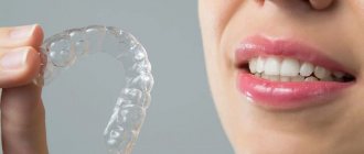
Aligners
In this case, the change in bite occurs due to the wearing of special aligners made of transparent biopolymer material.
The number of these devices often ranges from 10 to 40 pieces, which must be changed regularly during the treatment process.
Dentists note that this method is effective for correcting minor malocclusion pathologies, such as crowding and displacement of individual teeth, the need to equalize the length of some elements of a row, and eliminating teeth and diastemas. The duration of orthodontic treatment with aligners can be up to two years.
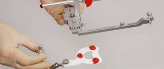
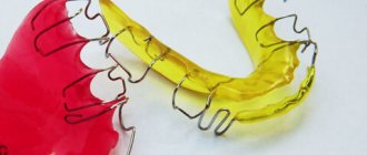
Plates
Orthodontic devices consisting of a palatal plane and a metal arch that exerts pressure on the protruding elements of the jaw row.
Dentists recommend this method of bite correction for a small number of crooked teeth and the absence of periodontal disease.
The plate must be worn at least 22 hours a day, removed when eating and performing hygiene procedures.
Orthodontic trainers
These devices, made of polymer material, are installed on two jaws and allow them to influence not only the teeth, but also the jaw muscles.
During the process of occlusion correction, the patient changes elastic trainers to more rigid ones as needed.
This allows you to get rid of such pathologies as shifting of individual teeth, increased tone of the chin muscles, and loose jaw contact. The duration of treatment with trainers is usually 1.5-2 years.
When it is not possible to correct a malocclusion using various devices, dentists resort to a more radical method - the method of surgical correction.
It is indicated when therapeutic and orthodontic treatment is ineffective. The essence of the method is to make an incision into soft and bone structures and move certain elements to the required position.
Surgical intervention can be used both for congenital anomalies in the structure of the jaw rows, and due to their deformation as a result of injuries.
Price
The cost of correcting a permanent dentition depends on the complexity of the malocclusion, the age of the patient, the number of teeth that need to be aligned, and the type of design used:
| Type of orthodontic device | Cost, rub. |
| Bracket system | From 5000 for metal to 90000-100000 for ceramic or sapphire |
| Aligners | 100000—180000 |
| Plate | From 4000 |
| Trainers | 6000—10000 |
It is worth understanding that prices may vary among different dental clinics. It depends on their location, prestige and fame.
Preventive actions
The formation of a physiologically correct permanent bite largely depends on the person himself.
Therefore, dentists recommend adhering to the following recommendations:
- to refuse from bad habits;
- adhere to proper nutrition to ensure that the body receives a sufficient amount of micro- and macroelements;
- promptly eliminate oral diseases to prevent tooth displacement;
- take measures to prevent the development of calcium deficiency, which negatively affects the condition of the teeth;
- carry out hygiene measures regularly and efficiently;
- Visit the dental office in a timely manner to identify developing diseases of the teeth and gums.
Additional information on the topic of the article is presented in the video.