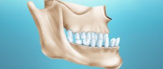
Human teeth are bony formations in the mouth that play a major role in the processes of processing food, the formation of speech skills and a smile.
Formula of adult teeth: decoding
Types of teeth and their number
Normally, an adult has 28-32 teeth, each of which has its own purpose. It is customary to divide all teeth into temporary (baby) and permanent (molar).
Each of the teeth belongs to one of four groups, depending on its functions.
Belonging to a particular group is determined based on the shape of the tooth, its internal structure and place in the dentition.
On a note! Teeth are the only organs in the human body that cannot be restored. Improper care and bad habits lead to tooth decay.
Types of teeth
According to the functions they perform, teeth are divided into four types:
- incisors - necessary for a person to easily separate pieces of food;
- fangs - designed to crush separated pieces;
- premolars - help to compress food as much as possible;
- molars - needed for grinding pieces of food.
On a note! The largest of all the teeth in the jaw are the molars.
The teeth form two arches above and below. Each of them contains 4 incisors, 2 canines, 4 small molars (premolars) and 6 large molars. Out of 6 molars, the norm can be only 4, since not all people have wisdom teeth.
Functions of teeth
The condition and health of the jaw elements has a direct impact on the digestive process. Thanks to the presence of healthy teeth, normal nutrition is provided, which affects the growth of the body and its normal development.
On a note! There are no medications in the world that can affect the shape of teeth, since it is genetically determined.
Names and formulas of teeth
When making a diagnosis, the dentist is guided by certain patterns and numbering of teeth. The numbers and letters reflect the order in which they are located, which makes it easier for dentists to make a diagnosis and determine the tooth that needs to be treated.
Number of tooth types
Special designations that are used to indicate the location of teeth are called a dental formula. Today, several recording systems for this scheme are actively used. The dental formula is the final result of jaw sanitation. Temporary teeth in the formula are represented by Roman numerals, and permanent teeth by Arabic numerals.
Teeth numbering
There is a universal dental chart, which is usually called the alphanumeric method. In the generally accepted formula, each type of tooth is designated in capital letters without indicating the quantity:
- I – incisors;
- C – fangs;
- P – premolars;
- M – molars.
When treating children, lowercase letters will be used instead of capital letters. Arabic numerals indicate the location of the tooth in the row.
Formulas of permanent and baby teeth
According to the schematic drawing, the formula looks like:
Teeth formula
If you decipher the record, it turns out that the patient has two pairs of incisors, one pair of canines on the upper jaw, two pairs of premolars and three pairs of molars. This formula works to maintain a permanent 32 tooth bite.
Reasons for creating a dental formula
Human teeth, despite their visual similarity, have certain differences. Each tooth is individual and designed to perform its function. Thanks to numbering in the dental field, examination of the patient's oral cavity is optimized to the maximum.
Dental formula
Clear and concise markings help to accurately record information in the patient's chart. In this case, the use of the same numbering system is observed.
This is necessary so that the new doctor can decipher the medical history and past treatments that were performed by another dentist.
Different dentists cannot use an individual calculation system, as this will change the patient's hospital history.
International two-digit Viola system
Numbering of teeth at dentists
In addition to the dental formula, dentists use special numbering. It starts from the middle of the row and moves in different directions to the end of the jaw. For example, doctors call molars “eights” or “sixes.”
The first number is assigned to the two front teeth, and the formations immediately behind them are given the second number. Both canines, which are located behind the incisors, are numbered “threes”. The teeth following them are written in the form of numbers 4 and 5. The last three molars in the row have further serial numbers: 6, 7 and 8.
Complete clinical formula
This numbering greatly simplifies the designation of teeth for dentists. But in addition to the number, the dentist needs to know in which segment of the jaw a certain tooth is located. To clarify records about the condition of the patient’s teeth, the entire jaw is virtually divided into four zones.
On a note! The order of counting teeth starts from the upper arch of the jaw, starting from the right edge, and is carried out clockwise.
Description of teeth
On the upper arc, tens are visualized from the beginning of the row, and the next zone at the top left is called twenties. The bottom of the jaw is also marked according to the same principle: the teeth on the left are called thirty, and those on the other side are called forties. When diagnosing the oral cavity, the number of the zone in which it is located is added to the number of the tooth being examined. So each tooth is assigned a number.
Complete formula of permanent teeth
The World Health Organization has developed its own form for recording dental formulas. In it, each tooth is designated by a number, as is each segment of the jaw. The entry looks like this:
Table 1. Dental formula according to the WHO method
Jaw Segment Number/Tooth NumberJaw Segment Number/Tooth Number
| 1 | 2 |
| 8 7 6 5 4 3 2 1 | 1 2 3 4 5 6 7 8 |
| 8 7 6 5 4 3 2 1 | 1 2 3 4 5 6 7 8 |
| 4 | 3 |
On a note! In the WHO formula, for convenience, the numbers increase clockwise.
Video - Anatomy of teeth
Numbering in pediatric dentistry
Due to the nuances of the anatomy of the children's jaw, numbering in dentistry for children is carried out differently.
A child’s milk teeth, which usually appear between 4 months and six months, erupt simultaneously with the formation of the roots of permanent teeth.
Because of this, both permanent and temporary teeth will be visible on the x-ray. The constants already have their own numbers, so children's teeth are counted from the next tens.
On a note! To count temporary teeth in children, there is a special formula in which the letter N indicates the number of teeth, and n indicates the child’s age. The formula is as follows: N = n – 4
X-ray of a child's teeth
Understanding the numbering of teeth and deciphering the formula of adult teeth will help the patient better understand their dentist. With the help of generally accepted values, the meaning of treatment becomes clear and the diagnosis recorded in the hospital record is clarified.
Source: https://glivec.su/2018/08/28/tablica-zubnaja-formula/
Why is tooth numbering needed in dentistry?
Dental units, regardless of their external similarity to each other, have certain differences and perform their own function. By optimizing the recording of the dental formula, specialists are able to clearly distinguish between dental disorders.
Unified tooth numbering allows you to enter each patient’s information into cards with the possibility of further study by another specialist.
A special case of this need is the transfer of a patient to a surgical office for the removal of a specific tooth. If individual recording options were allowed, individual dentists would not be able to transcribe the information in the medical record.
Morphological and functional characteristics of the bite
Human occlusion in dentistry is divided into two periods: temporary and permanent.
Temporary begins to form not yet at the stage of embryonic development. By the age of 6 months, a baby's teeth will usually begin to erupt. At the initial stages, they may have an incorrect location, but when the rest appear, the row aligns.
Due to acceleration in recent decades, early eruption has been observed. There are many cases where a baby was born with 1-2 teeth. Therefore, we can safely say that the timing of the eruption of baby teeth is individual for all children.
Important!
It is difficult to overestimate the functional significance of temporary occlusion, since only with the appearance of milk teeth can a child become familiar with solid food. Parents need to realize how important it is to introduce complementary foods in a timely manner. Solid foods promote the development of chewing muscles and the formation of correct jaw position.
The first permanent teeth appear between 5 and 7 years of age. At this age, rapid successive loss of milk begins. The formation of a permanent bite occurs gradually. Parents may notice that from the age of 3-4 years the child’s dental position changes - this begins a shift period. Gaps appear between the incisors - diastemas. Chewing teeth and fangs seem to be moving apart. This is due to jaw growth as the molars have a wider crown and need to fit in the place of the ones that fall out. The cutting surface begins to wear off, which becomes noticeable to the naked eye. This period in dentistry is called the mixed dentition in children, when the jaw prepares for the loss of temporary teeth and the appearance of permanent ones.
Complete clinical dental formula according to WHO
The WHO clinical dental formula implies the following use of defining values: the dentist divides the patient’s jaws along the axes of symmetry and numbers them from 1 to 8, depending on the number of units in the dentition. When the patient has no extreme molars, 7 values are sufficient.
Formula of permanent teeth
However, an expanded dentition requires full compliance. The order of establishing segments from the dentist looking at the patient is determined clockwise, starting from the left side of the upper jaw, and corresponds to the table below.
| Segment | Teeth positions | Segment | |
| 1 | 8, 7, 6 (molars), 5, 4 (premolars), 3 (canine), 2, 1 (incisors) | 1, 2 (incisors), 3 (canine), 4, 5 (premolars), 6, 7, 8 (molars) | 2 |
| 4 | 8, 7, 6 (molars), 5, 4 (premolars), 3 (canine), 2, 1 (incisors) | 1, 2 (incisors), 3 (canine), 4, 5 (premolars), 6, 7, 8 (molars) | 3 |
If we consider a position not distributed in the table, the record will be represented by a numerical value consisting of the segment number and the position of the tooth, for example - 33 (lower canine on the right, relative to the examining dentist).
For the milk series, segmentation by values is used - 5, 6, 7, 8. Teeth numbering is from 1 to 5. This is fully consistent with the Viola system.
Parents should not be surprised to find the 65th tooth next to the 24th in the child’s chart entry, since when replacing the deciduous series with a molar, the designations are replaced in accordance.
Names and arrangement of teeth in adults and children
All human teeth have their own names and are located according to a certain pattern. The formulas for primary (temporary) and permanent dental units are somewhat different, but have the same logic of construction.
A dental formula or diagram is a brief description of the dental structure of the human dentition using numbers and Latin letters. The letters are abbreviations from the Latin names of teeth accepted in medicine:
- I – these are dentes incisivi or incisors.
- C is for dentes canini or fangs.
- P stands for dentes premolars or premolars.
- M stands for dentes molars or molars.
The alphanumeric dental formula is common in many countries, including America, which is why it is sometimes called American. After the letters in the formula there is always a digital fraction, where the numerator indicates the number of teeth on the upper jaw, and the denominator on the lower jaw.
- The dental formula of a healthy adult with a normal bite and 32 teeth looks like this:
- This formula indicates that on each human jaw there is:
- 2 pairs of incisors;
- 1 pair of fangs;
- 2 pairs of premolars;
- 3 pairs of molars.
Dental formula in the absence of some teeth
It is not difficult to correctly number dental units with a healthy dentofacial apparatus; it is much more difficult to indicate the location of the teeth if a person does not have any molars, incisors, canines or premolars. In this case, the dentist indicates the number 0 opposite a certain group of teeth, rather than shifting the number series.
If the patient has any anomalies in the development of the dentofacial apparatus, for example, teething in the wrong place (hyperdontia, polydontia), the dentist prescribes an individual formula. In addition to numerical designations, the doctor generates a detailed report on the structure of a person’s dentition.
Description, structure, functions, name and location of human teeth
It is not without reason that human teeth have different shapes, structures and sizes. Each tooth performs certain functions, and its purpose is, one way or another, reflected in the name:
- The incisors are the front teeth that fall into the smile zone. They are quite sharp as their function is to tear or cut food, hence the name. Normally, every person has lateral and central (larger) incisors.
- Canines are cone-shaped teeth that can be used to tear and hold food. They are located immediately behind the incisors.
- Premolars are the small back molars located behind the canine teeth.
- Molars are the back teeth necessary for mechanical processing of food. Wisdom teeth refer specifically to molars.
The listed types of teeth are also found in animals. But in the process of their evolution, some dental units were slightly modified:
- elephants' upper incisors became tusks;
- Poisonous snakes have poison in their fangs.
Many animals have front teeth similar to human ones, but since they are called differently, they cannot always be identified with the usual incisors and canines.
The structure of human teeth
Human teeth consist of a crown and a root part. The first is located above the level of the gum, the second is inside it. The top of the crown is covered with enamel, the strongest tissue in the body. It protects the inner layer of the tooth – dentin – from external influences. Under the dentin there is a hollow chamber with nerves and blood vessels - the pulp.
Despite its strength, enamel is vulnerable to bacteria. If left untreated, caries can affect the dentin and pulp. When the pulp chamber, which contains nerve fibers, is damaged, pulpitis develops, accompanied by acute throbbing pain. If the infection spreads to the root of the tooth, it is highly likely that it will have to be removed.
The basis of the lower part of the tooth is the root canals, which also contain arteries, veins and nerve fibers. Through the apical foramen, all of these structures are connected to the main neurovascular bundle.
The lower part of the tooth is covered with dentin and cement, which is attached to the periodontium using collagen fibers. The roots of teeth are hidden in the alveoli - depressions in the jaw bone.
On what basis are teeth numbered according to different classification systems?
In addition to the Latin names of teeth, digital designations are used in dentistry. The numbering of teeth is based on the order in which they erupt. It starts from the front incisors (from the middle of the jaw) and runs to the left and right of them.
There are several generally accepted systems for numbering human teeth.
Universal system
Most often, dentists name teeth not by the Latin alphabet, but according to their location in the oral cavity (ordinal number). Moreover, using not Roman, but familiar Arabic numerals.
Names of teeth according to the universal classification system:
- two central incisors are located at number 1 and are called ones;
- the second incisors are numbered 2;
- the fangs are called triplets;
- chewing teeth or premolars are called fours and fives;
- molars are called sixes, sevens and eights.
According to the universal system of classification of dental units, the jaw is divided into 4 segments:
- top left;
- top right;
- bottom right:
- bottom left.
The further name indicates not only the serial number, but also the location of the dental unit in the human oral cavity.
The universal dental numbering system is the most popular and frequently used. It is used by dental therapists and surgeons in various countries.
The picture shows a diagram of the designation of teeth in the oral cavity of an adult according to the universal numbering system:
European system
The European Viola system is one of the newest and most advanced methods for naming human teeth. It is characterized by the division of the jaw into segments (two at the bottom and two at the top). Each segment is numbered (from 1 to 4).
Based on these numbers, each tooth receives a two-digit number. The first digit indicates the segment, and the second is the actual sequence number.
The Viola system is recognized internationally and is therefore popular all over the world. It is used in radiography, making panoramic images and allows dentists from different countries to exchange information about patients, overcoming the language barrier.
Haderup system
To designate dental units according to the Haderup system, Arabic numerals are used and segmentation into the lower and upper jaws is used:
- the “+” sign indicates that it belongs to the upper jaw;
- the “–” sign indicates the lower jaw.
The only disadvantage of such numbering is that it is necessary to additionally indicate whether the dental unit belongs to the left or right side of the jaw.
Zsigmond-Palmer system
The Zsigmond-Palmer system is considered the most imperfect, since it only indicates the numbers of teeth without their location. Standard Arabic numerals are used for numbering.
Anatomical (group) formulas
The anatomical norm of the dentition for an adult is 28-32, and for a child 20. At the age of 4 years, complementary molars erupt for the first time, and the entire set should be replaced with primary ones.
For an adult, the anatomical formula of teeth (full set) can be presented: I4/4C2/2P4/4M6/6=32
If we consider the milk series, it looks like this: i4/4c2/2m4/4=20 A child’s premolars are formed only at the age of 9-12 years, and wisdom teeth may not erupt at all.
The development of teeth and their formula are important for parents, who can promptly identify potential problems. If deviations from the described norm are detected, it is recommended to immediately contact a pediatric specialist for diagnosis.
Dental universal formula
The dental formula of a universal type is distinguished by the fact that in it the canines, as well as incisors and molars, are displayed in capital letters, and next to them the number of bone formations on both jaws.
Designations:
- I – incisors;
- C – fangs;
- P – premolars;
- M – molars.
Accordingly, their quantity is the same as indicated above and the same applies to the children's formula.
Adult formula:
Baby formula:
It is quite easy to decipher that an adult has 2 pairs of incisors and premolars, as well as 1 pair of canines and 3 molars, which in total gives 16 teeth on one jaw.
As for the child, he does not have 3 pairs of molars and therefore the total is 10.
How to find a tooth module?
To determine the specific module of a tooth in the jaw, it is enough to consider the proposed recording scheme using a standard formula, depending on the calculation method used.
In practice, recalculation of the jaw segment is performed starting from the front incisors, which are joined to each other.
In this order, you can determine a specific unit in the jaw, for example, the 63rd tooth of a child (according to WHO) for a person looking in profile will be located in 3 positions on the right. This principle is suitable for all types of dental formula and can be applied in practice.
Kuznetsov B.A. Key to vertebrate animals of the fauna of the USSR Mammals M.: Education. 1975
CLASS MAMMALS SUBCLASS PLACENTAL MAMMALS ORDER Carnivores
FAMILY FALID
A well-differentiated group of predatory mammals, the organization of which is adapted to obtaining animals that serve them as food by hiding them and lying in wait. Sizes range from the size of a domestic cat to the size of a tiger, whose weight is sometimes more than 300 kg. The body is elongated, slender, but strong. The head is rounded, with a short muzzle. The legs are rather short, digitigrade, with a rounded paw. There are 5 fingers on the forelimbs and 4 fingers on the hind limbs. All species, except the cheetah, have curved, sharp, retractable claws. The tail is of different lengths, it is evenly covered with hair. The skull is short and wide. The molars are laterally compressed, with sharp cutting crowns. The carnassial teeth are highly developed.
Dental formula:
I 3/3 C 1/1 RM 2-3/2-3 M 1/1 = 16(14) x 2 = 32(28)
TABLE FOR DETERMINING THE GENERUS OF THE CAT FAMILY
1(2) Body length with tail less than 175 cm. Tail length approximately equal to or less than 1/2 of body length. The condylobasal length of the skull is no more than 16 cm.
Cats
2(1) Body length with tail is more than 200 cm. Tail length is more than 1/2 of body length. The condylobasal length of the skull is over 16 cm. 3(4) The limbs are short, powerful, with retractable claws. There is no mane of coarse hair on the upper side of the neck. The diameter of the infraorbital foramina is greater than or equal to the distance from these holes to the edge of the orbits.
Panthers
4(3) Limbs long, thin. The claws are not retractable, rather blunt. A mane of coarse, standing hair stretches along the top of the neck. The diameter of the suborbital foramina is half the distance from these holes to the edge of the orbital sockets.
Cheetahs
GENUS OF CAT
There are 8 species in the fauna of the USSR.
TABLE FOR IDENTIFYING CAT GENUS SPECIES
1(2) The tail is short: its length is less than the length of the head. The final third of the tail is black. The body length of adults is usually more than 90 cm. The condylobasal length of the skull is more than 120 mm. The infraorbital openings are elongated vertically (subgenus Lнх).
Lynx
(Forest zone of the country from Belarus to the Far East, the Caucasus, mountains of Central Asia. Inhabitant of large forests. In the spring, after 67-74 days of pregnancy, females bring 2-5 kittens in the den. Hunts mainly at night. Eats various animals up to deer, and birds. Produces fur skin.) 2(1) The tail is longer than the head. The end of the tail is light or black only the very tip of the tail. Body length is not more than 90 cm, Condylobasal length of the skull is less than 120 mm. The infraorbital foramina are elongated in an oblique direction. 3(4) The upperparts are sandy yellow, the belly is white. The back of the ears is black. At the ends of the ears there are long tassels of elastic hair more than 3 cm long. There are black spots on the sides of the head. The postorbital processes of the skull are short: their length is less than the distance from their ends to the apices of the processes of the zygomatic arches (subgenus Caracal),
Caracal
(Deserts of Central Asia. A rare animal of our fauna. Lives in desert foothills and sandy deserts. During the day it hides in holes or dens under bushes. Gives birth to 2-4 cubs. Feeds on rodents and birds. Fur is of little value.) 4(3) The coloring is different. There are no hair tassels at the ends of the ears or they are less than 3 cm long. There are no black spots on the sides of the head. The postorbital processes of the skull are long: their length is greater than the distance from their ends to the apices of the processes of the zygomatic arches. 5(6) At the ends of the ears there are small tassels of elongated hair. The length of the tail with hair is approximately 1/3 of the length of the body. The color is one-color, without a pattern of dark spots (sometimes a dark stripe stretches along the ridge). The fur is rough. The condylobasal length of the skull is more than 104 mm (subgenus Chaus).
Jungle cat
(Eastern Transcaucasia, Eastern Ciscaucasia, lower reaches of the Volga, Southern Kazakhstan, plains of Central Asia. It adheres to tugai forests and reed thickets along the banks of rivers and lakes and at the seaside. It makes a den in these thickets. Kittens - 3-10 pieces - will be born in the spring. Main food - different birds and small animals. Fur is of little value.) 6 (5) There are no elongated hair tassels at the ends of the ears. The length of the tail with hair is approximately 1/2 the length of the body. The coloring is usually with a pattern of dark spots and stripes. The fur is soft. Condylobasal length of the skull is less than 104 mm. 7(8) There are 4-5 rusty transverse stripes on the chest. Frequent rust spots are scattered throughout the body. Two white stripes stretch from the eyes along the forehead and crown of the head. The bony palate continues beyond the line connecting the posterior edges of the last teeth by at least 1/2 the width of the palatal notch (Fig. 119, a) (subgenus Prionailurus).
Far Eastern cat
(Primorye and Amur region. Lives in mountain forests, hiding during the day in hollows and between stones. Estrus is in March, and childbearing is in May. Feeds on animals and birds. A rare animal of our fauna.)
Rice. 119. Skulls of Far Eastern (a) and forest (b) cats (top and bottom)
8(7) There are no transverse stripes on the chest. There are no rust spots on the body. There are no white stripes on the forehead and crown. The bony palate extends backward beyond the line connecting the posterior edges of the last teeth, less than 1/2 the width of the palatal notch (Fig. 119, b). 9(12) The color is almost uniform, with a small number of faint darker stripes on the back of the back; sometimes they are barely noticeable. Back with slight ripples. The anterior ends of the tympanic chambers extend beyond the posterior edges of the articular fossae. 10(11) Tail length less than 25 cm. The tail has wide blackish rings. The soles of the paws are bare. The ears are short, almost not protruding from the fur. The smallest distance between the tympanic chambers is approximately equal to the width of the palatine notch (subgenus Otocolobus).
Manul
(Central Asia, Kazakhstan, Tuva Autonomous Soviet Socialist Republic, Transbaikalia. Inhabits steppes and deserts. Lives in burrows and crevices of rocks. Nocturnal animal. Hunts mainly for various rodents. Reproduction has not been studied.) 11(10) Tail length more than 25 cm. Black rings on no tail. The soles of the paws are covered with hair. The ears protrude strongly from the fur. The smallest distance between the tympanic chambers is significantly less than the width of the palatine notch (subgenus Eremaelurus).
Dune cat
(Eastern Transcaucasia, lower reaches of the Volga River, plains of Central Asia, Kazakhstan. Inhabitant of semi-deserts and deserts. Lives in thickets of desert bushes, tugai and reeds, in the foothills, in oases. Lives in burrows. In the spring, females give birth to 3-7 cubs. Feeds animals, birds, lizards, insects. Hunted for their skins.) 12(9) Coloring with a clearly defined pattern of dark spots and stripes. The anterior ends of the tympanic chambers barely reach the posterior edge of the articular fossae (subgenus Felis). 13(14) The back and sides of the body are pale gray with a clear pattern of numerous dark specks and stripes. The end of the tail is pointed. The ends of the nasal bones do not extend beyond the ends of the premaxillary bones.
Steppe cat
(Eastern Transcaucasia, lower reaches of the Volga River, plains of Central Asia, Kazakhstan, Northern Kyrgyzstan. Inhabitant of deserts and semi-deserts. Lives in thickets of desert bushes, tugai and reeds, in the foothills and oases. Lives in burrows. In the spring, females give birth to 3-7 cubs .Feeds on animals, birds, lizards. It is hunted for its skin.) 14(13) The back and sides are brownish-gray; two dark stripes stretch along the back, and on the sides there are several blackish transverse stripes (sometimes they are faintly pronounced). The end of the tail is blunt. The ends of the nasal bones extend beyond the ends of the premaxillary bones.
Forest cat
(Ukraine, Moldova, Transcaucasia. Lives in forests, along floodplains. During the day, hides in hollows, under windbreaks, in rocks. There, females give birth to 3-6 cubs in the spring. Hunts at night for various animals and birds.)
KIND OF PANTHER
There are 3 species in the fauna of the USSR.
TABLE FOR IDENTIFYING SPECIES OF THE GENUS PANTHER
1(2) Body color is red with a clear pattern of wide black stripes. The body length of adult animals is more than 160 cm. The condylobasal length of the skull is more than 200 mm. The posterior ends of the nasal bones extend far beyond the line of the posterior ends of the maxillary bones (subgenus Tigris).
Tiger
(Preserved only in Primorye, Amur region, along the Amu Darya River in Central Asia; sometimes it comes from Iran to Turkmenistan and Azerbaijan. In Central Asia it lives in tugai forests along rivers, and in the Far East - in mountain forests. Reproduces in 2-3 years; there are 2-6 cubs in a litter. It feeds mainly on ungulates.) 2(1) Body coloring is red or grayish, with a clear or vague pattern of ring-shaped dark spots. Body length less than 150 cm. Condylobasal length of the skull up to 200 mm. The posterior ends of the nasal bones do not reach the line connecting the posterior ends of the maxillary bones, or only slightly extend beyond it. 3(4) Body color is red or yellow with a clear pattern of small (up to 5 cm in diameter) sharply defined black spots. The tail is thin and covered with low, lying hair. The fur is low and hard. Condylobasal length of the skull is over 180 mm. The length of the suture between the nasal bones is approximately 1.5 times the greatest overall width of these bones (subgenus Pardus).
Leopard
(Caucasus, Turkmenistan, Amur region, Primorye. Preserved mainly in mountain forests. The lair is made in rock clefts, caves, thickets of bushes. In April - May, after a 3-month pregnancy, females give birth to 2-5 cubs. Feeds on various animals and birds .) 4(3) The body color is smoky gray with a vague pattern of large, unclearly defined dark spots. The tail is luxuriantly pubescent. The fur is soft. Condylobasal length of the skull is less than 180 mm. The length of the suture between the nasal bones is only slightly greater than the greatest width of these bones (subgenus Uncia).
Leopard, or snow leopard
(Pamir, Tien Shan, Altai, mountains of the Tuva Autonomous Soviet Socialist Republic. Inhabitant of high mountain regions. Makes a lair in rocks, between scree stones, in caves. There are 2-4 cubs in a litter. Hunts for various animals and birds.)
CHEETAH GENUS
The only representative in our fauna.
Cheetah
(Turkmenistan. Desert inhabitant. In the spring, females give birth to 2-4 cubs. Feeds on various animals. A rare animal of our fauna.)
BackContentsNext
Why you can’t remove baby teeth prematurely
Many parents are calm about the removal of baby teeth because they expect permanent teeth to appear. However, dentists strongly do not recommend carrying out such a procedure ahead of schedule. If there is more than a year left before the eruption of permanent units, then removal is early. If possible, doctors try to carry out organ-preserving treatment, and if this is impossible, they recommend a temporary prosthesis for the child.
The functionality of the mammary units at the replacement stage includes preparing the jaw for the appearance of permanent ones. In the absence of these, the bone tissue of the jaw turns out to be underdeveloped. This leads to the formation of an incorrect bite after a replacement bite.
On a note!
Children who have had their primary incisors and canines removed prematurely require orthodontic treatment in 90% of cases. Therefore, they try to preserve even a filled tooth for as long as possible until its roots resolve naturally.
Timing of permanent teeth eruption
The time for the appearance of permanent dental units is individual for each child and depends on:
- heredity;
- quality of life and nutrition;
- health conditions;
- region of residence - in regions with a high fluoride content in water, eruption begins later.
The standard pattern for the appearance of molars involves the alternate appearance of incisors, premolars, canines and molars. The process is similar to the appearance of dairy products.
On a note!
However, each child may have characteristics that do not become a deviation from the norm. Typically, the formation of a permanent dentition begins at 6 years of age and is completed by 13 years of age.
Prevention of pathologies of mixed dentition
Changeable bite is a borderline condition between temporary and permanent. Since the structure of the jaw changes during this period, the child requires special attention. To prevent pathologies of mixed dentition, it is recommended:
- Organize a balanced diet. Due to the increased abrasion of the cutting areas, it becomes uncomfortable for the child to chew solid food. It is necessary to facilitate this process and change the menu for the baby to the appropriate one.
- Don't rush while eating. Due to the difficulty of chopping food, meal times may increase. We must be understanding of this and realize that it is more difficult for a child with a mixed bite than for an adult with a permanent one.
- Maintain oral hygiene. If damaged, the likelihood of caries formation increases, which in the future can lead to premature tooth loss and underdevelopment of bone tissue.
- Refuse forced tooth extraction if there is no indication for this.
- Regularly visit a dentist to assess the stages of formation of a mixed dentition. It should be remembered that in the initial stages it is easier to correct anatomical defects.
Possible deviations
When a child sees a dentist, the doctor draws up a dental formula. Each section of the same name has its own designation in Latin letters. The formula is entered into the personal card and allows you to calculate the total number of units.
At the first visit, complications may be detected in the formation of a mixed dentition:
- early mixed dentition can cause the formation of a distal one;
- since the canines are the last to complete eruption, they may take an incorrect position due to lack of space;
- with anatomical features of the jaw, a diastema appears between the front incisors;
- tooth retention, in which the whole tooth cannot be seen.
Important!
All deviations at the stage of mixed dentition lead to anatomical defects in the future. They, in turn, cause aesthetic and physiological suffering, become the cause of impaired diction and the appearance of complexes. Therefore, it is important to pay attention to the mixed bite and, if deviations are detected, promptly seek medical help.
