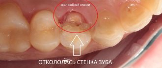
Tooth extrusion – what is this procedure? The essence of the method and indications for its use
Tooth extrusion is a separate dental procedure. Nowadays it is performed quite rarely, but in some clinical cases it is precisely this that makes it possible to preserve the root in case of severe destruction of the visible part of the tooth and subsequently, based on it, restore the crown using modern methods of restoration and prosthetics. Extrusion was first introduced to the world in 1973. The author of the technique was Dr. GS Heithersay, who proposed this procedure as an alternative to removing roots with fractures1. Read more about the essence of this method and how such treatment is carried out further in this article.
Extrusion Concept by Dr. Stefan Neumeier
After extraction of a molar, its resection, replantation and extrusion, a minimally invasive operation is allowed to install an implant in bone tissue, the volume of which is sufficient for this type of procedure. Further procedures with the implant are carried out after complete restoration of the alveoli and gums and have an aesthetically and functionally important clinical purpose. Two years after the installation of the implants, X-rays can be used to detect complete restoration of the bone tissue around them and the absence of signs of rejection.
INTRODUCTION
When a tooth falls out, the physiological load on the periodontium is no longer exerted, which leads to resorption of the alveoli, in most cases causing destruction of the vestibular wall of the tooth. If periodontal or osteolytic processes reach the tooth root, the probability of destruction of the vestibular bone lamella reaches more than 50%. The successful implementation of prosthetics on implants to replace a lost tooth is determined by a number of parameters. An important role here is played not only by complete osseointegration of the implant, but also, above all, by the presence of at least 1 mm of bone wall around the implant. Thanks to the latter, the likelihood of longer-term stability of soft tissues increases, thereby significantly reducing the risk of peri-implantitis [5, 8].
Many authors have proposed their own options for eliminating or minimizing the possibility of resorption after tooth extraction. The Socket Preservation method is a comprehensively studied and widely used method for preventing resorption processes. However, the safety of the bone is not guaranteed at all [1–4]. Thanks to augmentation methods, predictable and durable clinical results are achieved [7].
And yet, these methods often require serious surgical intervention and, accordingly, a high level of professionalism among specialists performing such operations [11, 12, 14]. The use of autologous material as a bone replacement is impossible in most cases, and the larger the defect, the lower the probability of predicting the outcome [11, 13, 14].
The concept of extrusion by Dr. Stefan Neumeier is a fairly gentle and minimally invasive method of surgical intervention, which allows not only to preserve the bone structure of the alveoli, but also to additionally build up hard and soft tissues. This concept represents the optimal option for long-lasting implant prosthetics. The concept involves first carefully removing the unrecoverable tooth, then resecting it, replanting it, and then extruding it [34]. The autologous bone obtained in this way ensures the preservation of the original volume of the alveoli and is durable. This creates ideal conditions for successful implant prosthetics, as well as for ensuring stable bone walls and soft tissue around the implant.
CLINICAL CASE DESCRIPTION
In November 2009, the patient was determined that tooth 36 could not be restored. The clinical picture shows a tooth with a crown without damage, the edges of the crown open to view and the periodontal complex without visible changes (Fig. 1).
Rice. 1
In this case, probing to determine the pathological gum pocket is not required; a wide layer of densely grown gum is observed in the cervical area. An X-ray from 1995 demonstrates incomplete filling of the root canals, resulting in periapical inflammation in the area of the mesial roots (Fig. 2).
Rice. 2
TREATMENT PROCESS
The tooth was carefully removed. In this case, special attention is paid to preserving the bone structure of the alveoli and marginal gums. When removed, the apical granuloma of the mesial root comes out along with the tooth (Fig. 3-4). The periodontium on the root surface remained undamaged. The tooth is placed in a physiological saline solution. Next, the apical side of the alveolus is scraped out with a sharp spoon and a surgical round bur.
Rice. 3
Rice. 4
In this case, the preservation of the integrity of the periodontal alveoli on the cervical girdle is carefully monitored.
At this stage, the alveolus is filled with blood. The extracted tooth is then resected so that the circumferential ligament is preserved around the root surface of the tooth by at least 2 mm. In multi-rooted teeth, the resection zone is located 2 mm below the root furcation. After complete removal of decay from the tooth cavity and root canals, the voids are filled both from the occlusal and apical sides with self-adhesive RelyX cement from 3M ESPE (Fig. 5). Then the tooth divides in the furcation zone, 2 segments arise, consisting of a clinical crown and root with 2 mm of the circular ligament apparatus. During extraoral treatment, the tooth is placed in a saline solution.
Rice. 5
Then, precise replantation of these treated root fragments into their alveoli is carried out.
In this case, due to the formation of a blood clot in the alveoli, the fragments arranged themselves spontaneously. With the help of composite and adhesive technology, these fragments are immovably fixed on adjacent teeth (Fig. 6-7). Re-suturing of periodontal fibers occurs after a few days, when complete immobility of the fragments has become apparent. Tires can also be used as an additional fixation.
Rice. 6
Rice. 7
10 days after replantation, extrusion of completely engrafted fragments can be carried out. At the preparatory stage, fiber pins are attached across the fragments using a fluid composite. A small amount of composite is applied to the ends of the posts to prevent the orthodontic bands from slipping (Fig. 8).
Rice. 8
After this, a small splint, previously prepared in the laboratory, is fixed on the occlusal surfaces of adjacent teeth using a bridge. This bridge is used as a support for orthodontic elastic bands, which are then attached to pins from the vestibular side of the tooth to the lingual side. Due to the accelerated extrusion process (several days), it is necessary to set the maximum tension of the rubber bands. In this case, orthodontic elastic bands with a diameter of 4.2 mm are used, since the patient should replace them twice a day. After 14 days, both fragments are firmly attached to the pins and extruded by 3-4 mm (Fig. 9-10).
Rice. 9
Rice. 10
During the process of accelerated extrusion, periodontal fibers are maximally stressed, and the stability of the fragments is significantly weakened. In the ninth week of the healing period, the fragments are re-fixed on adjacent teeth using a composite (Fig. 11).
Rice. eleven
On the control x-ray, complete regeneration of the bone structure of the alveoli is clearly observed with Lamina Dura still noticeable. The level of the intradental septum and bone in the furcation area can be completely preserved or even improved (Fig. 12). Probing of the gingival pocket showed the absence of any inflammatory processes.
Rice. 12
Before installing the implants, the newly adhered fragments are removed. Beneath them, the clinically restored bone structure of the alveolus and the completely preserved septum are noticeable (Fig. 13).
Rice. 13
The subsequent minimally invasive operation of implant installation is carried out without the use of additional augmentation methods and does not involve further surgical intervention (Fig. 14).
Rice. 14
A two-piece Aesthura classic implant from Nemris with a length of 11.5 mm and a diameter of 4.75 mm is installed. Primary stabilization of the implant in the bone is 35 Newton per centimeter. A Nemris prefabricated abutment with a high-load retention element is then screwed in as a basis for attaching the prosthesis (Fig. 14-15).
Rice. 15
After all procedures, a temporary screw-on crown, individually prepared in the laboratory, is installed in the patient’s oral cavity. Using a flowable composite, the protrusions of the crown, as well as its position, are corrected (Fig. 16-17).
Rice. 16
Rice. 17
When making a temporary crown, the areas of contact with adjacent teeth on the approximal and occlusal sides are noticeably reduced for sufficient primary stabilization of the implant, but this does not imply immediate restoration of the functional load of the crown (Fig. 18).
Rice. 18
The temporary crown was screwed in immediately after installation of the implant without the use of cement, which has a positive effect on the healing of the implant, since cement residues are difficult to remove from the just operated soft tissue. After just a few days, it is noticeable that there are no signs of rejection of the temporary crown (Fig. 19).
Rice. 19
After 3 months, the transfer is screwed in and hydrocolloid impression material is applied (Fig. 20).
Rice. 20
In the laboratory, the anatomical shape of the temporary crown can be repeated when making a permanent crown. After installation of a zirconium oxide crown, there are no signs of rejection; a well-formed alveolar process and a wide zone of keratinized gum are observed (Fig. 21-22).
Rice. 21
Rice. 22
The control x-ray after crown installation shows complete ossification of the bone around the implant (Fig. 23).
Rice. 23
The presence of attached vestibular gum and a wide zone of keratinized gum indicate that there is a sufficient amount of vestibular bone tissue on the implant. Thus, the durability of prostheses installed using implants is guaranteed, and the risk of peri-implantitis is significantly reduced. A control X-ray taken two years later, which shows no differences from the original image, confirms these predictions (Fig. 24).
Rice. 24
CONCLUSION
The clinical picture demonstrates the procedures associated with the installation of an implant to replace tooth 36, which has no visible differences from the neighboring teeth. A harmonious process of gum attachment and a pronounced bone structure are the result of installing an implant in bone tissue, the volume of which is sufficient for this type of procedure. Using extrusion methods, both bone and soft tissues are completely preserved, or more precisely, restored. Consequently, optimal conditions are created for the physiological integrity of the bone corresponding to the tooth being replaced. In order to preserve the structures of hard and soft tissues, it is necessary, first of all, to take into account the durability of prostheses installed using implants.
DISCUSSION
Socket- and Ridge Preservation methods are used to prevent resorption processes after tooth extraction. Numerous studies have shown that when using these methods, the set goal is not achieved, but only partial preservation of the structure of the alveoli is ensured [6]. Various augmentation methods have been proposed to correct alveolar tissue defects. Using these methods, predictable and long-lasting clinical results are achieved. At the same time, the use of augmentation methods often requires serious surgical intervention and, accordingly, a high level of professionalism and extensive experience among specialists performing such operations [11, 12, 14]. In addition, the larger the defect, the lower the probability of predicting the result [11, 13, 14].
The extrusion concept of Dr. Stefan Neumeier completely eliminates the consequences of tooth extraction for tissue, and also creates optimal conditions for further implantation. The concept is based solely on the biological patterns of tissue behavior in the oral cavity. The periodontium is of paramount importance here. Successful growth of the root segment is possible only if the connective tissues on the root segment and alveoli remain intact. When replanting a dental segment, one should not only avoid resorption of the vestibular bone lamella, but also additionally build up hard and soft tissues through extrusion of the segment [10].
The method is minimally invasive, absolutely painless for the patient and is used in practice by the dentist with the least amount of labor. The concept of extrusion, taking into account the results achieved through its use, is the optimal method for minimally invasive prosthetics on implants, guaranteeing high quality aesthetic and functional characteristics of structures and their durability. In practice, the scope of application of the extrusion concept is not limited to the preparation of the alveolar ridge for implantation. By minimizing or even eliminating the risk of resorption in tissues where teeth are missing, a higher level of aesthetic and functional characteristics of bridges is achieved. By extruding each root, deep subgingival defects can be moved into the cervical area to create a more favorable environment for crown placement or therapeutic procedures. In addition, the extrusion technique can be used to treat vertical periodontal pockets and marginal gingival recession for aesthetic reasons, thereby providing patients with treatment with the least amount of surgical intervention. In the future, they will definitely come up with other ways to put this method into practice. The extrusion concept of Dr. Stefan Neumeier is a painless and minimally invasive surgical method that involves gentle procedures for implant prosthetics in the event of tooth loss. The clinical results of the extrusion method prove that they are more than equivalent to the results of the widespread use of augmentation methods.
Source: dentalmagazine.ru
What is the essence of the procedure
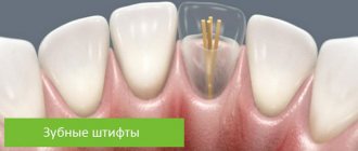
Tooth extrusion is the procedure of pulling a tooth or remaining root out of the gum in a vertical direction. According to this definition, extrusion allows a slight elevation of the root and creates more suitable conditions for a reliable and durable crown restoration. As part of this procedure, the tooth is usually pulled out by 3-4 mm. But to carry it out, it is important that at least the dentinal rim is preserved. Its thickness must be at least 1 mm, and its height must be at least 2 mm. If there is no such rim, the specialist may try to pull it out from under the gum.
Tooth extrusion is a procedure for pulling out a tooth or remaining root from the gum.
This procedure in some cases allows you to avoid root removal and subsequently restore the crown on it. However, if the root is damaged, its complete extraction followed by replacement with an implant will be the best solution. By the way, extrusion promotes the formation of bone tissue, so the procedure can also be performed as an alternative to artificial expansion of the jawbone for subsequent installation of implants.
Every year, the problem of prevention and treatment of hypersensitivity of hard dental tissues becomes more and more urgent due to the increasing influence of local and general factors. Dental hypersensitivity is a clinical condition manifested by a short-term painful reaction of exposed dentin in response to thermal, tactile, osmotic or chemical stimuli, which cannot be explained by any other known pathology [15]. As follows from the literature, the prevalence of dentinal hypersensitivity in the adult population varies from 4 to 74% [11, 25]. According to the latest data, in our country, 40-70% of the population experiences various forms of dental hyperesthesia [10]. According to some researchers, the incidence of dental hypersensitivity during periodontitis (tooth root sensitivity) is much higher - 85-95%. According to various authors, dental hyperesthesia occurs after whitening in 14-78% of cases [8, 35]. It is more common in patients aged 20-55 years. Dental hypersensitivity usually occurs more often and at a younger age in women than in men. Some authors believe that these differences are due to better oral hygiene and the consumption of more acidic foods by women. Most often, canines and premolars are affected, both on the upper and lower dentition. The cervical region of the vestibular surface of the teeth is most susceptible to hypersensitivity [11, 33]. A study by M. Addy (1990) showed that those teeth that are better cleaned with a toothbrush (less soft plaque, lower CP index) are more likely to have a predisposition to gum recession and the appearance of dentin hypersensitivity [11].
Etiology of dental hyperesthesia
Tooth hypersensitivity is caused by tooth exposure in the mouth due to loss of enamel and/or cementum. This pathology causes physical and psychological discomfort in the patient and is manifested by an acute short-term pain reaction [33].
The pain reaction can be caused by various stimuli of thermal, chemical or mechanical nature. The most common reaction is to cold, as well as sour and sweet foods.
J. Wagner et al. (2002) believe that sensitivity of the tooth root to cold liquid is the most common complaint of patients. Patients often report pain when brushing their teeth. Cold atmospheric air and air from the puster can also cause a pain reaction [13, 14, 16].
Pain can range from a certain level of discomfort to a very severe pain reaction. It is also related to individual tolerance, physical and emotional factors. Pain may be localized (one or two teeth) or generalized (multiple teeth), and in some cases it may involve all four quadrants [18].
The pain reaction may be accompanied by a pronounced emotional component. Therefore, in some cases, symptoms may disappear without treatment. In some cases, spontaneous disappearance of pain symptoms may occur due to natural remineralization, which ensures the closure of the dentinal tubules. However, pain may return over time as the smear layer is washed away by acidic foods and drinks, explaining the cyclical course of the disease [18, 25].
Various theories of the occurrence of dentin hypersensitivity have been proposed: odontoblastic, receptor, neuro-reflex, threshold and hydrodynamic. One of the earliest hypotheses was the dentinal receptor theory, according to which hypersensitivity was caused by direct stimulation of sensory nerve endings located in the dentine. However, microscopic studies have shown that there are no nerve cells in the sensitive part of the superficial dentin [22], so this theory is not widely accepted.
According to the odontoblast receptor theory proposed by R. Rapp et al. (1968), odontoblasts act as receptor cells, transmitting changes in membrane potential along synaptic connections [32]. This leads to pain in the nerve endings located in the area of the pulpodentinal border. However, this theory does not have clear evidence [36].
Pain caused by the movement of fluid in the dentinal tubules can be explained by the generally accepted "hydrodynamic theory" proposed by M. Brannstrom and A. Astron in 1964. According to this theory, the presence of damage to the enamel and/or cement in the cervical region with subsequent exposure of the dentinal tubules in the response to certain stimuli can cause movement of dentinal fluid within the tubules, which indirectly stimulates the pulpal nerve endings, causing pain. There is also evidence that if the pressure changes large enough, the resulting fluid flow can also initiate an electrical nerve impulse. The result is a sensation of sharp sharp pain, typical of sensitive dentin, which, as a rule, is experienced only during the action of the irritant or some time after its end. Histologically, sensitive dentin has dilated dentinal tubules that can be twice as wide as normal dentin. In addition, there is an increased number of dentinal tubules per unit area compared to normal dentin. As a result of doubling the diameter of the dentinal tubules, the fluid flow increases 16 times [7].
At the macroscopic level, sensitive dentin is no different from healthy dentin. The condition of the pulp in sensitive dentin is unknown. However, in a study by N. West (2000), it was suggested that there is minimal inflammation, since hypersensitivity persists for a very long time without developing into pulpitis [36].
Causes
Research results [18] have shown that dentin exposure may be a consequence of anatomical characteristics at the enamel-cementum junction and/or loss of cementum due to one or more of the following factors:
- insufficient or excessive brushing of teeth;
- poor oral hygiene;
— periodontal treatment (when removing above and/or subgingival dental plaque);
- the effect of acids of non-bacterial origin (present in food, chemical products, drugs) or endogenous acids during esophageal reflux or regurgitation of gastric contents;
— occlusal precontacts and excessive occlusal load [21, 23, 26, 27];
— physiological factors (extrusion of teeth, loss of antagonists) [24].
When teeth whitening, in particular professional, there is a release of macro- and microelements from the enamel, which leads to an increase in the permeability of the enamel and, as a result, sensitivity, and the less stable the enamel (i.e. in people with a high level of caries intensity), the higher the risk of this complication [7].
Dental hypersensitivity can often appear as a complication after large restorations on vital teeth. It manifests itself in the form of short-term pain that occurs when exposed to thermal irritants, and in more severe cases - the development of various forms of acute or chronic pulpitis.
Treatment of dental hypersensitivity
In 1935, L. Grossman developed requirements for an ideal treatment for dental hypersensitivity, which are still relevant today [19]. It should be fast acting, maintain the effect for a long time, be easy to apply, not irritate the pulp, not cause pain, and not stain the teeth.
Desensitizing agents can be classified:
— according to the method of action [17];
— method of application (at home or in the clinic);
— by chemical composition [34];
— according to the reversibility of the action [20];
- by release form: gel, toothpaste, mouth rinse or products applied to the tooth (varnishes, composite materials, glass ionomer cements, dentin adhesives and laser).
The main methods of treating dental hyperesthesia are based on stopping the hydrodynamic mechanism, i.e. on reducing the movement of fluid in the dentinal tubules in response to external stimuli. This can be achieved by:
— blockage of microspaces using desensitizers;
— reducing the volume of microspaces with the help of mineralizing agents.
Toothpastes are the most common form of medication for treating dental hypersensitivity due to their low cost, ease of use, and availability. They consist of various ingredients, one of which is a desensitizing agent such as strontium chloride, potassium nitrate, sodium citrate, formaldehyde, sodium fluoride, sodium monofluorophosphate and stannous fluoride [12, 28, 30, 31]. The mechanism of their action is based on the obliteration of dentinal tubules due to the precipitation of calcium phosphate on the surface of dentin [12]. Calcium is the most common ingredient in toothpastes. Many toothpastes contain abrasive compounds (calcium carbonate, aluminum, calcium phosphate, silicate, etc.), which can also cause obliteration of the dentinal tubules directly or through the formation of a smear layer [29].
A number of authors [20, 30] believe that the use of fluorides at home, as well as products containing potassium nitrate and strontium acetate in combination with fluorides, in the form of toothpastes and mouthwash solutions, can effectively reduce the increased sensitivity of teeth and also protect from caries. However, other authors [18] argue that, despite the widespread use of fluoride-containing products for home oral hygiene, their effectiveness for the treatment of dental hypersensitivity is very low.
Calcium preparations in the form of solutions and pastes for the treatment of non-carious enamel lesions were used by L.N. Sarap et al. (2006) [9].
According to some data, finely dispersed hydroxyapatate is used to mineralize dentin [10].
In the study by S.N. Garage (2001) demonstrated the ability of hydroxyapatite preparations to obstruct dentinal tubules and normalize hydrodynamic processes [4]. These processes increase with subsequent use of sodium fluoride.
E.V. Volkov and E.V. Borovsky developed a mineralizing agent BV, containing two components, the sequential use of which leads to the formation of brushite-type crystals (CaHPO4 + 2H2O). The latter, in turn, lead to obstruction of the dentinal tubules. The disadvantage of using this product is the high solubility of the mineral sediment, which makes the effect of its use short-lived [2, 3].
In a study by A. Poitevin et al. (2004) showed the effective use of casein phosphopeptide in the treatment of hypersensitivity of teeth. Phosphopeptide casein retains calcium and phosphate in an amorphous, non-crystalline state, which in turn contributes to the clogging of open dentinal tubules.
According to A. Vavilyuk, after teeth whitening, damage to the enamel surface occurs, and the use of amorphous calcium phosphate promotes remineralization of the enamel [1].
In a study conducted at TsNIIS, using immobilized alkaline phosphatase, it was demonstrated that tooth mineralization occurs most effectively in the presence of glycerophosphate in comparison with other calcium phosphate substrates [5].
Research by S.V. Kramara, A.A. Gonibova showed that the use of fluorohydroxyapatite effectively increases the resistance of hard dental tissues [6].
Based on the above, in the treatment of hypersensitivity of teeth, it seems promising to study the clinical effectiveness of the use of various gels containing calcium compounds.
Indications for extrusion
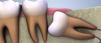
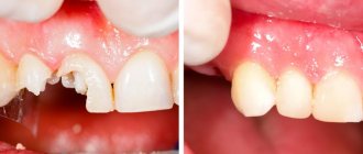
As mentioned above, this method is indicated for severe destruction and fracture of the crown of the tooth, as well as for a root fracture, but only if the fracture is in the gum. For reference: according to ICD-10, the root fracture is assigned the number S02.5. Among other indications for the use of this method, dental experts highlight retention. This diagnosis is made when the coronal part does not completely erupt or remains in the gum. This situation is especially typical for wisdom teeth.
“They performed extrusion on me. There was a tooth that was constantly being destroyed. The doctors increased it several times, but it became so dark and chipped all the time. When I once again came to the appointment, they again removed all the composite, they cleaned something there for a long time, and then they said that’s all, I can’t build it up anymore. Instead of removing it, they suggested pulling out the root a little. I agreed. The procedure went well and there was no pain. In the end, they installed a crown, and it’s holding up so far.”
Ira, review on the website www.32top.ru
This procedure can be life-saving if the carious cavity is localized below the gingival level. By removing the affected surface, it is possible to prevent the influence of harmful microorganisms on the mucous membrane and its inflammation.
Traction is also prescribed for retention
Another indication, which was already mentioned above, is insufficient thickness and height of the jaw bone to securely fasten the implant. Extrusion of the tooth root allows you to free up some space between it and the alveolus, which will promote the growth of bone tissue cells and connective epithelium.
Types of orthodontic tooth movement
Depending on the direction of the acting force, the movement of the teeth can be inclined-rotational (see Fig. 149,150), body (see Fig. 152), which also includes vertical, i.e. dento-alveolar lengthening or shortening (see Fig. 153-156) and rotational (rotation around the longitudinal axis, see Fig. 157).
One of the most common types of orthodontic movement is oblique-rotational. During this movement, the tooth does not move body-wise, when parallelism with its original longitudinal axis is maintained, and the crown with part of the root tilts in the direction of the acting force, while the apical part of the root moves in the opposite direction. Thus, 4 zones of tissue transformations are formed: two pressure zones (see Fig. 149.6 - zones 1 and 4) and two traction zones (see Fig. 149.6 - zones 2 and 3). This type of movement includes vestibulo-oral tilt (torque or inclination) and mesiodistal (angulation).
With this type of movement, a certain place in the tooth root does not move and rotation occurs around it (see Fig. 150, a - 0). The location of the rotation axis depends on a number of circumstances: the place of application of force to the crown, its clinical height, the magnitude of the acting force, and the anatomical structure of the socket. But most often the axis of rotation is located between the middle and apical third of the root (see Fig. 149, b, c and 150, a). At this point, the periodontal fissure retains its original size, periodontal tissue is not compressed and, therefore, no movement occurs. If you trace from this point “0” (see Fig. 150, a) towards the neck and apex of the tooth to the points of contact of the root with the wall of the socket, then you can clearly see that the degree of compression of the periodontium gradually increases (see Fig. 150, a - 2 and 6).
When studying the topographic relationships of the tooth in the alveolus in a transverse section in the cervical area (see Fig. 150, b), it is clear that the root is in contact with the wall only in a small area (see Fig. 150, b - 1). In adjacent areas (Fig. 150, b - 2) very slight compression of the periodontium can be seen, and at point 0, which is the center of resistance or rotation, it is completely absent.
For the body movement of the tooth, it is necessary to create such a force that its resultant passes through the center of rotation or, at least, in the immediate vicinity of it (Fig. 142,152). The solution to this issue can be twofold: first, move the point of application of force closer to the center of rotation, which is difficult to do directly in relation to the root, but you can lengthen the rigid fastening from the vestibular side of the apparatus used towards the apex of the root, creating a couple of forces; the second is to create, by combining two apparatuses, a pair of oppositely acting forces of equal magnitude. For example, if, during retrusion of a front tooth, a force is applied to any removable apparatus in the cervical area to move it labially, then a second force can be created closer to the cutting edge on the vestibular side, directing the force to the oral side.
Tissue changes during vertical movements of teeth are subject to general laws, namely, when there is a load along the axis of the tooth in the direction of the root apex, bone tissue is resorption at the bottom of the socket, and with the opposite effect - tooth extension - new bone is formed there. It should be noted that during intrusion, it is undesirable to carry out simultaneous extrusion of the neighboring one, since the latter will predominate.
Dental alveolar lengthening (“extension”, extrusion) under the influence of orthodontic appliances is most often planned for the lateral teeth in the treatment of a deep bite or for the anterior teeth in the treatment of an open bite, as well as in the elimination of supra- or infraocclusion of individual teeth. Under the influence of traction force, new bone is built in the area of the crest of the alveolar process (i.e., at the edges of the socket), the bottom of the alveoli (see Fig. 153, 154) and its entire internal surface.
Extrusion of single-, double- and multi-rooted teeth occurs according to the same laws; only in the last two groups of teeth is the formation of new bone at the dome of the interroot septum clearly visible (see Fig. 153, b - IV). In all cases, bone is also formed at the edges of the alveoli. Tissue transformations are identical both during tooth extraction with mechanically operating devices and with functional guides; the difference can only be quantitative. After extrusion, as a rule, minor correction of the gum contours and sometimes the bone socket is required.
Dentoalveolar shortening (immersion, impaction, intrusion) can be straight, purely vertical, or inclined, depending on the point of application of force. The practical application of this type of tooth movement occurs in relation to the anterior teeth when treating a deep bite and in relation to the lateral teeth when correcting an open bite. With increased pressure (the action of the orthodontic apparatus), the periodontium is compressed mainly in the area of the root apex (pressure zone) and less along the rest of the inner surface of the socket, and according to this, bone tissue resorption occurs (Fig. 156).
A special situation is created in the interroot septa, on the surface of which a wide zone of pressure appears and not only their dome, but also the entire surface is resorbed (see Fig. 156, IV). On the frontal histological section in the area of two incisors, one of which was subjected to intrusion (Fig. 155), the septum near the latter was compressed. No changes were noted on the cancellous bone beams lying immediately below the circular ligament, but a small number of osteoclasts were noticeable on the deeper ones. According to the movement of the septum, the periodontal space of the displaced tooth on the traction side was one third wider than that of the neighboring one. The cervical ligament of the tooth being moved is lowered, while that of the symmetrical tooth is elevated.
Contraindications to the use of the method
Like any other dental procedure, extrusion has its contraindications. For example, it can only be carried out if the adjacent soft tissues and teeth are in satisfactory condition, since they will act as supports for fixing the traction structure.
The procedure will not be possible if the roots are too short or severely curved, and the condition of the canals and crown does not allow the installation of at least one of the elements of the corrective system: ligatures, a button or a pin with a hook.
Therefore, before prescribing extrusion, the doctor must assess the condition and length of the roots, their location. If the patient is diagnosed with an occlusion pathology in which there is no free space for traction, the procedure will not be possible. And one more thing: the method is indicated exclusively for pulpless teeth, so the nerves will first have to be removed if this has not been done previously.
For which patients is extrusion contraindicated?
The procedure is carried out only if the periodontal tissues, as well as the teeth located adjacent to the one that needs extrusion, are in good condition. This is important, since it is on them that the pulling structure will rest.
It will not be possible to carry out extrusion if the patient has severely curved roots of the causative tooth, if the condition of the canals and the coronal part does not allow the installation of one of the elements of the traction system (ligatures and buttons, or a pin with a hook).
Before the procedure, the condition and length of the roots must be assessed - if they are too short, then this is a contraindication. Lack of free space due to bite pathologies or improper occlusion is another reason why doctors will not perform this manipulation.
Important! Extrusion is carried out only on pulpless units, so before the procedure the nerve will definitely have to be removed if this has not been done previously.
Methods of carrying out the procedure
There are two main methods of extrusion. Let's look at each type in a little more detail:
- orthodontic is a more gentle method, which is often combined with correction with a brace system. This method is indicated for impacted teeth, but it requires more time and patience to achieve results. Pulling is carried out by installing an appropriate structure,
- surgical is a rather radical method, which is fraught with serious complications if errors are made on the part of the doctor. But if everything is done correctly, the result can be obtained much faster. The method does not require long-term wearing of the traction system. We will talk further about how extrusion is carried out in this case.
In the meantime, it should be noted that after surgical traction, the patient is given a temporary restoration for the period of tissue and ligament restoration. It allows you to quickly restore aesthetics.
Types of extrusion
Doctors perform two types of extrusion – orthodontic and surgical.
The orthodontic method is more gentle and can be combined with wearing braces; it is often used if there are impacted elements, but it requires more time and patience from the patient, as well as more visits to the clinic.
The surgical method is more radical, and if performed incorrectly, complications can occur. But with a technically purely performed operation, the result can be achieved faster. At the same time, you do not have to wear orthodontic devices, which during the treatment period violate the aesthetics of the smile and cause some discomfort.
During the period of tissue healing and ligament restoration after surgical extrusion, the patient is given a temporary restoration that makes the smile attractive - this is impossible with the orthodontic method.
Preparatory stage
Before the procedure, regardless of the chosen method, it is important to undergo a full diagnostic examination to ensure that the patient has no contraindications. You will need to take an X-ray or undergo a computed tomography scan, and a procedure performed by a prof. hygiene to remove plaque and deposits, as well as remove the nerve from the causative tooth if the crown is preserved and depulpation has not been performed previously.
Before the procedure, the patient should undergo a computed tomography scan
How is extrusion performed using the orthodontic method?
In this case, traction is carried out using a special orthodontic system. Its design includes the following components:
- arch or bracket - one of these parts is fixed on elements adjacent to the causative tooth using a composite or special orthodontic rings. If the patient already has braces, the bracket is not installed;
- a screw or a special pin with hooks is attached to the causative tooth, that is, to the remaining crown. If there is not enough hard tissue for this, the pin is fixed in the root canal using dental cement,
- traction – they combine the above-described structural elements and create a force effect.
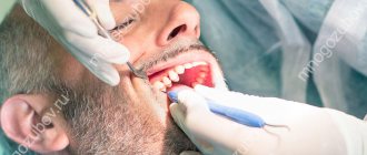
The photo shows the surgical method.
The system is fixed under local anesthesia, since to install it the doctor has to cut the fibers that connect the gum to the root. This manipulation is called fibrotomy. Due to this, the specialist gives the root mobility in order to speed up the pulling process and prevent it from returning to its original position.
Features of the orthodontic method
Orthodontic tooth extrusion is carried out using a special structure, which consists of the following elements:
- a bracket or arch that is secured to two adjacent units: it is secured using composite materials or orthodontic rings. If the patient is already undergoing basic orthodontic treatment and is wearing braces at the time of extrusion, then installation of a bracket is not required,
- hook pin or screw: These are attached to the tooth that needs traction. The screw is fixed on the remaining crown, but if this is not possible due to lack of tissue, then a pin is used, cemented in the root canal,
- rubber bands, or rods: they connect the first and second elements of the system to each other and exert a force effect.
The installation procedure must be carried out under local anesthesia, since at the end the doctor performs a dissection of the fibers connecting the gum to the root - fibrotomy. This manipulation allows you to achieve root mobility, speed up the process, and also eliminate the possibility of the root returning to its original position.
Duration of treatment
After installing the traction structure, you will need to visit a doctor once a week to monitor the process and tension of the traction (if necessary). On average, a stretch of 1 mm1 occurs per week. Accordingly, to extend the root by 3–4 mm, you need to make 3–4 visits to the clinic, and you will have to wear the device for about a month.
However, after pulling, the treatment process does not end, since the root and the surrounding ligaments need time to recover - on average, this takes about 2 more months. During this period, a temporary restoration is carried out with removal from the bite (to eliminate the chewing load) and splinting.
Recommendations for the treatment period
It is necessary to carry out oral hygiene not only in the morning and evening, but also after meals. It is better to chew on the side of the jaw opposite to the one where the pulling structure is installed. In case of rubbing of the mucous membrane by the system, it is necessary to use orthodontic wax.
How long does treatment last?
After the procedure, the patient will have to visit the orthodontist once a week so that he can monitor the process and adjust the tension of the rods. Usually, in one week, in this way it is possible to extend the root by about 1 mm. Therefore, to stretch it by 3-4 mm, you need to visit the doctor about 3-4 times, and the process itself can take up to a month.
But the treatment does not end there. It will take some time for the root and ligaments to recover. This process usually takes a couple of months. In the meantime, the patient is undergoing a temporary restoration with the removal of the causative element from the bite and its splinting - this is necessary to relieve the chewing load from it.
Expert recommendations during treatment
During the period of traction, orthodontic experts recommend increasing your oral care. You should brush your teeth not only in the morning and evening, but also every time after eating. It is also recommended to chew on the opposite side and try to eliminate any mechanical factors that could interfere with the healing process. If the structural elements rub the mucous membrane, you should resort to orthodontic wax.
Orthodontic wax will help protect the mucous membrane from damage
Features of the surgical procedure
This method is most often used in cases where the crown part of the tooth is so damaged that it can no longer be restored in the classical way, or if it is necessary to re-prosthetize / replace the old restoration material. This procedure can also be performed if the patient wants to get results faster and without the need for temporary “aesthetic loss”.
This operation is not classified as complex, but it requires appropriate experience and skill from a specialist. For pulling, the doctor uses a special scalpel, elevator and forceps. You could say he creates a dislocation and then does not completely remove the root, after which he fixes it with sutures.
The photo shows the surgical method of the procedure
Recommendations after surgery
As mentioned above, after the operation the tooth is covered with a temporary restoration. About a week after surgery, the sutures are removed. Over the next 2-3 weeks, the patient must adhere to a special diet - it is recommended to eat only soft, warm food, not too cold or hot. You will also need to strengthen your oral hygiene and treat soft tissues using an antiseptic solution, for example, Chlorhexidine, as prescribed by your doctor.
To treat the mucous membrane, you can use Chlorhexidine
1.5-3 months after the operation, the root acquires sufficient stability, which allows endodontic treatment and prosthetics to begin. Some experts insist that it is better to install a permanent prosthesis only 1-2 years after traction in order to prevent the development of complications. It will take quite a long time for the periodontal ligaments to fully recover.
A balanced decision on braces
Before deciding to install a braces system, you need to take into account all the nuances. Treatment may take a long time, and to achieve the desired result, you will need to strictly follow everything that the attending physician recommends.
It will also be necessary to carefully maintain oral hygiene in order to prevent caries and prevent the formation of plaque. Since metal braces cannot be removed, you will need to spend more time removing food debris than regular teeth brushing. However, the result is worth such diligence. Smooth, healthy teeth are the key to a radiant smile becoming your constant companion.
By visiting Adent dentistry, you will receive complete information regarding all brace systems in modern dentistry.
The post Metal Braces appeared first on .
]]>
How impacted elements are pulled out
Extrusion in this case has its own characteristics. It can be carried out in two different ways, but each of them necessarily includes both surgical and orthodontic stages. These are the two methods:
- delayed - first the doctor dissects the gum and exposes the crown. At the next appointment, 2-3 days later, he attaches a special orthodontic button and rods to the impacted element, after which he installs brackets on the adjacent supporting elements (if there are braces, there is no need for brackets),
- one-step – in one visit, the doctor cuts the gum, fixes the orthodontic system and applies sutures.
With the one-step method, the structure is installed immediately.
During rehabilitation, the specialist prescribes antibiotics and antiseptics to the patient. Again, you will have to adhere to a special diet and exclude any traumatic factors. Pulling in this way can last up to 1 year or even longer.
Advantages and disadvantages of the method
The main advantage of the method is the ability to preserve the root and subsequently carry out prosthetics based on it. Here is a list of other obvious advantages of this procedure:
- the ability to avoid expensive treatment, for example, prosthetic bridges or implantation,
- the ability to extend the life of a living tooth for up to 5-10 years and even more,
- the ability to avoid traumatic surgery to build bone tissue before installing implants.
The main thing is that an experienced, highly qualified doctor undertakes the treatment, since much here depends on the quality and accuracy of the manipulations performed. If a specialist makes mistakes, they can result in resorption and a decrease in root stability, and the development of ankylosis.
Among the disadvantages, patients highlight the duration of treatment and the inconvenience associated with it. Partly, it is the fact that pulling takes quite a long time, during which you have to adhere to strict dietary restrictions and be careful not to accidentally damage the system. But the result of such a correction can be considered the preservation of a living root, the opportunity to avoid more expensive and, by the way, no less lengthy implantation.
1Persii L.S. Orthodontics. Diagnosis and treatment of dental anomalies: a guide for doctors, 2004.
Installation of metal braces
The size of braces is selected individually. It depends, first of all, on which tooth the system is intended for. In order not to make a mistake with the direction of the groove, special markings are applied to the finished bracket system. Depending on the client’s wishes, the bracket system can be installed both on the outside and on the inside of the dentition.
Before fixing metal braces, the surface of the teeth must be prepared:
- clean from tartar and plaque,
- treat with a fluorine-containing compound,
- restoring tooth enamel.