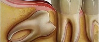
Impacted – this is a tooth that has not erupted and is located in the bone tissue, covered by the gum. Dystopic These are teeth that have an abnormal position - that is, they are shifted towards the cheek or tongue, rotated around their axis or tilted to the side. At the same time, some teeth can be either impacted or dystopic - that is, they are located in the bone, while being turned to the side relative to the correct vertical position. This mainly concerns “wisdom teeth” or molar eights, which are located at the very end of the dentition.
Why do wisdom teeth grow incorrectly?
Wisdom teeth, or the outermost teeth in a row, usually appear between the ages of 18 and 25. And they grow, as a rule, with serious disturbances. Only the lucky ones can boast of straight and evenly grown “eights”. Frequent problems of wisdom teeth are retention and dystopia, in most cases even both ailments at once.
The abnormal growth of wisdom teeth is explained by the fact that, firstly, they grow in adulthood, when the bone tissue is fully formed, it is very dense and hard - naturally, it is incredibly difficult to “break through” it. Secondly, modern man does not need eighth teeth - our ancestors used them to chew raw meat. Wisdom teeth don’t even have guides—baby teeth—so they have to make their own way through hard bone.
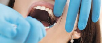
In most cases, wisdom teeth must be removed, especially if they do not appear above the gum and interfere with neighboring teeth (press on the roots of their neighbors on the side). It is also very important to check the condition of impacted wisdom teeth if you experience discomfort when opening and closing your jaw, pain in the ear, or an increase in body temperature.
Removal of impacted and dystopic tooth
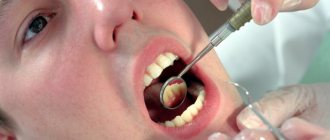
An impacted wisdom tooth can be in a straight or rotated position (dystopia) - in this case it will put pressure on the roots of its neighbor. To avoid complications, such ungrown teeth must be removed. How is the operation performed? In several stages:
- mandatory administration of anesthesia: tooth extraction is a painful operation that requires serious tissue anesthesia,
- the gum is cut - access is created to the impacted tooth,
- a hole is created in the bone using a bur,
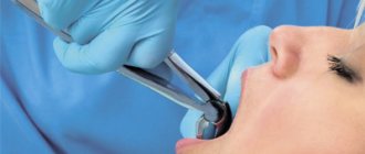
- tooth extraction: the tooth is removed immediately using forceps, or is first divided into parts using a bur and then removed from the bone tissue,
- the bone is restored: in some cases, the cavity vacated by the tooth is filled with artificial bone material, or with the patient’s own blood plasma - this measure is necessary so that empty space does not form in the bone, which can be occupied by neighboring teeth. If they begin to shift, then the entire dentition will gradually “creep” - gaps will appear between the teeth. In order to understand whether the hole will need to be filled with any material, you need to consult a doctor,
- the gum is sutured: after all manipulations are completed, the gum is returned to its place, sutures are placed on top.
The operation to remove an impacted / dystopic tooth lasts about 30 minutes to an hour. This is a fairly ordinary operation, which, when performed professionally, will not lead to any complications. In some cases, if there are a lot of uneven roots, it may take longer. The doctor can determine the approximate time of the operation based on the results of examining the image.
Complications after wisdom teeth removal
Unerupted teeth are sometimes located quite deep in the bone, so extracting them can be quite a difficult procedure and can lead to certain complications. For example, a nerve injury in the lower jaw, resulting in numbness of the lips, tongue and part of the cheek (goes away on its own after a few weeks or months), rupture of the nasal sinus (a runny nose or sinusitis forms). With complex removal of wisdom teeth, there is a high probability of fracture of the jaw, adjacent teeth or dentures. In order to avoid such complications, it is best to have wisdom teeth removed by an oral and maxillofacial surgeon who has extensive practical experience in this field.
Care after wisdom teeth removal
After removal of impacted wisdom teeth, minor pain, bleeding from the wound, and rarely, suture separation may occur. Tissue swelling and pain when chewing food and opening the jaw are also observed. If the pain does not go away within 2-3 days (at this time it is necessary to take painkillers - these can be Ketarol, Ketanov, Nise, etc., depending on the prescription after the operation), you must consult a doctor. In general, rehabilitation takes from 1 to 5 days. After a few days, you must visit a doctor for a follow-up examination of the results of treatment and removal of stitches.
Types of dental retention
Orthodontists distinguish 2 characteristic types of abnormal eruption of impacted teeth:
- partial or semi-retention, in which a small part of the dental unit protrudes above the gingival hood. The coronal apex of the tooth is detected by a dental probe;
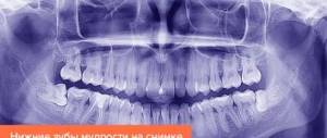
- complete retention, when the entire tooth and its crown part are immersed in the mucosa and completely covered with periodontium. Can only be accurately diagnosed by targeted X-ray examination
The location of the coronal part and root of a non-sprouted or partially sprouted dental unit, with its retention, can be:
- vertical or corresponding to the axis of location of the rest of the dentition. This type of arrangement occurs due to thick and very dense mucous membranes surrounding the tooth or a small intermediate place in the dentition;
- horizontal or parallel to the location of the jaw arches. The inclination of an undeveloped dental unit can be: sagittal, oblique or transverse. Accompanied by loosening of adjacent teeth;
- buccal-angular or inclined towards the cheek;
- medial-angular or located with an inclination forward;
- lingual-angular or inclined towards the tongue;
- distal-angular or sloping backward.
Depending on the depth of occurrence, impacted dental units can be located:
- in the depths of the jaw bone and under the mucosa. This type of localization of retention is complex and requires serious surgical intervention;
- in the soft gingival tissues of the periodontium. Most typical for impacted teeth.
What is the risk?
The best option is to begin treatment for dystopia in adolescence, when the jaw is not yet fully formed. This will make it possible to fully normalize the position of the teeth and, in turn, achieve aesthetic results.
When visiting a dentist as an adult, the treatment will take longer, but it is still worth it.
If you do not contact specialists with this problem in a timely manner, this will lead to a number of negative consequences , namely:
- a dystopic wisdom tooth causes destruction of the adjacent one and provokes the occurrence of periodontitis;
- an incorrect bite is formed;
- the chewing process is disrupted, which in turn leads to diseases of the gastrointestinal tract;
- speech defects occur;
- injury to the oral mucosa occurs;
- it becomes difficult to carry out full hygienic procedures;
- Accelerates the formation of tartar on damaged teeth;
- There is a tendency towards facial asymmetry.
Causes of impacted teeth
The main causes and risk factors leading to the development of retention:
- Individual characteristics of the masticatory apparatus: a thick, dense layer of gingival tissue, thick walls of the mucous membrane surrounding the crown of the erupting tooth, weak germinal force.
- Gum tissues that are too loose, preventing the tooth root from being in its normal position.
- Features of embryonic development with incorrect formation of tooth buds or incorrect location of their axis. As they germinate, they may encounter a previously erupted tooth.
- Dislocation or complete loss of primary teeth as a result of trauma in early childhood and displacement of the adjacent one in its place.
- Existing supernumerary, additional tooth buds (hyperdotnia) between the canines and upper incisors, in the lateral sections, in the frontal area of the alveolar process on the lower jaw.
- Failures and delays in the mechanism of replacing baby teeth with permanent ones.
- Delays in resorption of the root of a baby tooth.
- Abnormal conditions in the process of phylogenetic jaw development, congenital cleft palate or upper lip.
- Bite pathologies, narrowing of the dentition.
- Congenital or acquired rickets.
- A condition of chronic fibrous inflammation of the tissues surrounding the tooth.
- Reduction of functions, weakening (reduction) of the masticatory apparatus in the process of evolution, leading to a decrease in the distal alveolar processes of the jaws, which causes a lack of space for teeth.
- Pathologies of the endocrine glands and disorders in metabolism and the functioning of the endocrine system.
- Reduced immunity as a result of acute or chronic infectious and some general diseases.
- Lack of calcium in the body, diseases of the skeletal system.
- Hereditary causes, disturbances in the structure of genes responsible for the formation of tooth buds and their further tissue differentiation.
- Long courses of chemotherapy, stopping or completely inhibiting the process of tooth germination.
What it is?
In dentistry, dystopia is understood as a phenomenon such as the incorrect position of a tooth germ as a result of a violation of embryonic development or due to genetic and external factors.
Most often, wisdom teeth and canines are susceptible to this phenomenon..
A person suffering from this disease feels very awkward in society. He tries to smile as little as possible and talk with strangers, because, for example, with the abnormal position of the fangs, the impression of a “vampire” smile is created.
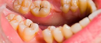
In addition, the disease negatively affects the health of the entire oral cavity . It can lead to changes in the shape of the jaw, making cleaning difficult, which in turn gives rise to periodontal disease and caries.
Possible complications
When there is an impacted tooth in the oral cavity, dental problems arise over time, which can be used to determine the presence of an impacted tooth:
- the appearance of swelling and soreness of the gums and soft tissues around, aggravated by palpation, an inflammatory process in them;
- redness of the gums, pain reaction in it;
- numbness of the face that occurs due to the pressure of a tightly fitting (not erupted) tooth on the nerve endings;
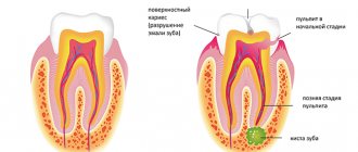
- pulpitis and caries of neighboring teeth;
- the appearance of an odontogenic cyst;
- resorption (absorption) of the roots of teeth located next to the impacted tooth and constantly compressed by it;
- the onset of inflammation of the periosteum (flux);
- emerging gingivitis, pericoronitis, abscess;
- periostitis, osteomyelitis of the jaw bone and surrounding soft tissue;
- problems with the functional muscles of the face, violation of its aesthetics, difficulty opening the mouth, speech impairment;
- bad breath;
- painful, uncomfortable condition when chewing, inflammation of the mandibular nerve;
- disturbance of taste sensations;
- headaches, trigeminal neuralgia;
- Body temperature may rise and weakness may occur.
Signs and symptoms
In most cases, you can recognize the presence of an impacted tooth yourself at home.
The main signs for this are:
- soreness in the gum area, which can radiate towards the ear, temple and along the location of the trigeminal nerve;
- regular injury to one area of the mucosa;
- swelling, numbness and hyperemia of the periodontium;
- with slight eruption, gingivitis or pericoronitis may begin;
- protrusion of a limited area of the gum;
- displacement or loosening of adjacent teeth;
- discomfort while eating or opening the mouth;
- the presence of a cyst or purulent formation may be observed;
- During the inflammatory process, the general condition worsens: the temperature rises, chills, headaches, and weakness appear.
When is it necessary to remove an impacted tooth?
Removal is indicated if:
- there is repeated acute, subacute or chronic pericoronitis;
- signs of progressive painful periostatitis, caries, periodontitis, pulpitis appeared on impacted or adjacent teeth and their surrounding tissues;
- destruction of the dentin tissue of the root occurs, resorption of the roots of neighboring teeth, which are under pressure from the impacted dental unit;
- neuralgic pain occurred as a result of a chronic inflammatory process;
- a follicular cyst has formed;
- a tumor (odontoma, ameloblastoma) has formed around an unerupted tooth;
- there is no place for it in the dentition;
- destruction of the tooth neck has occurred;
- necessary orthodontic treatment requires additional free space;
- a medial diastema developed as a result of a transversely impacted tooth.
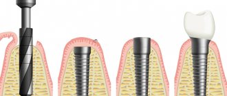
Removal technique
The surgical operation to remove an impacted tooth lasts from 25 minutes. up to 3-4 hours. Main removal steps:
- organizational measures for local anesthesia of infiltration type or general anesthesia;
- a complete (extensive) incision or patchwork incision of the soft mucous tissues and periosteum, creating access to the coronal part of the tooth;
- peeling of mucous tissue, exposure of the bone bed;
- preparation and formation of the surface of an impacted tooth for removal. Using a special bur, in order to gain access to the roots of the tooth, a hole is drilled and sawed out in the bone tissue;
- direct tooth extraction, extraction. Can be done with elevators, simultaneously with pliers and elevators, or with a cutter with a straight tip. First, the upper part of the tooth is cut and removed, then its root and everything else. The release of all parts of the tooth and their extraction from the socket are performed by dislocation and at right angles. This makes it possible to break all periodontal bonds;
- The entire surface of the open wound and the resulting voids are well cleaned of bone tissue residues, and washed with antiseptic solutions. If the area of the affected area around the tooth was extensive and a strong inflammatory process was observed, then turunda soaked in iodoform is placed in the hole;
- suturing, suturing damaged tissue with suture material, catgut.
In case of partial retention, a mucosal incision is not necessary. The surgeon, using forceps, swings the crown part of the tooth in a pendulum-like motion, and then pulls it out. Impacted teeth located on the lower jaws are more difficult to remove.
The removal operation can also be performed with a laser beam (laser). This is a less traumatic and faster procedure. It takes place without drilling, the rehabilitation period is simple and short, the complications that arise are minimal, and the risk of wound infection is significantly reduced.
Contraindications
Surgery to remove an impacted tooth is contraindicated in the following cases:
- state of hypertensive crisis;
- acute stages of heart disease or diseases of the nervous system;
- existing viral or infectious diseases:
- existing diseases of the blood and hematopoietic system;
- in the last phase of menstruation in women;
- if less than 14 days have passed since the operation to terminate the pregnancy.
Diagnostic methods
In case of partial retention, a visual examination and instrumental examination may be sufficient for diagnosis. To clarify the position of the tooth, targeted radiography is used.
In some cases, orthopantomography and computed tomography are prescribed to obtain a comprehensive picture.
Care after removal of an impacted tooth
After surgery:
- in the first 15-20 minutes. after removal, you need to firmly press the gauze swab prescribed by the doctor, this will stop the bleeding;
- make sure that the blood from the wound does not flow too intensely, otherwise consult a doctor;
- You should abstain from food for a long time (at least 4 hours). After you start eating, and until the wound heals completely, you should not eat hot or soft food;
- if severe swelling of the mucous membrane appears, you can apply a cold compress to the cheek (for a short time);
- to avoid the occurrence of an inflammatory process, it is impossible to heat the hole from the extracted tooth;
- the protective blood clot formed in the hole cannot be removed, it prevents infection from entering it;
- on the second day after removal, you can apply a cotton swab soaked in retinol acetate (vitamin A) or rinse your mouth with a decoction of sage, chamomile, calendula, weak solutions of furatsilin, potassium permanganate or other antiseptic drugs;
- severe pain can be relieved with painkillers.
Complete healing of the socket tissue occurs in 3-5 weeks. It is important to monitor the healing process and notice any dental problems that arise. In case of suppuration of the surgical suture, you must consult your doctor.
Conservative methods of treating retention
In dental practice, orthodontists try to preserve an impacted tooth that has not sprouted. An impacted tooth is not removed:
- The tooth in the gum is positioned correctly, it does not displace neighboring teeth or interfere with them.
- If the interference with complete tooth eruption is minimal and it is easy to eliminate the cause of the infringement of the tooth germ.
- If there is a horizontal area of at least a few mm behind the crown part of the tooth.
- It is possible to carry out high-quality anti-caries treatment.
- In case it is needed as a support, for partial or complete prosthetics.
- When an X-ray examination did not reveal cysts and various neoplasms.
- When the tooth is healthy and there are no serious inflammatory processes both in it and in its surrounding tissues.
- If it is involved in the bite and is involved in the chewing processes.
How to stimulate tooth eruption
When an X-ray image shows that the impacted tooth is positioned correctly (vertically), but its roots have not yet formed, then after eliminating the cause of its infringement, special physical procedures can be performed to stimulate the eruption process. To activate and restore blood circulation, accelerate metabolic processes in the area of tooth germination, use:
- vacuum massage of the gums with a special device to tone and strengthen the mucous membrane around the impacted tooth;
- light finger massage and rubbing of the gums. To enhance the effect, you can apply extracts and tinctures of calendula, propolis, eucalyptus, and sage;
- electrical stimulation or irritation with electric current with the simultaneous administration of vegetotropic drugs. Can be performed by pulsed or galvanic current;
- vacuum stimulation (massage) using the method of focal dosed vacuum;
- electrophoresis with adrenaline, lidase, ronidase, humic acids;
- ultraphonophoresis with calcium chloride;
- vibrovacuum technique;
- stimulation with low-intensity red or infrared laser radiation;
- exposure to low-frequency ultrasound;
- wearing special removable irritating dentures (raich therapy).
Orthodontic treatment
If there is not enough space for the erupting tooth, ordotontic treatment is performed to expand the dentition.
When the roots of an impacted tooth have already formed, independent tooth eruption is impossible. In these cases, various methods of orthodontic traction and alignment are used with simultaneous correct placement of the tooth in the dentition.
- Dental alveolar lengthening (extrusion) using a transparent aligner.
- Installation of bracket systems, push-button clamps, tubes. If the tooth is completely impacted, then such orthodontic operations are carried out in several stages: the mucous membrane is dissected, the flap is folded back, the impacted tooth is exposed, then braces or buttons are installed. A single or double jaw traction can be installed. After installing the brace system, the excised flap of tissue is placed in place and the wound is sutured. In the case of superficial retention, a light dissection of the soft tissue is performed and the bracket button is glued to the impacted tooth. Using nitinol and hard steel arcs, traction begins.
The issue of preserving or removing an impacted tooth should be resolved individually, for each specific case and exclusively by an orthodontist or dental surgeon.
Sources used:
- Bone grafting in dentistry and maxillofacial surgery: monograph. / A.S. Pankratov, M.V. Lekishvili, I.S. Kopetsky. - M.: Binom, 2011.
- Guide to surgical dentistry and maxillofacial surgery (set of 2 books) / Edited by V.M. Bezrukova, T.G. Robustova. - M.: Medicine, 2000.
- Wikipedia article
Prevention
It is necessary to take care of the prevention of dystopia even before the birth of her baby. To do this, the expectant mother should eat properly and avoid exposure to various provoking factors (stress, infectious diseases, illegal drugs, etc.)
And throughout your life, you must not forget that an abnormal tooth may creep in among your healthy counterparts. That is why important methods for preventing dystopia are:
- balanced nutrition for newborns without lack of important nutrients in the diet;
- timely introduction of solid foods;
- elimination of bad habits during the newborn period (finger or pacifier sucking);
- prevention of jaw injuries;
- timely removal of baby teeth;
- replacement of missing teeth;
- strict adherence to oral hygiene;
- timely visit to the orthodontist.