The one-stage technique mainly includes one-stage implantation protocols with immediate loading, but in some situations, simultaneously with tooth extraction, a classic implant can also be installed using a two-stage technology. According to many clinical studies, it is one-stage implants installed in the socket of an extracted tooth that protect bone tissue from atrophy and can reduce treatment time with a guaranteed high long-term result of up to 96%1.
Indications
- teeth to be removed are rotten, mobile, destroyed by carious processes,
- single restorations,
- multiple restorations,
- tooth injury,
- simultaneous tooth extraction and implant installation.
Contraindications
- emergency tooth extraction when it is not possible to prepare for implantation,
- complex tooth extraction,
- general contraindications to implantation and surgery.
Implantation methods after tooth extraction
Dental implantation after extraction is carried out according to two protocols:
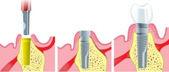
- One-stage - involves installing an implant immediately into the socket of the extracted tooth. There are techniques that allow not only the installation of an implant, but also the fixation of a crown on it.
- Delayed - requires complete healing of the hole and restoration of bone tissue after removal. The bone is restored in 2-5 months depending on the health of the patient. Only then is implantation carried out.
Insert ceramic teeth
Inserting ceramic teeth will be the right decision if you need to restore your front teeth, which are clearly visible when communicating with people around you. But for the restoration of chewing teeth, ceramic crowns are not suitable: ceramics is a fairly fragile material and a crown made from it may not withstand the heavy load that is constantly placed on the chewing teeth when chewing food.
The high aesthetics of ceramic crowns determines the expensive prices for this type of orthopedic structures. But the patient gets a beautiful and natural smile: you can also evaluate the results of dental restoration with ceramic crowns from the photos that we have posted below. Ceramic crowns can be placed on both the abutment tooth and the implant.
When it is possible to perform implantation - the doctor chooses after a complete diagnosis
It is necessary to study the condition of the bone tissue, its size and density. Based on the results, the doctor makes a decision - delayed or immediate implantation. Our Center is equipped with diagnostic equipment, so results can be obtained within 15 minutes . But this is only a preliminary assessment; the final decision is made after tooth extraction.
Levin Dmitry Valerievich
Chief physician, Ph.D.
Orthopantomogram or OPTG is an important component of preparation for dental implantation
Article navigation
- Orthopantomogram - the essence of the study
- Indications for OPTG
- What types of photographs are there?
- Preparing for the examination
- How is the procedure performed?
- Are there any restrictions
- Acceptability during pregnancy
- Potential dangers and possible complications
- How to decrypt a photo
Question for a specialist
An orthopantomogram is a separate type of x-ray examination that allows you to obtain a panoramic image of the patient’s entire dental system. It reveals detailed information not only about the condition of the teeth of both jaws, but also about the maxillary sinuses and other important anatomical structures. This is a modern, affordable and effective way to conduct a diagnostic examination, making it possible to identify all problems at once. True, the detail of OPTG is always slightly lower than that of targeted images and computed tomography results. But today we will take a closer look at the orthopantomogram, find out what it is, how it is done and what it shows. Well, let’s answer the most important question – is it needed before implantation.
Indications and contraindications for immediate implant placement
Indications
- Mechanical damage to the root - fracture, dislocation
- Restoration of frontal units
- Therapeutic treatment did not bring results
- Sufficient bone volume
- No contraindications
Contraindications
- Deficiency, looseness of bone
- Inflammation at the root apex
- Enlarged socket after removal
- Inflammatory processes of the oral cavity, gum pathologies
- Reduced immunity, systemic diseases
- Pregnancy, breastfeeding period
Preparing for the examination
The procedure does not require any specific preparation; it can be carried out directly on the day of contacting the dentist or diagnostic center. However, before sending a person for OPTG, the specialist is obliged to conduct a visual examination of the oral cavity and collect a complete anamnesis, that is, take into account all complaints and possible pathological phenomena in the maxillofacial area. After the initial diagnosis, all that remains is to settle some formalities: the doctor will ask you to sign a consent to conduct the examination.
How to do dental implantation immediately after extraction
After a tooth is removed and an implant is immediately placed, the process of osseointegration occurs faster. The hole with the artificial root begins to heal, the blood clot turns into bone tissue that surrounds the implant in the early stages of healing. This promotes accelerated osseointegration of the implant with the jawbone, ensuring reliable fixation and stability.
Stages:
Diagnostics
Full diagnostics, choice of design in the absence of contraindications.
Tooth extraction, artificial root installation
Local anesthesia or sedation is used. A bed is formed in the hole, an implant is installed, and the space around is filled with osteoplastic material. After the procedure is completed, the gums are sutured. A temporary removable prosthesis is installed. Engraftment lasts 4-6 months.
Prosthetics
After osseointegration of the implant with the jaw bone, the gum is opened, and a gum former is fixed to the implant for 10-14 days to create a gum contour. Then the former is replaced with an abutment, and a crown made from the impression is installed.
In rare cases, it is possible to fix the crown to the implant immediately after its installation. At the same time, the crown is removed from the bite so that the implant does not move during chewing. This method is recommended for the anterior parts of the jaw in order to quickly restore the defect. Not suitable for lateral use due to active chewing loads.
Stages of treatment
The duration of one-stage implantation is about 4-6 months. Restoration of bone tissue after tooth extraction is not required; bone tissue augmentation as a separate procedure is also not performed. Therefore, after installing the implants, a temporary prosthesis is immediately fixed, and after a few months, when the implants and bone tissue have completely fused, a permanent dental crown is installed.
Galina Vladimirovna, 57 years old
“I had my natural teeth removed and implants were immediately placed. Within a week my smile was back. I really like the way I look and the way I talk. My diction hasn’t changed, I’m comfortable. I am very pleased and grateful to the doctors!”
- we give SMILES, HEALTH and new LIFE
- smile and enjoy every day
- start CHEWING immediately
- forget about the unpleasant smell and pain
- complete the entire treatment in a WEEK
watch a video with the patient
Preparation for implantation (from 2 days to 2-3 weeks)
When a tooth is removed and an implant is installed at once, sometimes there is very little time left to prepare for treatment, since most often teeth that are in very poor condition have to be removed. However, implantologists pay great attention to this stage, since it is necessary to conduct a thorough examination of the patient’s oral cavity and body, to exclude possible contraindications that could cause rejection of the structure.
Preparation for immediate implantation is absolutely standard and includes taking a series of blood tests, taking an X-ray or panoramic photo of the jaw, as well as dental treatment and removal of deposits. If there are acute problems with the body, we can refer the patient to an appointment with a highly specialized doctor to clarify the diagnosis, assess the state of health and carry out appropriate treatment
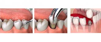
Installation of implants and bone grafting (1-2 hours)
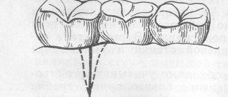
Installation of an implant with simultaneous tooth extraction is carried out in a fairly simplified form: additional tissue incisions and bone drilling are not required to form a bed for the implant. The tooth is removed from the socket as carefully as possible, with minimal trauma to the bone tissue. Next, the cavity is thoroughly sanitized. If necessary, an x-ray is prescribed so that you can make sure that the cavity is empty and there are no pieces of instruments or parts of the tooth left inside it. Next, a dental implant is fixed in the hole, and the space around it is sprinkled with artificial bone tissue. The gums around the implant are sutured.
Temporary prosthetics (3-6 months)
Depending on the initial situation, both one-piece and two-piece implants can be used for simultaneous implantation. Both of them can be instantly loaded with a temporary prosthesis.
Three types of structures can be used as temporary dentures (they are selected based on the quality of bone tissue, the degree of primary fixation of the dental implant and the number of missing teeth):
- removable immediate denture (for front and side teeth),
- lightweight plastic crown (only for front teeth with single restorations),
- acrylic prosthesis with plastic crowns (with complete edentia).
A resin crown is created that is slightly shorter in length than the adjacent teeth in the row. This allows you to significantly reduce the load on the prosthesis, and hence the implant itself. At the same time, the appearance of the smile is maintained at the highest level. From the point of view of further aesthetics, a single crown is a more preferable option for single restorations, since it allows you to shape the position of the gums.
It is important! Installation of a single crown is possible only on single-rooted teeth, which do not bear much pressure, and they mostly perform aesthetic functions - these are incisors and canines. If you chew food with the restored tooth, the implant simply will not take root, since it will shift due to the load.
In case of complete edentia, when it is necessary to restore the entire dentition, several implants are installed at once. If there are teeth that need to be removed, some of the implants can be fixed into their sockets. In this case, a fixed prosthesis with a metal base, acrylic base and plastic crowns is fixed on top. It firmly connects all installed implants, which prevents them from moving. Read more about immediate loading methods >>>
Permanent prosthetics (1-2 weeks)
Permanent prosthetics are the final stage of treatment. It can be started only after the doctor is convinced that the implants have completely fused with the bone tissue - measurements are carried out with special devices and x-rays.
The temporary crown is removed, impressions are taken - the data is transferred to a dental laboratory, where an individual prosthesis is created. The dental technician must take into account the position of neighboring and antagonist teeth located on the opposite jaw. The manufacturing process takes about 1-2 weeks.
Metal-ceramics or zirconium can be chosen as materials for permanent dentures - these crowns are more aesthetic, stronger and more durable.
Advantages of the method
- shortened treatment times,
- no bone grafting required
- instant restoration of aesthetics,
- no bone tissue augmentation is carried out,
- lower cost of treatment,
- The service life of installed implants is more than 20 years.
Disadvantages of the method
The only disadvantage of one-step implantation is that it is not always possible to carry out it. You need to prepare for treatment: assess the condition of the body, exclude contraindications, select an implant model depending on the volume and quality of bone tissue. But the removal of your own tooth quite often has to be done urgently, which does not make it possible to conduct a full examination and understand whether it is possible to install an implant at the same time.
Advantages and disadvantages of one-stage dental implantation
pros
- One operation is performed, waiting time is reduced
- Simplified installation process
- Minimal loss of mucous and bone tissue after surgery
- If the technology is followed, the percentage of successful operations is 99.6%
Minuses
- Contraindicated in chronic inflammation, diseases
- Not performed if there is insufficient bone
- Strict rehabilitation period, frequent visits to the implantologist
For a favorable outcome, entrust the operation to a surgeon with extensive experience in this protocol.
What types of photographs are there?
- film - this type of OPTG appeared first. Initially, models of devices were used that involved the use of a special film. It was placed on one side of the patient's head, while the X-ray tube was on the other side. After the procedure, the image was transferred to film, after which it was processed and developed,
- digital – a modern version of the examination involves recording images on a cassette in digital format. The obtained data is loaded into a special computer program, and the doctor can carefully study the already processed image from the computer screen.
Is it possible to place an implant if a tooth has been lost for a long time?
Whether it is possible to place an implant if the tooth was removed a long time ago is decided only by the surgeon after a complete diagnosis. Already 3 months after removal, the bone is so absorbed that it may not be enough to install an implant and augmentation will be required. The recommended implantation period is no later than 2-3 months.
In case of bone atrophy, osteoplasty is performed:
- Guided bone regeneration . Most often performed on the lower jaw, bone materials, biomembranes, and bone growth stimulators are used. It is used if the width and height of the patient’s bone is not enough for implantation. If bone tissue atrophy is insignificant, it is possible to perform NCR simultaneously with the installation of implants. Otherwise, it is carried out in a separate preliminary stage. It takes 3-4 months for the grafted material to fuse with the bone.
- Sinus lift. Intervention in the bone structure of the upper jaw in the area of the maxillary sinuses. Using instruments, tissue is punctured to raise the bottom of the sinus. The resulting space is filled with osteoplastic material. The operation can be closed or open. A closed sinus lift is performed for minor bone atrophy simultaneously with the installation of implants. Open - for moderate and critical atrophy in a separate stage.
Make an appointment with an implantologist
Insert teeth with metal-ceramic crowns
You can inexpensively replace teeth with metal-ceramic crowns. Such dental prostheses have a metal base and are covered with a layer of ceramic mass on top. This is done so that the finished crown has a natural and aesthetic appearance.
Metal-ceramic crowns are used in both traditional prosthetics and implantation, however, if you need to insert a front tooth, this option of dentures is not suitable. The fact is that the metal from which the base of the crown is made oxidizes over time and this reaction can cause the gums to appear blue in the area where the crown is installed. This cyanosis will be clearly visible when smiling or talking, which of course will affect the level of aesthetics.
If you want to see what a smile will look like if you replace your teeth with metal-ceramic crowns, look at the photos we publish below.
Price
The Center for Private Dentistry Dr. Levin provides a case payment system.
- When implanting Nobel Biocare PMC Select or Groovy, the cost includes the surgical stages, including the design model, surgery, anesthesia, and all necessary procedures. The price is indicated taking into account the preparatory stage.
- Removal is paid separately, the price includes surgery, inflammation relief, socket rehabilitation, consumables, anesthesia.
- The case for installing a metal-free zirconium dioxide crown includes all stages - impressions, manufacturing, installation.
- Sinus lifting and any bone tissue augmentation operations are paid separately.
How is the procedure performed?
We have figured out why an orthopantomogram is done in dentistry, and now we will look at the progress of the procedure. Diagnostics is completely painless for the patient, and the operation of the device itself takes no more than a minute. In this case, the patient immediately receives the examination results. Immediately before the procedure, the specialist will ask you to remove all metal items and jewelry and put on a protective vest. The doctor will indicate the place where you need to stand during the diagnosis. After that, he will raise the working area of the device and fix it at head level.
“I took a panoramic photo before implantation - this is the most harmless procedure I have ever undergone in dentistry, especially compared to the operation itself! I don’t understand the reason for the excitement of some patients, after all, they probably underwent fluorography, everything is similar here, only they examine not the chest, but the jaw. Everything really takes no more than a few minutes. Moreover, the doctor immediately showed me the result on the computer screen, gave some comments, gave me the picture, and I also wrote it down on a flash drive, just in case. In general, don’t be afraid, there’s nothing scary there at all...”
Mila1989, Moscow, from correspondence on the woman.ru forum
After this, the person’s head is fixed using a special plate. At one end it is attached to the equipment, and its second free end is opposite the mouth. A sterile tip is fixed on it so that the patient can clamp it in his teeth - this helps ensure the immobility of the jaw apparatus during the examination and eliminate any distortions.
When all the equipment settings are ready, the specialist will definitely warn the patient about the start of the examination, and also remind him of the importance of remaining still. After turning on the tomograph, the plates will gradually rotate around the head, making two revolutions. Next, the doctor will be able to evaluate and show the patient the result on the computer screen, and will offer to print the image or record it on a digital medium.
Alternative options for tooth restoration after extraction
After removal, doctors recommend restoring the tooth in a short time. This is caused by bone tissue atrophy, which begins in the absence of chewing load, which complicates recovery in the future.
If for some reason the patient is not ready for implantation, alternatives can be offered:
- Bridge prosthesis. It is a non-removable structure of interconnected crowns. For installation, two supporting teeth are depulped (nerve removal, canal filling) and ground down for crowns. At the same time, the load on the support increases, mobility develops, which is why the bridge will need to be replaced. The bridge structure is not able to completely prevent bone tissue atrophy, but it prevents rarefaction, displacement, and curvature of the dentition. The rate of bone atrophy and the development of mobility depend on the patient’s health.
- Removable dentures . They represent a structure made of artificial gum with soldered crowns, attached to adjacent teeth with hooks. The supporting teeth are ground down if the fastenings are metal. The prosthesis has a range of mobility and requires careful care, since food particles can get under it. Removable dentures do not stop atrophy, but they do prevent displacement of adjacent units.
Implantation prevents all problems, including bone atrophy. An implant is an artificial root on which a crown is placed. Neighboring teeth are not ground down or damaged. The chewing load is maintained. The survival rate of dental implants is 98%. Implantation is considered the most reliable way to prevent bone resorption.
Review of modern technologies that allow you to insert teeth
Just 30 years ago, there was only one way to replace lost teeth—prosthetics, and patients were offered crowns made of heavy and unesthetic materials—various metal alloys. And no one was embarrassed by the sight of metal teeth in the smile area. Nowadays, you can insert teeth in such a way that no stranger will even guess that you have a crown or prosthesis in your mouth, and not your natural teeth - to do this, just choose crowns made of metal-ceramics, ceramics or zirconium dioxide.
Teeth can be replaced using dentures or implantation. Both technologies are universal and applicable both for single restorations (in the absence of 1-4 teeth) and for completely toothless jaws.
It is important to understand that implantation does not exclude the stage of prosthetics, however, the crown in this case is not placed on the supporting tooth, but on the abutment of the implant implanted into the jaw bone.
How can you insert teeth in your case? The answer to this question will depend on how many teeth you are missing:
1. If you have teeth, but are severely damaged, you may be offered prosthetic crowns or immediate implantation.
2. If only one tooth is missing in a row, it can be inserted either by installing a denture (bridge) or by implantation. The latter method will be preferable, as it will allow the tooth to be inserted without affecting healthy teeth. The implanted implant will serve as a support for the crown.
3. If a large number of teeth, or even all teeth, are missing in the rows, they can be inserted using removable dentures or dentures on implants.
IMPORTANT: On the Internet you can find many reviews from people who decided to have teeth inserted and subsequently encountered various complications - from pain to prosthesis breakage or implant rejection. There is only one reason for such reviews - poor quality treatment. Therefore, if you decide to have teeth inserted, do not be lazy to spend time carefully choosing the clinic where you will have your teeth inserted.
Both during prosthetics and implantation, you may be offered to insert teeth with different types of dentures and crowns. Let's look at the features, pros and cons of all possible options.
Pros of 3D photography
- Accuracy. The images obtained by this method, due to their high resolution, allow the specialist to accurately determine the main parameters of the clinical picture in terms of both hard and soft tissues.
- Rapidity. It only takes a few minutes to take a 3D image.
- Flexibility. If you have previously received implants, you can also undergo the procedure with them.
- Minimum contraindications.
Contact Dentika and get a free consultation before the procedure of obtaining an image for implant installation. The doctor will tell you in more detail about all the features of implantation and together with you will develop the optimal solution to the problem.
Acceptability during pregnancy
Panoramic tomography is extremely undesirable for patients during pregnancy - in such cases it is performed only if there is an urgent need. The procedure poses an increased risk during the first trimester. In later stages of pregnancy, X-ray radiation will not have such a severe effect, but can still cause certain disturbances in the functioning of internal systems. For example, the thyroid gland is especially sensitive to the effects of radiation.
Safety
According to the established SanPiN requirements, the annual exposure rate should not exceed 1000 microsieverts. Modern X-ray equipment is safe - the table below shows the radiation exposure during one image and the permissible number of procedures per year.
| Radiation exposure per 1 procedure | Safe number of procedures per year | |
| Sight shot | 1-3 µSv | 500 |
| OPTG | 13-17 µSv | 80 |
| CT | 50-60 µSv | 20 |
| Type of diagnosis | Radiation exposure per 1 procedure | Safe number of procedures per year |
| Sight shot | 1-3 µSv | 500 |
| OPTG | 13-17 µSv | 80 |
| CT | 50-60 µSv | 20 |
X-ray diagnostics are not recommended during the first and last trimester of pregnancy.
How to decrypt a photo
In good clinics and specialized centers, the radiologist often provides a detailed description of the image, which identifies all the identified problems. However, such an approach to work is not found in all medical institutions today. Usually, the patient is given only an envelope with an image, and its detailed study and analysis is carried out by the attending physician.
If you try to “read” the results of an X-ray examination yourself, then, if you wish and have some skill, you can assess the position of your wisdom teeth, detect inflamed areas, and identify a decrease in the level of bone tissue. In addition, on the Internet today you can find a lot of sources with visual examples of dental problems shown in panoramic photographs of different patients - they can be used for comparison with your results.
Dental experts say that minimal radiation exposure allows up to 100 procedures to be safely performed throughout the year. Of course, such a number of images is not necessary even before complex treatment or extensive implantation. Typically, the procedure is carried out before and after the implantation of artificial roots, and several control images may be required to assess the success of placement and implantation.
Chibisova M.A. Digital and film radiography in outpatient dentistry, 2004.
Author: Bespalov R. D. (Thank you for your help in writing the article and the information provided)