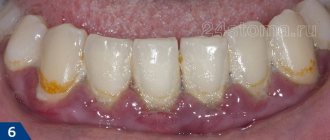
One of the types of gum inflammation is catarrhal gingivitis. The disease develops due to the adverse effects of external and internal factors and is characterized by the following symptoms:
- gums become red, swollen, itchy;
- gingival papillae swell;
- When chewing hard food and brushing your teeth, blood appears;
- there is an unpleasant taste in the mouth;
- gums hurt when pressed;
- there is a general feeling of malaise.
With catarrhal gingivitis, the teeth remain stable. But if, in addition to the symptoms described above, their mobility is observed, it means that periodontitis has already begun.
Types of catarrhal gingivitis
The disease can be local or generalized. In the first case, the gums are inflamed in the area of one or two teeth, in the second, both jaws can be affected at once.
Downstream in dentistry there are two forms:
- Acute catarrhal gingivitis. Usually it occurs once and, if professional treatment is carried out in time, does not recur. This disease often develops in young children during teething; it also often accompanies infectious diseases (diphtheria, measles, acute respiratory infections, etc.). Sometimes the symptoms of acute catarrhal gingivitis precede the appearance of herpetic stomatitis or are a reaction to any allergen. In the absence of medical intervention, the disease progresses to the next (chronic) stage.
- Chronic catarrhal gingivitis. This form is characterized by a sluggish course with periodic exacerbations. The gums are constantly inflamed, bluish in color and bleed at the slightest injury. There is a feeling of swelling in the gums, and sometimes an unpleasant odor appears from the mouth. With exacerbation of chronic catarrhal gingivitis, these complaints intensify.
Symptoms
The symptoms of the disease depend on the degree of inflammation. Redness of the gums that does not go away for a long time is the first “bell” to visit the dentist. Swelling of the gums, bleeding gums, bad breath, pain and burning are all signs of the disease.
Already during the initial examination, the doctor will notice a change in the color of the gums, the presence of plaque or tartar.
In severe cases of the disease, the temperature may rise, the body's condition may deteriorate, and drowsiness may occur.
Diagnostics
As a rule, dentists diagnose the disease during the first examination. To clarify the diagnosis, the following methods are used:
- Schiller-Pisarev test - assesses how rapidly the inflammatory process progresses;
- Silnes and Loe index, Green and Vermilion index - determines the density of dental plaque;
- probe test - evaluates bleeding.
It is also possible to use the following techniques: biopsy, study of soft tissues, rheoparodontography, analysis of intercellular fluid from the gums.
Treatment
Treatment of catarrhal gingivitis begins with eliminating external factors that maintain the inflamed state of the gums. We perform replacement of fillings and dentures, removal of plaque and tartar. In most cases, this is enough to get rid of the disease.
If these measures do not help to completely get rid of the disease, medication treatment comes into play. These may include antibiotics, anti-inflammatory drugs and multivitamins. If the development of the disease is associated with general diseases of the body, it is necessary to contact specialized specialists.
An effective method of treating chronic catarrhal gingivitis is gum massage. Blood flow speeds up metabolism, which contributes to a rapid improvement in their condition.
Treatment of acute gingivitis, like any other form, includes the above procedures and training by specialists in proper gum care. Which is important to avoid recurrence of the disease.
Prevention
To prevent recurrence of the disease, you must follow the care recommendations:
- regular use of dental floss;
- rinsing the mouth with antiseptics;
- thorough cleaning of the oral cavity, preferably using specialized toothpaste;
- avoiding injury to the oral cavity.
If symptoms appear, you must immediately contact the clinic for timely diagnosis and treatment. Mild forms of gingivitis are much easier and less expensive to cure.
Cost and duration of treatment
The cost of treatment directly depends on the degree of advanced disease. This includes:
- the number of procedures that will need to be performed during treatment;
- medications used to treat the causes of gingivitis;
- possible replacement of fillings and dentures.
Gingivitis is a very unpleasant disease that is almost impossible to cure at home. Only a dentist can correctly diagnose and prescribe effective treatment.
At the first signs, contact the clinic to eliminate the unpleasant symptoms and causes of catarrhal gingivitis. Call to make an appointment.
Treatment of catarrhal gingivitis
When treating catarrhal gingivitis, it is important to urgently eliminate local factors that support the inflammatory process. At the Doka-Dent clinic in Moscow, therapy includes:
- removal of dental plaque;
- treatment of caries;
- replacing old fillings;
- plastic surgery of frenulum (according to indications);
- re-prosthetics, etc.
Sometimes these measures are enough to stop inflammation.
Local drug therapy can also be included in the treatment - periodontal applications, rinsing with antiseptic solutions, applying medicinal dressings to the gums, etc. In some cases, multivitamins and anti-inflammatory drugs are prescribed for oral administration.
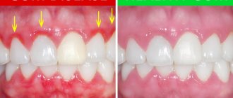
Acute and chronic gingivitis require mandatory treatment. Don’t wait for the problem to worsen, come to the Doka-Dent clinic. Qualified dental care gives a positive result, proof of this is the reviews of our patients and photos before and after treatment.
Clinical examination
In case of transition to a chronic form :
Mild severity (I): 1st dispensary group - examination by a doctor once a year.
Moderate severity (II): 2nd dispensary group - examination by a doctor 2 times a year.
Severe degree of disease (III): 3rd dispensary group - examination by a doctor 3 times a year.
If the treatment is effective: POSSIBLE RESULTS
- no complaints of pain in the gums;
- no bleeding gums;
- gums pale pink;
- dense;
- painless on palpation;
- There are no dental deposits.
If the treatment is ineffective: POSSIBLE RESULTS
- bleeding gums continue;
- the gums are swollen;
- the presence of dental plaque is noted;
- further destruction of the dentogingival attachment is possible;
- formation of periodontal pockets;
- atrophy of the alveolar bone - the occurrence of localized or generalized periodontitis.
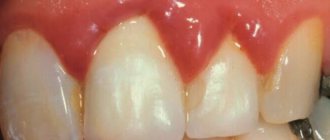
Gums are normal
Normal gums have a characteristic appearance. It is convenient to describe it using several simple criteria:
| Sign | Norm |
| Color | Pale pink, coral, salmon (if we are talking about fair-skinned people) |
| Surface | Lumpy (“lemon peel”) |
| Circuit | Pointed or trapezoidal interdental papillae |
| Consistency | Dense |
| Bleeding | No |
A few words about each of the criteria:
Gum color
The color of the gums is noticeably different from the color of the mucous membrane of the alveolar process. The alveolar mucosa is more red, smooth and shiny. All due to the characteristics of its epithelium and connective tissue:
— the epithelium of the mucous membrane of the alveolar process is thin, non-keratinizing, there are no epithelial outgrowths;
- looser connective tissue contains more blood vessels.
(you can read more about the structure of the gums in the article “Structure of the periodontium”)
The black arrow indicates the gums, the white arrow indicates the mucous membrane of the alveolar process.
Gum surface
A lumpy surface is characteristic of the attached gingiva (the marginal gingiva is smooth). Microscopically, these are gingival depressions (arrows in the figure) and elevations due to the different arrangement of the connective tissue layer.
Gum contour
The contour, or shape of the edge, of the gum may be different. It can be more flat or, conversely, arched on the vestibular and lingual sides. This depends on the shape and inclination of the tooth crown: the more convex the crown, the greater its inclination, the more pronounced the curvature of the gums.
The gum contour is flatter in the upper central incisors, more curved in the canines.
The interdental papillae are also different in shape: the less space there is for them (the teeth are denser), the “sharper” they are; the more, the, on the contrary, they are wider:
Wide interdental papilla Pointed narrow interdental papilla
Gum consistency
Healthy gums are dense in consistency. This is all thanks to the abundance of strong collagen gingival fibers in its connective tissue.
Gingival fibers
Bleeding gums
There are also vessels there that are not pathologically changed. That is why there is no bleeding of the gums when brushing your teeth at home or probing them by a dentist.
| Gum probing |
Now let's figure out how the gums change when inflammation occurs - gingivitis. First of all, why does it arise?
Prevention
Prevention is quite simple and everyone, if desired, can protect themselves from inflammatory gum diseases. You just need to follow a few simple rules:
- using electric toothbrushes;
- maintaining oral hygiene (toothpastes, gels, rinses);
- to give up smoking;
- dental gels with metronidazole, for example Metrogyl Denta;
- ambazon, 2,4-dichlorobenzyl alcohol, amylmetacresol in the form of lozenges;
- use of toothpastes with triclosan;
- use of calcium supplements;
- rinsing the mouth with solutions containing chlorhexidine, hydrogen peroxide, ethanol, thymol, cineol, methyl salicylate, menthol, methylparaben, benzalkonium chloride, fluorides and xylitol. Recent scientific research has also shown the beneficial effects of mouthwashes containing essential oils.
The above preventive measures are also used for treatment. The use of dental floss is not advisable.
Diagnostics
Gingivitis must be distinguished from periodontitis and periodontal disease. The main feature that distinguishes gingivitis from other periodontal diseases is that the inflammatory process affects only the gum tissue, the remaining structures (periodontal ligaments that hold the tooth in the jaw and bone tissue) remain unchanged. Normally, the depth of the dentogingival junction is 1-1.5 mm; with the destruction of the dentogingival junction, periodontal pockets (4 mm or more in depth) are observed, which are a sign of periodontitis.
With gingivitis, there are no periodontal pockets, but there are false periodontal pockets that form with the hypertrophic form of gingivitis, and in general with any swelling of the gums. Along with this symptom, gingivitis is not characterized by periodontal pockets, exposure of the necks of the teeth, or their mobility - these signs indicate damage to the bone apparatus. For differential diagnosis, radiography is used - changes in the height of the interalveolar septa are not typical for gingivitis.
Among the advanced technologies, genetic testing can be noted, which can show all the risks by performing a test for genetic polymorphism in individuals predisposed to increased production of interleukin-1 (IL-1), which is a mediator of inflammation.