Causes of gingivitis
After eating food, plaque forms on the teeth. In the absence or insufficient oral hygiene, the plaque becomes denser and bacteria begin to actively multiply in it. Pathogenic microorganisms penetrate the gums and cause inflammation. There are many provoking factors that increase the risk of developing the disease.
External reasons include:
- insufficient hygiene, infections - inflammatory processes occur due to food debris, bacteria from dirty hands, toothbrushes, toys, pacifiers, spoons, in the presence of childhood infectious diseases - scarlet fever, chickenpox, rubella and similar pathologies;
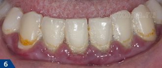
- tartar is a compacted plaque that is deposited in periodontal pockets, injuring and infecting soft tissues;
- advanced caries is a source of infection;
- poor-quality dental treatment - injuries to the mucous membranes, incorrectly installed fillings, dentures (bulge and injure the gums), insufficient antiseptics, implant rejection;
- injury to soft tissues when cleaning with hard brushes or hard food;
- chemical injuries from medications, hot spices, food acids, and other substances;
- poisoning with heavy metals (lead, mercury, etc.);
- cigarette smoke reduces the level of local immunity, impairs tissue nutrition, salivation, and acts as an irritant;
- impaired nasal breathing contributes to dry mucous membranes, which means increases the risk of infection.
Pathology can be caused by internal causes:
- anomalies in the development of teeth, jaws, malocclusion - injuries to the mucous membranes when chewing food;
- eruption of teeth - injury from the inside;
- diseases of the gastrointestinal tract, helminthic infestations lead to a deficiency of nutrients, vitamins, decreased salivation, and immunopathological reactions;
- vitamin deficiency, especially lack of vitamins C, E, A, group B;
- decreased immunity, chronic respiratory diseases, hepatitis, immunodeficiency, tuberculosis;
- diabetes mellitus, hormonal disorders during pregnancy, thyroid disease, decreased production of sex hormones.
The answer to the question whether gingivitis is contagious cannot be unambiguous. Inflammation in the oral cavity is most often provoked by staphylococcus, streptococcus, candida fungi, herpes virus, and E. coli. However, provoking factors are required for the development of the disease. If a person has a strong immune system and regularly takes care of his teeth, then pathology will not arise.
Symptoms and signs
The pathology can be easily detected by traces of blood on the toothbrush - the main sign of gingivitis. The more pronounced the inflammatory process, the more severe the bleeding of the gums. In advanced cases, bleeding can occur even without mechanical impact, especially after waking up from sleep. Depending on the type of disease, symptoms may vary.
The disease is characterized by:
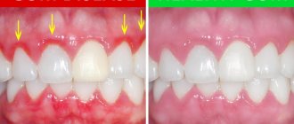
- swelling, redness, swelling, bleeding gums;
- pain due to mechanical impact, eating hot, cold or spicy food;
- unpleasant odor that cannot be eliminated by hygiene products;
- ulcers, ulcers (for some types);
- symptoms characteristic of intoxication - fever, nausea, loss of appetite, weakness.
If left untreated, gum pockets may form. The infection penetrates deeper, affecting the periodontium. This threatens the development of severe complications and tooth loss.
Symptoms of catarrhal gingivitis
The first symptom, characteristic of both acute catarrhal gingivitis and its chronic form, is bleeding gums. It occurs at the slightest pressure or injury, and brushing your teeth cannot be done without it. Eating food, even soft foods, also causes bleeding. Many patients constantly feel the taste of blood in their mouth.
Acute catarrhal gingivitis can also be suspected if there is severe redness and swelling of the gums, pain in the gum area. In the vast majority of cases, the pathogenesis of gingivitis is limited to this. But sometimes an acute process can cause fever, muscle pain, weakness, lethargy, etc.
If you do not take action and hope that the unpleasant sensations will somehow go away on their own, it is likely that instead of acute, chronic catarrhal gingivitis will develop. Pain and bleeding will become constant, bad breath will appear, the gums will gradually begin to change shape and structure and gather in ridges at the edge. In this case, the formation of tartar increases.
If gingivitis occurs in one place, as a result of abnormal crowding of the teeth or improper installation of the prosthesis, but the patient does not go to the dentist, over time, generalized gingivitis may occur throughout the jaw.
Diagnostic methods
A visual examination by a dentist may be sufficient to assess the disease. If symptoms are severe, the doctor may refer the patient for an x-ray. This is necessary if infection of the pulp, periodontal and bone tissue is suspected.
In the presence of ulcers or chronic inflammation, a microbiological analysis is performed, which identifies the type of bacteria and their resistance to antibiotics. If a generalized form is diagnosed, then additional diagnostics are carried out by relevant specialists: ENT doctor, gastroenterologist, immunologist, and other specialists. The patient donates blood for general tests, sugar, HIV infection, syphilis.
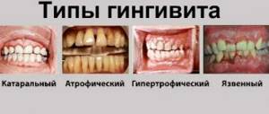
Types of gingivitis
The disease is classified according to its course, localization of the inflammatory process, severity, pathological changes in tissues, and causes of development. How to treat gingivitis depends on the type of pathology, so differential diagnosis is important.
Forms for localization of pathology
For correct diagnosis and correct treatment, an important indicator is the prevalence of the disease.
Gingivitis can be:
- Localized - inflammatory processes affect only a small area of soft tissue near one or several teeth.
- Generalized - all gums and mucous membranes of the mouth become inflamed. Often associated with other infectious and systemic diseases.
Catarrhal gingivitis
It can be acute or chronic. Serous inflammation of the gums - formation of exudate, swelling, hyperemia, bleeding, pain. The most common form of pathology - up to 90% of all clinical cases.
Acute form
It is more often diagnosed in children, adolescents, and young people under 30 years of age. It develops quickly and has pronounced symptoms. The gums bleed heavily, become painful, become loose, and occasional small ulcers may appear. After treatment they recover completely. If left untreated, the disease may become chronic.
Acute necrotizing ulcerative gingivitis
The ulcerative-necrotic form is otherwise called Vincent gingivitis. Has a severe course. The symptoms are pronounced. The disease is accompanied by necrosis of the interdental papillae, increased temperature, enlarged regional lymph nodes, and symptoms of intoxication. Separately classified according to ICD-10.
Chronic form
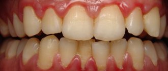
Inflammatory processes have a sluggish course, symptoms are mild or absent. Bleeding occurs regularly when brushing your teeth. May be accompanied by an unpleasant odor and metallic taste. During an exacerbation, the mucous membranes become red, swell, and pressure is felt from the inside. Usually there is a dense plaque and tartar on the teeth.
Types of chronic course:
- Desquamative - abundant desquamation of the epithelium of the gum mucosa, severe redness of the surface layer of soft tissue.
- Ulcerative - formation of ulcers, burning, itching, bleeding.
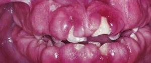
- Hyperplastic (hypertrophic) - the volume of the papillae increases, false pathological periodontal pockets are formed, blood and purulent contents are released, the mucous membranes acquire a purple-bluish tint. The gum grows, covering the crown of the tooth. Hyperplasia is diagnosed in three stages of severity; in the third, more than half of the crown is closed.
- Simple marginal - occurs more often than other species. It has standard clinical symptoms - bleeding, swelling of the gum tissue, pain when chewing, and unpleasant odor. Usually diagnosed in childhood with insufficient oral hygiene.
- Atrophic - characterized by reduction (dystrophy) of the gums. Symptoms are mild or absent, since no inflammation is observed. Over time, the interdental papillae disappear, exposing the neck and roots of the tooth. It occurs in old age, less often in young people with anomalies in the development of the dental system or incorrect orthodontic treatment.
Hormone-modulated gingivitis
Hormones and their ratio in the body do not directly affect the development of the disease. However, hormonal imbalances can become a provoking factor in the presence of dental plaque or lack of proper hygiene.
Depending on the causes of hormonal imbalances, there are:
- gingivitis in pregnant women;
- pubertal at puberty;
- when taking contraceptives.
The classification of gingivitis also includes types such as lymphocytic, plasmacytic, viral, herpetic and other varieties.
Pathogenesis of gingivitis
In the pathogenesis of gingivitis, there are several stages that transform into one another:
- initial stage;
- early stage;
- stage of stable damage;
- progressive stage.
The initial stage of gum inflammation develops in the first 2-4 days. Polymorphonuclear leukocytes begin to fight dental plaque microorganisms. Let me remind you that these include basophils, eosinophils and neutrophils. In our case, neutrophils predominate:
They penetrate from the blood vessels into the connective tissue of the gums, the epithelium of the gingival sulcus, and the attachment epithelium. They displace and compress the collagen fibers around the vessels, releasing collagenase, which destroys them. In response to the appearance of such a large number of immune cells, capillaries dilate and blood flow increases. And, as a result, more exudate and extravascular proteins are released from the gingival sulcus - the amount of gingival fluid increases.
This stage is also called subclinical gingivitis, because outwardly the gums look absolutely healthy. Pathological changes are visible at the microscopic level:
Neutrophils (NF) in blood vessels near the junctional epithelium (JE) and even between its cells. There are no neutrophils in the gingival crevicular epithelium (GED)
1 - healthy gums. Connective tissue (CT) contains fibroblasts, blood vessels, and dense collagen fibers. 2 – after a few days of plaque accumulation. Many immune cells in connective tissue, epithelium (CE). Dilated blood vessel (CV) in the center
The body's response determines how the initial stage will end. Recovery and return to a normal state or transition to the next stage - the stage of early damage.
The initial stage becomes early after about 1 week of plaque accumulation. Other immune cells - macrophages, lymphocytes - begin to migrate into the inflammation zone, forming an infiltrate. Around such infiltrates, as well as around vessels in the initial stage, collagen is destroyed:
The appearance of lymphocytes of small (ML) and large (BL) sizes. Light spaces around them are missing collagen
Moreover, its production by fibroblasts decreases. The worst conditions are for circular (4) and dentogingival fibers (1,2,3 in the figure below).
The connective epithelium and blood vessels grow: epithelial cords appear, between which there are capillary loops. Therefore, the color of the gums changes and erythema occurs. The first clinical sign also appears - bleeding gums.
Over time (according to various sources, from 2-3 weeks to even 1-2 months), the process enters the stage of stable damage (developed). This period can already be characterized as a chronic inflammatory process in the gums. Many immune cells of previous stages (neutrophils, macrophages, lymphocytes) are destroyed. From them there is only a trace in the form of expanded intercellular spaces, granular residues and lysosomes. They are replaced by others - B lymphocytes and plasma cells. The amount of collagen continues to decrease.
Plasma cell degeneration, cellular debris (debris)
Epithelium of the gingival sulcus with increased intercellular spaces and lymphocytes
The blood vessels change significantly: they are overloaded. As a result, their fragility increases, blood circulation in them slows down, and venous stagnation occurs. The color of the gums becomes darker and cyanotic. Plus, red blood cells leave the capillaries into the connective tissue, the hemoglobin in them breaks down, this enhances the staining.
Strands in the epithelial attachment can become very pronounced. So much so that, continuing to grow in the direction of the connective tissue of the gums, they can partially destroy the basal lamina (with the help of which the epithelium is attached to the connective tissue).
The stage of sustained damage can have several scenarios:
- With one of them, the process remains stable for several months or years.
- In the other case, it is more active and causes progressive destructive lesions.
- But the most favorable path is tissue restoration thanks to successful therapy. This happens due to the return of healthy periodontal microflora, which causes the inflammatory response of immune cells to disappear.
Some authors also distinguish stage 4 of gingivitis: progressive damage. But this is already periodontitis. This stage is left and considered, because gum inflammation with the involvement of other periodontal structures (ligament, bone) does not disappear anywhere. But this is a completely different story, so we’ll limit ourselves to three.
For clarity, we can summarize the results with a table:
| Stage | Time (days) | Blood vessels | Connective and sulcus epithelium | Predominant immune cells | Collagen | Clinical signs |
| Initial | 2-4 | Dilation, inflammation, increased permeability | Infiltration of polymorphonuclear leukocytes | PMN (mainly neutrophils) | Destruction around blood vessels | Increased amount of gingival fluid |
| Early | 4-7* | Proliferation | The same + epithelial strands, atrophy zones | Lymphocytes | Destruction around infiltrates | Erythema Bleeding (probe) |
| Persistent damage (developed) | 14-21* | Same + blood stagnation, fragility | Same + progression | Plasma cells | Progressive destruction | Changes in color, size, texture, surface, etc. |
* other authors note the duration of stage II is 1-3 weeks, stage III – 1-2 months.
I hope I was able to lift the veil of mystery about the occurrence and development of gingivitis. With this knowledge, it will be easier to understand where all the clinical signs, or symptoms, of gum inflammation come from.
The inflammation itself can occur and manifest itself in different ways. Due to these features, various forms of gingivitis are distinguished.
Treatment methods
With early diagnosis of the disease, gum health can be completely restored in 7 to 10 days. If gingivitis is not cured in a timely manner, long-term therapy will be required. Treatment methods depend on the type, severity of the pathology, causes of development, and age of the patient.
The complex of treatment measures includes:
- professional hygiene in dental clinics to clean teeth from deposits and stones;
- sanitation of the oral cavity to remove pathogenic microorganisms and their metabolic products;
- taking vitamins, antihistamines, antioxidants and other medications prescribed by a doctor;
- rinsing the mouth with solutions for antiseptics, relieving inflammation, accelerating tissue healing;
- surgical operations for hypertrophic, atrophic forms (excision or augmentation of gums);
- correction of hygiene procedures - replacing a hard brush with a soft one, consulting a dentist on proper oral care and nutrition.
For inflammatory gum disease, traditional medicine methods can and should be used. How to rinse your mouth with gingivitis? Doctors recommend using decoctions of chamomile, sage, calendula, and oak bark. If there are no herbs, you can prepare a soda solution. Rinse should be done after every meal.
Acute gingivitis
The diagnosis of “acute gingivitis” is based on the collected medical history, physical examination data, and the results of additional research methods. During an examination for catarrhal gingivitis, the dentist identifies hyperemic, swollen gums. The papillae are tense and bleed when touched lightly. The integrity of the dental epithelial attachment in acute gingivitis is preserved; there are no periodontal pockets. In the desquamative form of acute gingivitis, along with swelling and bleeding in the gum area, areas of desquamation of the surface epithelium are diagnosed.
Ulcerative gingivitis is characterized by the formation of ulcers. The gingival margin is corroded and the interproximal spaces are gaping. Regional lymph nodes in patients with the ulcerative form of acute gingivitis are enlarged. The Schiller-Pisarev test for acute gingivitis is positive. After applying iodine-containing preparations, the color of the mucous membrane changes from light yellow to dark brown. The intensity of staining correlates with the severity of inflammation. The PMA index in acute gingivitis is positive. When the interdental papillae are affected, PMA is 25%; when the marginal part of the gums is involved in the pathological process, PMA is 50%. If the PMA index is more than 50%, this indicates the presence of inflammatory changes in the alveolar gum area.
When assessing the level of hygiene in acute gingivitis, high numbers of Green-Vermillion indicators are revealed, which indicates an unsatisfactory hygiene index. On a targeted radiograph and orthopantomogram, there are no pathological changes in the bone tissue of the alveolar process. In the ulcerative form of acute gingivitis, using bacterioscopic examination, it is possible to detect in the surface layer of the mucosa, along with the resident microflora of the oral cavity, an increased content of fusobacteria and spirochetes. The deeper tissues contain exclusively pure culture of fusispirillary symbiosis. When conducting bacterioscopy in patients with the catarrhal form of acute gingivitis, an increased number of actinomycetes is detected.
It is necessary to differentiate acute gingivitis of infectious origin not only from periodontitis, but also from a secondary inflammatory process that develops with blood diseases, pathologies of the endocrine system, and disorders of the functioning of the gastrointestinal tract. To exclude symptomatic acute gingivitis, consultations with specialized specialists are indicated: hematologist, endocrinologist, gastroenterologist.