3123
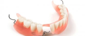
Edentia or absence of teeth in both jaws is a fairly common situation that can occur not only in elderly patients, but also in fairly young people.
This pathology requires immediate elimination, both due to the lack of aesthetics of the oral cavity and due to the possibility of developing a large number of complications.
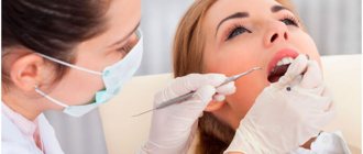
However, to choose the right treatment tactics, a specialist needs to correctly study the structural features of the patient’s jaw with missing teeth, which is greatly simplified when using the classifications of toothless jaws available in dental practice.
General overview
Classifications of edentulous jaws play an important role in dental science. They allow specialists to adhere to uniform terminology and features of determining existing anomalies in the structure of the dentofacial rows.
Thanks to generally accepted classification criteria developed by renowned scientists and doctors of medical sciences, orthopedic specialists are able to accurately plan further treatment and determine in advance what problems may be encountered during therapeutic measures.
Types and features
There is still no single comprehensive classification of toothless jaws. This is due to the fact that in addition to the marginal forms of jaws named in the known groups, there are many transitional types that have certain structural features.
Currently, the most popular are five groups of toothless jaws, named after the developers.
What is the purpose of relining a removable denture and the material used.
Let's calculate here how much dentures with suction cups cost.
At this address https://www.vash-dentist.ru/protezirovanie/semnyie-p/poliuretanovyie-zubnyie-idealnyiy.html we will discuss the pros and cons of polyurethane dentures.
According to Schroeder
According to Schroeder's classification, the upper jaw rows with missing teeth can be divided into three types, which is due to different levels of bone depletion in the alveolar area:
- I jaw involves insignificant thinning of the tooth-bearing area.
In this situation, the cusps of the jaw and the areas of the upper row, designed to hold the teeth, are clearly visible, and the palatine vault is deep. The folds of the mucosa and the areas of muscle attachment are located quite high. According to experts, this type of jaw row is the most desirable for installing a prosthetic structure, because its elements do not interfere with the attachment of artificial teeth. - Type II is recognized when there is an average level of thinning of the alveolar process and its not very clear expression.
The patient has moderate depth of the palatal plane. The transitional fold is shifted towards the alveolar ridge. When attaching a prosthesis for this type of jaw, there are risks of reducing the quality of its fixation as a result of spasms of facial muscles. - Type III is indicated by an excessive level of atrophy of the bony base of the jaw.
Alveolar ridges and tubercles are completely smoothed. The palate takes on a flat shape. The mucosal fold is located low in the same plane as the palate. When placing dentures, this jaw shape causes the most difficulties, which is associated with high mobility of the structure as a result of the anatomical features of the elements of the dentition.
According to Keller
According to experts, the lower jaw causes more difficulties in prosthetics than the upper jaw. This is due to its anatomical and physiological characteristics.
To simplify the process of restoring elements of the lower jaw row, the Keller classification was developed, which assumes that the patient may have one of four types of jaw:
- The first type of lower dentition suggests slight atrophy and equal smoothing of the alveolar parts.
This creates an ideal basis for fixing the prosthetic structure and prevents it from moving forward and in different directions.The attachment of the folds of the mucous membrane and muscles is located in the base of the alveolar region.
Dentists note that this option occurs quite rarely in patients, mainly with the simultaneous extraction of teeth and a slow process of thinning of bone tissue.
- Second jaw shape characterized by a uniform and clearly defined atrophic process occurring in the alveolar area.
The ridge stands out slightly against the background of the floor of the oral cavity, but has a rather sharp surface, which complicates the procedure for fixing the prosthesis.The muscles in this case are attached in the area where the alveolar ridge is located. Due to the anatomical structure of the jaw, the use of a prosthesis often causes pain and discomfort due to the possibility of its displacement.
- Third jaw type dentists identify in patients with early extraction of lateral teeth. It is characterized by thinning of the alveolar process in the area of premolars and molars, while maintaining the volume of bone tissue in the central sections.
Prosthetics with this classification option is considered acceptable, since in the lateral sections of the dentition there are smooth surfaces suitable for fixing artificial molars.In addition, maintaining the alveolar cusp in the central section prevents artificial teeth from sliding forward under load during chewing.
- The fourth form of jaws without teeth according to Keller’s classification involves severe atrophy of the alveolar area in the area of the frontal incisors.
At the same time, in the lateral areas of the dentition, bone tissue is preserved much better. Fixation of the prosthesis in this case is not very reliable, since the structure may lose stability and shift.
According to dentists, fixing the prosthesis on the lower jaw is acceptable for each of the Keller classification options, however, for the second and fourth types of dentition, it causes many difficulties associated with the structure of the oral cavity.
According to Oksman
The famous Soviet doctor of medical sciences I.M. Oksman presented his own version of the classification of the upper and lower jaw rows, in which all teeth are missing.
In his opinion, the upper dental line can be divided into the following types:
- The first type involves the presence of a high alveolar process and tubercles. The surface of the palate in this version is clearly defined, the muscles are attached quite high.
- In the second type, the decrease in bone thickness occurs evenly and is much more noticeable. The palate has less depth than in the previous version, and the mucosal surface membrane is attached to the central sector of the alveolar part.
- The third type of jaw has a significant rate of atrophy of the alveolar region, which occurs evenly in all its areas. The palatal surface appears flat, and the mucous membrane is fixed on the ridge.
- The fourth type corresponds to unmeasured atrophy of the alveolar areas of the upper jaw. Pathological signs of changes in the dentition include the previous three types.
The edentulous lower jaw has 4 varieties based on the stage of bone atrophy. Each species has characteristic anatomical features:
- First type. The alveolar process has a large height, the fold of the mucous membrane and the areas of attachment of the frenulum are located low.
- Second type. The change in the density of the alveolar tissue occurs evenly and has an average degree of severity.
- Third type. The alveolar part is practically not expressed or completely absent. The jaw itself is often deformed.
- Fourth type. Thinning of the bone develops spasmodically in different parts of the row as a result of tooth extraction scattered over time.
Clinical and laboratory stages of manufacturing a clasp prosthesis and methods of fixation.
In this publication we will talk about the features of making removable dentures.
Here https://www.vash-dentist.ru/protezirovanie/semnyie-p/tselesoobraznost-primeneniya-mikro.html all the most important things about nylon microprostheses.
According to Kurland
Classification developed in 1953 by V.Yu. Kurlyandsky, takes into account not only the level of reduction in bone tissue thickness during edentia, but also the change in the location and fastening of muscle tissue.
According to this systematization, four groups of toothless jaws were identified:
- Group 1 involves protrusion of the alveolar process above the level of muscle fixation;
- Group 2 is characterized by thinning of the bone tissue in the area of the process and body of the jaw, as well as their placement at the level of muscle attachment;
- Group 3 indicates severe atrophy of the jaw areas located below the muscle attachment site;
- Group 4 involves thinning of the bone in the area where molars and premolars were previously located;
- Group 5 atrophic process affects the bone tissue at the location of the front teeth.
According to Doynikov
The classification of toothless jaws according to Doynikov echoes the grouping proposed by Schroeder, but has some differences based on the uneven thinning of areas of bone tissue:
- 1 type On both jaws there is a clear expression of the alveolar processes and ridges. The mucous membrane is located evenly on the palatal plane and has good compliance. The folds of the mucous membrane are located a short distance from the top of the ridge.
- Type 2 The patient is diagnosed with a moderate degree of atrophy of the jaw tubercles. The depth of the palatal plane is slightly reduced compared to the previous form, and the torus is quite well defined.
- Type 3 The alveolar areas of the dentition are not visible, the size of the jaw body and tubercles is sharply reduced compared to the normal variant. The palate is flat and the torus is quite wide.
- Type 4 The severity of the alveolar process is observed only in the frontal region of the dental line. The lateral areas are subject to severe atrophy.
- Type 5 The anterior region of the jaw is susceptible to atrophy, while in the lateral areas the bone density is maintained.
Schroeder classification
According to this scientist, the classification of toothless, namely the upper jaws, can be made according to the degree of reduction of the part of the jaw on which the teeth are located. He identified several types, let's consider them.
- The very first, experts consider this case the most suitable for installing a prosthesis. In this case, there are practically no physical abnormalities that would prevent the installation of a prosthesis. An undoubted advantage is that the jaw tubercles are very well defined.
- With this type, according to the scientist, the arch of the palate is quite clearly expressed and it was still possible to preserve the alveolar tubercles and process. But in the described case, the transitional fold will be very close to the top of the process, and if the muscles of facial expression are overstrained, the prosthesis will most likely move.
- Sharp atrophy will be the main characteristic feature of the third type. The palatal bottom is completely flat, and the fold is located in the same plane with the palate. Installing dentures on this type of jaw is the most difficult.
Classification of toothless jaws according to Schroeder. Visualization.
The anatomical features of the jaw of the lower row are very different from the jaw above. Making and using prosthetics is therefore a little more difficult.
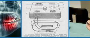
Impressions
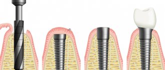
The impression is a reverse imprint of the surface of the soft and hard tissues of the oral cavity, which are located in the area of the prosthetic bed.
Their implementation contributes to the production of diagnostic and working models, which serve as the basis for casting prosthetic structures.
There are several types of prints.
Anatomical
It is removed using standard impression trays and a large amount of dental plaster. Has high edges.
Functional tests are not used in this case, as a result of which the condition of the tissues bordering the prosthetic bed is not taken into account.
Functional
To make this type of impression, a personal spoon and special functional tests are used, with the help of which the mobility of the folds of the mucous membrane is reflected.
The edges of the impression are slightly lower than those of the previous type, and the border of the manufactured prosthesis covers the mucous membrane by no more than 2 mm.
Based on pressure on the oral mucosa, functional impressions are divided into three types:
- unloading – removed using a plaster mass using minimal pressure on the mucous membrane;
- compression - used when the mucous membrane is highly pliable, and is performed under pressure using silicone, gypsum or thermoplastic mass;
- combined - allow you to compress areas of the mucosa with high compliance, without overloading areas with low compliance.
Mucous membrane of the prosthetic bed
During preparation for prosthetic replacement of a toothless jaw, in addition to its type, specialists pay attention to the characteristics of the mucous membrane located in the prosthetic bed.
There are three main types of mucous membrane:
- Normal has moderate pliability and a high degree of moisture. The color of the mucous membrane is light pink. This option is optimal for prosthetics.
- Hypertrophied has increased friability and a high content of intermediate substances. It is characterized by a good degree of moisture, however, due to increased compliance, mobility of the fixed prosthesis is often observed.
- Atrophied - has a high density and a low degree of moisture. The color is usually whitish. On the maxillary process, the mucous membrane is attached to the periosteum. This option is the most unsuitable for prosthetics.
Doynikov classification
The scientist's classification is based on the unevenness of atrophy. This classification system is very similar to the classifier that Schroeder proposed to use.
Doynikov identified 5 features of the classification:
- Both jaws have ridges that are pronounced and also have alveolar processes. The mucous membrane is very pliable, and it covers the palate evenly.
- The jaw mounds have an average degree of destructiveness. In this case, the average and depth of the sky.
- There is no alveolar segment and process. The palatal floor is quite flat.
- Only in front there is an alveolar process, and the lateral areas have significant atrophy.
- The alveolar ridge is very noticeable in the lateral parts, and in the front there is severe atrophy.
This classification is very convenient for the doctor because a large number of cases are covered. But specialists working in our time use all types of classifications in their work.
conclusions
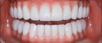
Dentists are unanimous in their opinion that in case of complete edentia, prosthetics should not be delayed . Long-term absence of teeth over time leads to irreversible changes in the anatomy of the dentofacial rows:
- thinning of bone tissue;
- changes in pliability and complete atrophy of the mucous membrane;
- disorders in the functioning of the temporomandibular joint;
- development of inflammatory processes in the oral cavity;
- impossibility of proper nutrition;
- violation of diction;
- deformations of facial tissues and muscles.
Therefore, dentists note that one of the guarantees of restoring the aesthetics and functionality of the dentition is regular preventive examinations, which allow timely identification of existing violations and elimination of them.
If you find an error, please select a piece of text and press Ctrl+Enter.
Tags classification of toothless jaws prosthetics removable dentures
Did you like the article? stay tuned
Previous article
Methods for identifying and eliminating senile progeny
Next article
The mechanism of formation of primary occlusion and possible deviations
Classification according to Courland
There is also the case that jaws are separated using the Kurlyandsky method. He applied his classification based on the reduction in bone tissue, but also due to the changes that occurred in the place where the muscles are attached. The scientist identified 5 types of atrophy of the lower jaw.
- In the first case, patients are identified whose process protrudes further than the place where the muscles are attached.
- The location of the alveolar process at the same level together with the place of muscle attachment.
- Atrophy of parts that are lower than the places where the muscles are attached.
- The bone tissue becomes thinner where the chewing teeth were (in the lateral area).
- Damage to the tissues of the places where the front teeth were present.