Effective treatment of stomatitis in adults
Treatment of stomatitis in adults largely depends on the type of this disease, as well as concomitant diseases that provoke the appearance of ulcers and inflammation in the mouth. The most interesting thing is that the diagnosis of stomatitis is still carried out visually: the patient undergoes tests only if other, more serious diseases are suspected. Effective therapy for stomatitis is possible, but only if the type of disease is correctly determined. Recovery usually occurs within one or several weeks, but there are also more severe forms when long-term rehabilitation is required for complete recovery. Let's look at the main types of disease and drugs for the treatment of stomatitis in adults.
Free consultation on the cost of treatment in our dentistry
Leave a request and the clinic administrator will contact you within 15 minutes!
The cause of herpetic stomatitis can be not only a malfunction of the body’s general immunity, but also a deterioration of local immunity, in which the oral mucosa becomes sensitive to the effects of pathogenic microflora. Local immunity is reduced under the following circumstances:
- With poor oral hygiene, due to which hard and soft plaque begins to accumulate on dental surfaces and soft tissues;
- For untreated dental diseases;
- For tonsil infection;
- When you inhale air not through your nose, but through your mouth. It leads to dehydration of the mucous membrane, which is harmful because bacteria will penetrate the tissue more easily and quickly. Treatment methods
Since this form of stomatitis is caused by the herpes virus, the basis of treatment for the disease should be special antiviral drugs. However, these remedies show the greatest effectiveness in the initial stages of the development of the disease, until the blisters turn into ulcers. In the later phases of development, treatment with antiviral drugs will be ineffective.
Treatment of herpetic stomatitis
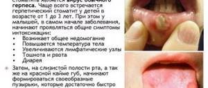
Treatment of stomatitis after detection can be carried out many times (once a year or more often), since once settled in our body, the herpes virus remains there for life. Herpes stomatitis is characterized by the accumulation of reddish transparent blisters on the lips and inside the mouth, so it is quite easy to detect. Especially often, herpetic stomatitis manifests itself against the background of decreased immunity, vitamin deficiency, hypothermia and stress. Remember that herpetic stomatitis in adults is contagious, so you should avoid tactile contact.
Types of drugs
- Antiviral agents.
- Immunostimulants (as a preventative measure).
- Ointments (including to eliminate visual manifestations of the disease).
Stomatitis - symptoms and treatment
During diagnosis, it is very important for the doctor to collect a detailed medical history. Ask the patient about concomitant and past diseases, find out the allergy status. Stomatitis often manifests itself in diseases of the gastrointestinal tract (gastritis, colitis), diabetes, HIV infection, after radiation therapy, and bronchial asthma. It is also important to find out whether the patient is worried about this disease for the first time or whether he has already encountered such a problem. If this is not the first time the patient has consulted a doctor or has tried to undergo treatment on his own, then it is necessary to ask what means and medications were used for the treatment and whether it brought results.
When examining the oral cavity, it is necessary to pay attention to dental plaque, the presence of sharp edges of crowns, pathological abrasion of teeth, and the condition of orthopedic structures. All this can be a source of chronic or acute injury to the mucous membrane. Aphthae occur at the site of exposure to a traumatic factor.
Candidal stomatitis can be diagnosed by characteristic clinical signs, as well as by the results of laboratory tests. First of all, a microscopic analysis of the material taken from the lesion is carried out, its seeding is carried out taking into account the degree of contamination (infection) of the affected tissue [5]. Fungi of the genus Candida are found in large quantities in the smear. They also do intradermal tests with a yeast allergen and carry out serological studies (detection of certain antibodies or antigens in the blood serum of patients) - agglutination reactions (gluing together of antigens with the help of antibodies) and complement fixation reactions (an immunological reaction for determining antibodies by a known antigen or antigens by a known antibody) [4].
For viral stomatitis, diagnosis is based on the use of special studies:
- virological - to determine a specific virus in the blood;
- serological - to determine antibodies in the blood to certain microorganisms;
- cytological - to determine the structure of cellular elements;
- immunological.
Changes characteristic of an acute inflammatory process are detected in the blood. Histological examination reveals acantholysis of epithelial cells (a degenerative change in the spinous layer), and an acute inflammatory process is expressed in the underlying mucous membrane [5].
With herpetic stomatitis in the first days of the disease, as well as during relapses, the herpes virus is easily isolated from the contents of the vesicles [5]. At the beginning of the disease, antibodies to the virus are not detected. As symptoms worsen, the number of antibodies to the virus increases. After clinical recovery, the herpes virus remains in the body, as a rule, for life [4].
Differential diagnosis
It is necessary to differentiate a traumatic ulcer from ulcers due to tuberculosis, syphilis, and neoplasms. It is important to know that with prolonged chronic injury, the transition of a traumatic ulcer into a malignant neoplasm is possible. If, after eliminating the traumatic factor, rapid healing occurs, then this is a traumatic ulcer. In a malignant course, the neoplasm is painless and does not heal after eliminating the traumatic factor. Cytological and histological studies reveal atypical changes in epithelial cells.
With a tuberculous ulcer, Mycobacterium tuberculosis is found in the scraping. Hard chancre (ulcerative formation) in syphilis differs from a traumatic ulcer by the presence of a dense infiltrate around the ulcer, smooth edges, a smooth bottom, enlargement and hardening of regional lymph nodes (scleradenitis) and the absence of pain symptoms [7]. Histological examination reveals Treponema pallidum (the bacterium that causes syphilis).
Treatment of candidal stomatitis
A type of stomatitis that is caused by a special type of fungus – Candida albicans. Very often, candidal stomatitis begins with glossitis, which is why its second name is stomatitis on the tongue. Treatment in this case requires fairly prompt treatment, since in adults it is quite painful and is accompanied by a burning sensation in the mucous membrane and sore throat. On the inner surface of the lips and cheeks, as well as on the tongue, characteristic foci of inflammation appear, covered with a white cheesy coating.
Treatment methods
- Treatment of common diseases (if detected).
- Antimicrobial therapy.
- Preventive rinses.
Symptoms of stomatitis
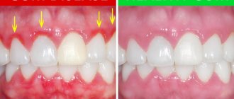
Light redness of the oral mucosa is the first symptom of stomatitis. Over time, they swell and a burning sensation appears. If treatment is not started at this stage, the redness is replaced by small oval or round ulcers, grayish or white, with a red halo and a film on top. Moreover, the tissue around them looks absolutely healthy. Mouth ulcers are very painful and make eating difficult. They appear on the inner surface of the cheeks and lips, under the tongue. In most cases, mild stomatitis manifests itself as one ulcer.
The appearance of several ulcers of larger size and depth, which sometimes merge into one, are signs of more severe forms of stomatitis. The appearance of ulcers is accompanied by fever, inflammation of the lymph nodes, general deterioration of health, headache, loss of appetite and constipation. Acute stomatitis is accompanied by severe pain in the mouth, which interferes with eating and speaking. In addition, there may be excessive salivation, a coating on the tongue, a bright red mouth, irritability, and vomiting after eating.
Treatment of aphthous stomatitis
A distinctive feature of this type of stomatitis is the appearance of so-called aphthae on the oral mucosa. These are small round ulcers with redness at the edges, the touch of which causes unpleasant painful sensations. Often occurs as a result of the activity of microorganisms (in particular, staphylococcus). Treating aphthous stomatitis at home is only recommended if it is not caused by a more severe illness. Often aphthous stomatitis occurs with problems with the immune system, liver and gastrointestinal tract.
Treatment methods
- Allergy therapy.
- Antiseptic therapy (treatment of aphthae with anti-inflammatory solutions, gels and ointments).
- Dental treatment and sanitation of the oral cavity (after dental treatment, stomatitis occurs less frequently than in the presence of carious lesions).
- Immunotherapy.
- Diet.
Diagnostics
The clinical picture of the disease is very characteristic. The oral mucosa is pale, with a bluish tint, dilated blood vessels are clearly visible, and there is a small amount of discharge on the mucous membrane, reminiscent of thick saliva. Necrotic films of dirty white or yellowish color are located in various places on the mucosa. With necrotizing stomatitis type I, necrotic films are easily removed with a cotton swab or tweezers, the mucous membrane does not bleed. With type II stomatitis, necrosis is deep, dead tissue sits tightly on the mucous membrane, which bleeds heavily even at the slightest touch. In severe cases, abundant purulent discharge, deep ulcers appear, muscles are exposed, bones are destroyed, and teeth fall out.
Treatment of ulcerative stomatitis
Ulcerative stomatitis very often occurs due to poor oral hygiene and bad habits (especially smoking). Often this type of stomatitis at its onset is confused with periodontitis and gingivitis, since it is characterized by inflammation of the gums, a grayish coating around the teeth and death of soft tissues. Often accompanied by fever and fever. One of the few forms of stomatitis, which often requires treatment by a doctor, and in the most advanced cases, surgical intervention.
Treatment methods
- Antibiotic therapy.
- Antibacterial treatment of affected areas.
- Surgical intervention (in severe cases, removal of areas of dead tissue and gum grafting).
What is stomatitis?
Stomatitis is an inflammation of the oral mucosa; in English literature it is often called “mouthrot” - rotting of the mouth. The process can be local or generalized, acute or chronic.
Causes of stomatitis in snakes.
Stomatitis is not a primary disease and develops against the background of immunosuppression caused by: - hypothermia and the stressful state of the animal; — hypovitaminosis caused by poor, improper nutrition (primarily a lack of vitamin C, then A); — other diseases that reduce the animal’s immunity. Often stomatitis begins with mechanical injuries to the oral cavity (snake hitting the head against glass when throwing, damage caused by food rodents, etc.).
Signs by which the presence of the disease can be determined.
- at the beginning of the disease, the oral mucosa is pale, with a slight bluish tint, and there is slight swelling; - on the surface of the mucous membrane a small amount of discharge is visible, resembling thick saliva. In the future, the discharge will acquire a purulent character (pus in reptiles most often has the appearance of cottage cheese with a slight yellowish tint, in advanced stages it is mixed with blood). - Focal stomatitis often spreads in the submucosal layer between the gum and skin or throughout the submucosal layer of the pharynx and tissues of the neck and head, leading to oropharyngeal cellulitis (the “edematous” form of stomatitis), characteristic of snakes and monitor lizards. - inflammation of the oral mucosa in snakes is often accompanied by refusal of food, general exhaustion and lethargy. The photo below shows classic stomatitis. The characteristic shade of the mucous membrane and curdled purulent discharge are clearly visible.
What happens if stomatitis is not treated?
Bacterial lesions in reptiles, as a rule, do not go away on their own, and untreated stomatitis will eventually turn into gingivitis (inflammation of the gums), oropharyngeal cellulitis (inflammatory swelling of the submucous membrane of the head and neck) and osteomyelitis (inflammation of bone tissue), which often ends fatal.
The photo below shows a reticulated python with osteomyelitis during treatment:
How to open a snake's mouth
In order to open the snake's mouth, you will need a medical spatula or something similar to it: a plastic card or a ruler. With our left hand we fix the head, with our right hand we carefully insert the “tool” under the upper lip of the snake and move it into the mouth, holding it horizontally and shaking it. Having sensed a foreign object, the snake itself will open its mouth.
How to treat stomatitis in snakes.
Treatment of stomatitis consists of treating the oral cavity and (in some cases) administering medications that affect the entire body (vitamins or antibiotics). Dead tissue of the oral cavity, blood clots and pus are removed with a gauze swab soaked in hydrogen peroxide, after which the mucous membrane is irrigated with some antiseptic (an aqueous solution of furatsilin, potassium permanganate, etc.) Then the mucous membrane is treated with an antibacterial drug. In this case, a 0.5% solution of dioxidine in combination with the drug Septefril (Decamethoxin) has proven itself very well. It is more convenient to rinse with dioxidine using a large syringe without a needle, fixing the snake’s head with its nose down so that the rinsing solution flows freely from the mouth without entering the trachea. The Septefril tablet must be crushed into powder and sprinkled on the affected areas. The effect will be greater if you put a little powder into the protective film shells of the teeth, gently rubbing the gums with your finger, exposing the teeth. Cleaning the oral cavity (removing dead tissue, etc.) should be done once a day, and treatment with antibacterial drugs should be done twice a day. It is very desirable in the treatment of stomatitis to use vitamin C intramuscularly at a dose of 50 mg/kg on the first day of treatment, and in the next 5 days, 25 mg/kg. If there is no positive dynamics within a week, then the animal must be shown to a veterinarian to determine further treatment tactics. Author : collective intelligence and generalized user experience. Used literature : 1. S.V. Kudryavtsev, V.E. Frolov, A.V. Korolev “Terrarium and its inhabitants”, Publishing house “Forest industry”, Moscow, 1991 2. Vasiliev D.B “Veterinary herpetology: Lizards”, Moscow, 2005
Treatment of bacterial stomatitis
This type of stomatitis is also called prosthetic stomatitis, since it occurs due to unsatisfactory care of the orthopedic structure, when many pathogenic microorganisms accumulate in the area of contact between the prosthesis and soft tissues. After treating foci of inflammation, it is necessary to carry out complete cleaning and antibacterial treatment of the prosthesis or replace it. This type of stomatitis should not be confused with allergic stomatitis, which develops against the background of an allergy to the prosthetic material. In this case, the prosthesis must be changed to a hypoallergenic one.
Treatment methods
- Anti-infective therapy.
- Mouth rinse.
Prosthetic stomatitis –
If you use removable dentures and have periodic outbreaks of stomatitis, this may be related. With prosthetic stomatitis, usually only redness of the mucous membrane of the denture floor occurs (i.e. in the area of the prosthetic bed). The formation of ulcers and necrosis is usually not typical, but it is possible, and, as a rule, this happens more often in the toxic-allergic form of denture stomatitis, which develops when there is an excessive content of monomer in the plastic of the denture (Fig. 23).
Allergic prosthetic stomatitis: photo
Allergic prosthetic stomatitis –
Allergic denture stomatitis is a toxic-allergic reaction to an excess of one of the plastic components - the monomer. Moreover, an allergy to the monomer, as such, is extremely rare. Much more often, such a patient’s reaction to plastic appears due to the incompetence of the dental technician, who does not comply with the proportions of the ingredients from which the plastic is made (24stoma.ru).
If the technician poured more monomer than necessary, then you can be sure that you will get such a toxic-allergic reaction. Moreover, redness of the mucous membrane can occur not only under the denture, but also on any other part of the mucous membrane (for example, cheeks, lips, tongue) that come into contact with the plastic of the denture. However, in dental clinics, in order not to redo the prosthesis, they will certainly convince you that it is your body and your allergies that are to blame.
Allergy to dentures: what to do as a rule (in 95% of cases), replacing a low-quality denture with one made without excess monomer completely solves the problem. Of course, the clinic must remake the prosthesis at its own expense. If the clinic refuses, you can conduct an independent examination of the prosthesis for monomer content (the Consumer Rights Protection Society will tell you where this can be done).
Bacterial denture stomatitis –
Bacterial denture stomatitis occurs in cases of unsatisfactory hygienic care of dentures, when a lot of microbial plaque and tartar accumulate on the surface of the denture. Such dentures usually smell very unpleasant. Remember that dentures (like teeth) need to be cleaned after every meal, but in no case should this be done with regular toothpaste or powder.
If microbial plaque is not regularly removed from the prosthesis, a tightly attached bacterial film appears on it. You cannot scrape it off yourself, because... the use of abrasive agents will scratch the denture, which will cause bacteria and food debris to stick to it even more quickly. How to get rid of stomatitis in this case - you can clean the denture at home only with the help of special disinfectants (see the link below), or in an ultrasonic bath. You can also go to a dental clinic for this, where they will clean and polish it for you.
→ Cleaning and disinfection of prosthetics at home
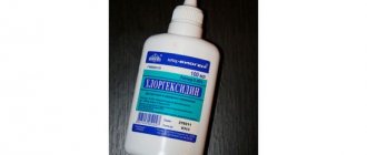
Drug treatment of the mucous membrane under the denture - after cleaning the denture, you will need a course of antiseptic rinses with Chlorhexidine 0.12-0.2% (2-3 times a day) and treatment of the mucous membrane under the denture with Cholisal-gel (2 times a day). Moreover, it will be better if you apply a thin layer of gel not to the mucous membrane, but to the entire inner surface of the prosthesis and put it on. The course of treatment is usually 10 days. But remember that the treatment will not be effective if you do not disinfect the prosthesis.
Treatment of allergic stomatitis
This type of stomatitis is caused by the body's immunological response to contact with an allergen. The role of the latter can be played by anything: food products, new oral hygiene products, dentures, animal hair and much more. At risk are patients who are predisposed to allergies or suffer from various autoimmune diseases. A distinctive feature of allergic stomatitis is the acute onset of the disease, accompanied by an increase in temperature, severe pain and putrid odor from the mouth, which does not disappear even after thorough brushing of the teeth.
Treatment methods
- Identifying the allergen and eliminating it (or minimizing contact).
- Antihistamine therapy.
- Relief of inflammation with corticosteroids.
- Taking analgesics to relieve pain.
- Local antiseptic drugs
- Diet.
How to treat stomatitis?
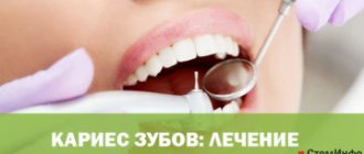
Any treatment for stomatitis begins with a professional hygienic cleaning procedure, during which tartar and soft plaque are removed. Almost any dental clinic in Moscow provides a similar service. Teeth affected by caries are treated. The mucous membrane is treated with antiseptic rinses. During the day, rinse the mouth with a warm solution of chamomile or calendula decoction. With timely treatment, catarrhal stomatitis disappears in 5–10 days. For ulcerative or aphthous stomatitis, local treatment is combined with general treatment. In addition to professional oral hygiene, antiseptic procedures are carried out in the clinic where stomatitis is treated.
If the presence of herpetic stomatitis is suspected, additional antiviral therapy is carried out. In case of oral candidiasis, treatment is prescribed with antifungal drugs. If the cause of stomatitis is some other disease, for example, the stomach or intestines, then it is necessary to begin to treat the root cause. During the treatment process, you should definitely follow a diet limiting spicy, hot, cold, sour and rough foods. A side effect of taking medications can be a green coating on the tongue.
Treatment of stomatitis in children
Treatment of childhood and adult stomatitis follows the same methods. At the same time, some drugs used for adults are replaced with those that are better suited for the child’s body. First of all, we are talking about antibiotics, antiseptics and drugs to enhance immunity. Children are often prescribed more gentle methods, which include the use of herbs and natural tinctures for rinsing. At an early age, dentists recommend preventing stomatitis using traditional medicine.
| Age | Type of disease |
| From 0 to 3 years | Candidal stomatitis, or childhood oral thrush. This type of stomatitis is especially common in infants. |
| 1 – 4 years | Aphthous and herpetic stomatitis caused by external infections and mechanical trauma to the oral cavity. |
| Children of primary and secondary school age | Allergic and aphthous stomatitis. |
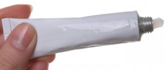
Types and categories of ointments for stomatitis
A variety of active ingredients will eliminate any cause of inflammation of the mucous membrane. Ointments against stomatitis are divided into the following types:
- Universal. They relieve inflammation and are needed both for ulcerative stomatitis and for aphthous and catarrhal stomatitis;
- antifungal. For the treatment of candidal stomatitis;
- antiviral. Needed for the treatment of herpetic stomatitis;
- antibacterial, used for aphthous or traumatic stomatitis.
It is important to know! None of the ointments will help if the inflammation of the mucous membrane is caused by allergies. It is not the infection that is to blame here, but internal disorders.
Stomatitis ointment for children
Not every ointment is suitable for a child. Therefore, when fighting inflammation of the mucous membranes in children, you must always consult a pediatric dentist and study the instructions for any product: the dosage can vary greatly.
In addition, it is better to replace ointment for stomatitis in children under one year old with solutions: they are contraindicated for the youngest. Among the products that are acceptable for use for children, it is worth noting:
- Actovegin (antibacterial);
- acyclovir (for herpetic stomatitis);
- Viferon (also an antiherpetic agent);
- Zovirax (like the two previous ointments - antiherpetic);
- lidochlor (relieves pain);
- nystatin (for fungal stomatitis);
- Cholisal (for bacterial stomatitis).
But all of them are suitable only for babies over one year old. By the way, at this age most often stomatitis is candidiasis, so pay attention to fungal remedies.
Treatment of stomatitis at home
I would like to immediately note that treating stomatitis with folk remedies at home is possible, but you need to understand that in many cases it is impossible to do without a visit to a specialist and the use of antibiotics. Ignoring these points can lead to worsening of the disease and longer rehabilitation. In addition, on the Internet you can find a whole lot of “revolutionary” recipes for stomatitis, so you need to be able to separate the wheat from the chaff and understand which remedies can really help, and which are the fruit of the inflamed imagination of “couch healers”. Below we publish the most common methods of preventing and treating stomatitis with folk remedies, but please note that only an experienced specialist can draw up an optimal treatment plan.
In the case of children, treatment of stomatitis with folk remedies at home is used quite often. This is especially justified as a preventive measure, since the child’s body is more susceptible to the influence of the external environment. The most popular remedies are considered to be all kinds of decoctions, for the production of which chamomile, calendula, burdock, propolis and blackberry leaves are used. A homemade ointment is also used to treat stomatitis, which is made from novocaine, egg white and honey. Be that as it may, if your child shows signs of stomatitis, it is best to immediately take him to a specialist who can make the correct diagnosis and plan treatment.
Folk remedies
Treatment of stomatitis at home should be carried out as prescribed by a doctor, who will take into account the causes of the disease and the general condition of the body. After eliminating the cause, he will prescribe rinses, ointments, gels, and multivitamin preparations.
- Hydrogen peroxide. Rinse your mouth with 3% hydrogen peroxide diluted in the same amount of water 2-3 times a day for one to two weeks.
- Blue iodine. Dilute blue iodine in a 1:1 ratio with warm water. Rinse your mouth with a glass of water 3 times a day. Apply gauze bandages moistened with blue iodine to the affected areas for 5 minutes. Treat stomatitis with this method 3 times a day.
- Propolis tincture. After eating, rinse your mouth with hot water and then hydrogen peroxide to thoroughly clean out the sores. Some additionally dry them with warm air using a hairdryer. Then lubricate the affected areas with alcohol tincture of propolis diluted with water and dry again with warm air so that a film forms more quickly.
- Myrrh oil. As legend has it, ancient Greek warriors did not go on a campaign without a paste of myrrh resin. Thick oil prevents the spread of infection, treats various skin lesions, helps with coughs and colds, and has an anti-inflammatory and antiseptic effect for bacterial and viral lesions.
- Silver water. Used to treat stomatitis in children and adults. Rinse your mouth with warm water several times a day. To prepare silver water, place a silver item in water at room temperature for a day.
- Honey. Brew 1 tsp. green tea with a glass of boiling water in a teapot, leave for 45 minutes, strain. Melt 1 tbsp in a water bath. honey, add to green tea infusion. Rinse your mouth several times a day with warm infusion to treat stomatitis.
- Blueberry. Brew 0.5 liters of boiling water with 3-4 tablespoons. blueberries, cook for 15 minutes over low heat with the lid closed, let cool. Rinse your mouth with a decoction for stomatitis or irritation of the mucous membrane.
- Get 1 tsp. freshly squeezed onion juice, 1 tsp. Kalanchoe juice, add 3 tbsp to the juice mixture. water. Rinse your mouth several times a day.
Treating stomatitis with home remedies
| Facilities | Description |
| Hydrogen peroxide | Rinsing with a solution of hydrogen peroxide helps reduce discomfort and has an antiseptic effect. |
| Baking soda | Treatment of stomatitis in adults with soda is recommended when the disease is detected in the initial stages. A solution of the substance (a spoon in a glass of water) is recommended for rinsing. |
| Decoctions of chamomile, rose hips and calendula | They have disinfecting properties and contain vitamins and nutrients to maintain oral health. |
Causes of stomatitis
The mechanism of stomatitis is not yet fully understood. But scientists are inclined to believe that the root cause of its development is the reaction of the human immune system to various irritants. At some point, the immune system ceases to recognize the potential threat of internal and external factors, which causes its atypical reaction, as a result of which “aggressive behavior” of lymphocytes is observed. The attack of lymphocytes against irritant molecules leads to lesions of the oral mucosa.
A variety of factors can provoke an atypical reaction of the immune system. The most likely of them are the following irritants:
- Pathogenic microorganisms that live in the mouth.
- Improper oral hygiene.
- Various damage to the mucous membrane, for example, burns from eating too hot food or mechanical injuries from seeds, nuts, crackers and other hard foods.
- General dehydration due to high fever, blood loss, vomiting, diarrhea, or thirst.
- Poor quality treatment of teeth and gums.
- An allergic reaction to dental structures in the mouth - braces, implants, crowns, bridges, etc.
- Long-term use of medications.
- A diet depleted of beneficial vitamins and elements.
- Smoking.
- Malignant formations of the oral cavity, respiratory organs or undergoing a course of chemotherapy.
- Hormonal imbalances in the body, for example in pregnant women or children during puberty.
- The presence of chronic diseases or allergies.
- Severe stress.
Interesting to know! Frequent stomatitis in adults can be caused by the use of toothpaste containing sodium lauryl sulfate, a substance added to oral care products to form a thick foam. According to recent studies, it dehydrates the oral mucosa and makes it vulnerable to various types of irritants. Patient observation data confirms the fact that avoiding the use of sodium lauryl sulfate paste can reduce the risk of developing stomatitis in adults by 81%.