198
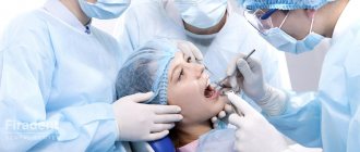
When starting to treat a tooth with an ambiguous condition, the dentist is always faced with a dilemma: try to preserve it, despite its poor condition, or remove it.
Both options have serious reasons. The principle of tooth preservation professed in domestic dentistry requires the doctor to fight for every tooth to the end.
On the other hand, a slow infection in a tooth infects neighboring units, and sometimes the entire body, causing much more harm to a person’s health than the absence of one tooth.
The essence of the method
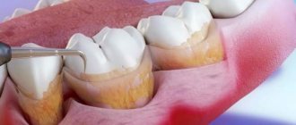
Coronoradiculular separation is a dissection of the coronal part of the tooth in the area where the grooves are located. Manipulation is classified as a preserving organ or process.
It is justified if pathological phenomena are observed in the area of bifurcation of a multi-root unit. The organ is dissected and problematic fragments are amputated.
After the wound has healed, stump inlays are installed in place of the removed tissue. The purpose of the operation is to save the tooth by eliminating its affected fragments.
Technique
The separation technology involves the use of two methods - mechanical and physiological. With coronoradicular separation, the first method is used, which is faster for patients and with minimal discomfort.
The operation takes place in the following sequence:
- Introduction of anesthesia (conduction or infiltration).
- Dissection of the mucosa.
- Peeling it off to get a sufficient view of the bifurcations.
- Sawing the crown part with a disk or bur without damaging the partition between the roots.
- Removal of destructive hard tissues.
- Grinding the edges of the crown.
- Carrying out curettage in the bifurcation zone with the introduction of osteoregenerative agents.
- Medicinal and antiseptic treatment.
- Hemostasis.
- Fixation of tooth fragments with a splint or noracryl with adjacent elements.
- Placement of mucosal flaps in their original position.
- Fixation with sutures.
After the operation, it is advisable to immediately prepare the roots for the installation of crowns, and take impressions from two tooth fragments to make crowns.
Indications
Coronoradicular separation is used in the treatment of mandibular molar units. Indications for the operation are limited:
- periodontal changes in the bifurcation zone - against the background of lysis of the apex of the septum between the root parts of the tooth;
- deep perforation of the bottom of the organ - this anomaly occurs in the process of incorrect treatment;
- perforation of the cavity bottom , which was a consequence of the development of abnormal destructive processes;
- the appearance of additional canals – we are talking about connecting fragments of the tooth cavity and periodontium in the bifurcation zone.
The essence of the technique
Coronoradicular (coronary-radicular) separation or, in other words, premolarization refers to tooth-saving operations. At the same time, it can be considered as a preparation procedure for prosthetics, and as a treatment for periodontal diseases.
In a word, this is a multi-purpose and rather complex operation, although it is often presented as simple and obvious, which in our opinion is a simplification.
Premolarization consists of dividing (separation) the double-rooted molars of the lower jaw into two separate teeth, each of which is a half-crown with one root. The crown is divided vertically, approximately in the middle, the roots are divided in the bifurcation zone.
Reference . Coronary-radicular separation is performed only for mandibular molars. This is due to the fact that, unlike the upper ones, they have two roots. And this is a prerequisite for the operation.
The term “premolarization” means that after separation the molars become similar to the premolars of the NP, which have only one root.
Teeth with serious damage in the bifurcation area, but with a satisfactory condition of the roots, which can still continue to serve their owner, are subject to coronoradicular separation.
The nature of the lesion in the bifurcation zone can be different:
- interradicular granuloma or cyst;
- periodontal damage with the spread of inflammation and necrotization to the dentin of the tooth in the bifurcation area;
- the formation of pockets with granulations in this area;
- perforation of the bottom of the pulp chamber, etc.
It is necessary that the affected area is in the bifurcation area and, after removal, leaves healthy roots and parts of the crown.
Although the requirements for the crown are not as stringent as for the roots. In extreme cases, a significantly damaged crown can be replaced with an artificial stump on intracanal pins. The main thing is that the roots are healthy.
As a result of separation, the affected bifurcation area is partially removed, partially treated, and the double-rooted tooth is divided into two single-rooted ones.
Subsequently, the separated parts of the crown are covered with a common (soldered) artificial crown, and the tooth can continue to be used as an abutment or regular chewing one.
Why is a split tooth dangerous, and how to get rid of the anomaly.
Come here to find out whether fused teeth need treatment.
At this address https://orto-info.ru/zubocheliustnye-anomalii/zubov/kak-ispravit-vyisokoe-i-nizkoe-polozhenie-otdelnyih.html read how to correct the high position of individual teeth.
Contraindications
As for the prohibitions on performing this method of dental surgery, their list is much longer than direct indications.
The operation is impossible for the following objective reasons:
- Atypical localization of birufication is if it is located in unacceptable proximity to the upper part of the roots. In this clinical case, amputation of too large a part of the organ is inevitable, after which it will no longer be possible to return it to its former usefulness and functionality.
- Periodontitis - regardless of the stage of its course.
- Acute osteomyelitis.
- Too much mobility of the tooth - we are talking about the third degree of pathology, when, against the background of these processes, severe destruction of the alveolar tissue occurs, more than 50% of the length of the root itself.
- Visible destruction of the coronal part of the jaw row unit - in combination with the adjacency of the subgingival root region affected by caries.
In such a clinical situation, it is better to restore the structural integrity of the tooth with filling composites and core liners. - Severe diseases of the body associated with disruption of its vital functions - diabetes, heart failure, renal dysfunction, liver pathologies, poor blood clotting.
- Age restrictions - in very elderly people, the effectiveness of the operation is quite low due to the natural disruption of the structural composition of hard bone tissue.
Indications for removal of salivary gland stones and relapse rate.
Read the following article about the need for surgical tooth repositioning.
Here https://www.vash-dentist.ru/hirurgiya/operatsii-na-zubah/kogda-opravdano-sekvestrektomii.html we will tell you the purpose of sequestrectomy for osteomyelitis.
Other tooth-preserving methods
In addition to coronoradicular separation, other tooth-preserving operations are often performed in dentistry. These include:
- Replantation is the return of a dental element after medical treatment to its socket. It is performed when it is impossible to perform conservative therapy, as well as in case of dislocation due to severe trauma.
- Root amputation is the extraction of the root up to the furcation while preserving the dental crown.
The procedure is only applicable to maxillary molars. During amputation, after detachment of the mucosa, the root cut off by the bur is removed, and the hole is filled with osteoplastic mass. Once the hole has healed, the crown is used in prosthetics as a support. - Hemisection – removal of part of the root and the entire coronal part.
The operation is performed on mandibular molars when the pathology is located in the area of only one root. The essence of the procedure is the resection of some part of the crown and extraction of the affected root. The remains of the root are subsequently used as support for the bridge.
Each of these methods is safe for the patient, occurs without consequences with minimal discomfort, and is performed under local anesthesia.
Preparing for surgery
All preparatory manipulations should begin only after a complete diagnostic history of the pathology has been collected.
To obtain an informative clinical picture, the patient is prescribed:
- X-ray examination is a standard procedure during dental surgery;
- computed tomography – makes it possible to see in a three-dimensional image the location of the anomaly, its shape and size, and also to develop an optimal scheme for the operation;
- a general blood test is necessary to understand the general physical condition of the patient’s body, as well as the degree of negative impact of the processes occurring inside the dental cavity.
After the diagnostic stage of treatment has been completed, all inflammatory manifestations in the oral cavity should be eliminated - if this is not done, then there is a high risk of wound infection, and this is fraught with serious complications.
It is mandatory to remove food fragments by rinsing the oral cavity with antibacterial compounds and antiseptics, and the surgical field is soaked with them especially thoroughly. At the same time tartar is removed so that during the separation process its pieces do not fall into the open wound.
Preparatory stage
Preparatory manipulations begin only after diagnosis and choice of treatment method. In order to assess the condition and choose a treatment algorithm, the doctor prescribes the following types of studies:
- X-ray examination necessary to assess the condition of the tissues and determine the area of intervention;
- tomography to localize pathology and develop a treatment regimen;
- general blood analysis.
Next, an examination of the oral cavity and professional cleaning of plaque and stone are carried out. The surgical field is impregnated with antibacterial compounds, which is necessary to reduce the risk of infection.
Operation stages
The algorithm for performing coronoradicular tooth separation according to the medical protocol is as follows:
- selection of a drug for administering anesthesia is carried out by testing the patient’s tolerance to a specific drug;
- selection of necessary tools and auxiliary devices;
- preparing the drill and handpieces for surgical manipulation;
- antiseptic treatment of specialist hands;
- administration of anesthetic;
- cavity incision of soft mucous tissues in the affected area, incision of the periosteum in the root dental projection;
- detachment of a mucous tissue fragment, formation of a periosteal flap, exposure of the approach to the alveolar ending;
- cutting down bone fragments using a bur above the root zone, the root of which is planned to be removed;
- separation of the coronal part is also performed using a drill;
- amputation of pathological parts of the organ - coronal zone and root;
- smoothing sharp edges with a level;
- removal of granulant - performed with a curettage device similar to a spoon;
- antiseptic treatment of the separation site, thorough drying of the wound;
- applying a patch fragment to a permanent place;
- suturing the outer surface.
In addition, during the procedure, the surgeon will prepare the root area and take an impression for subsequent prosthetics and installation of an artificial crown.
Progress of the operation
Premolarization begins after local anesthesia and antiseptic treatment of the oral cavity. Access to the operation area is through the vestibular surface of the gum in the projection of the causative tooth.
Surgical intervention is carried out in the following order:
- incision of the mucous membrane and periosteum, detachment of the flap;
- curettage of the interroot area - removal of inflamed, necrotic tissues and granulations, antiseptic treatment of the wound;
- vertical section of the crown and bifurcation zone using a drill;
- smoothing of sharp edges of separated teeth, antiseptic treatment;
- filling the bone wound with bone substitute materials (according to indications);
- application of a flap, suturing the wound.
After the healing process is complete and the alveolar ridge is restored, which may take several months, the clinical crowns of the newly formed teeth are covered with a general artificial crown.
It should be noted that depending on the condition and shape of the crowns of the separated tooth, different options for their prosthetics are possible. In the simplest case, as already mentioned, a common artificial crown is placed on both parts of the tooth.
In case of significant destruction of the crown, it is possible to use a stump pin tab, the “legs” of which are fixed in the separated root canals.
Need for curettage
Curettage is a necessary manipulation during surgery. It is this that allows you to most effectively clean periodontal gum pockets from accumulated granules and eliminate the development of infectious complications. It is carried out using an open patchwork method.
If you refuse the procedure, the patient faces postoperative bleeding, inflammatory pathologies and severe pain.
Features of mandibular osteosynthesis surgery, indications and contraindications.
In this post, we will talk about the signs that indicate the need for wisdom tooth hood removal.
Follow the link https://www.vash-dentist.ru/hirurgiya/operatsii-na-zubah/loskutnoy-na-desne.html if you are interested in reviews about flap surgery on the gums.
Preoperative stage
Coronoradicular separation is planned in advance.
First, the patient undergoes the necessary examination, consisting of the following procedures:
- Taking an anamnesis is recording all the diseases the patient has at the time of treatment.
- In-depth examination of the body - general urine and blood tests, biochemical blood test, test for HIV infection, test for syphilis and viral hepatitis. Based on the results obtained, it becomes clear whether surgery is possible or not. If other diseases are mentioned in the medical history, additional tests are performed. If serious contraindications are discovered during the examination, the patient will be offered surgery after undergoing an appropriate course of treatment.
- Taking allergy tests for drugs used during surgery.
- X-ray of the oral cavity.
- Computed tomography - allows you to see the location of the damage, its shape and size.
- Preparation of the oral cavity - professional cleaning with certain means; treatment of teeth growing nearby; removal of roots that are no longer suitable for restoration; filling of dental canals.
- Organization of psychological mood.
Postoperative period
The recovery period after this method of separation lasts about 7–8 days, and if everything is carried out within the framework of the protocol, it proceeds quite calmly.
The first day after the anesthesia wears off, the patient may be bothered by painful manifestations at the site of the operation. They are treated with targeted painkillers.
Every day, throughout the entire stage of rehabilitation, the patient undergoes antiseptic treatment of the affected area, and on the sixth day the sutures are removed.
The main thing that is required from the patient is careful adherence to oral hygiene and nutrition , based on bringing products to a puree-like consistency.
In addition, high and low temperature changes should be avoided . Clean your teeth carefully, trying not to touch the area where the sutures are applied.
Temporary dentures can be made a month after separation, permanent crowns are installed no earlier than 4-5 months in the absence of any complications.
Preparatory activities
Preparation aims to accurately assess the condition of the tooth, draw up a treatment plan, and bring the tooth into a state in which it can be separated without any risk.
Assessment of the condition of the tooth involves diagnostics, including standard dental examinations:
- studying the anamnesis and interviewing the patient;
- examination of the oral cavity and physical examination (probing, palpation);
- X-ray examinations (sighted images, orthopantomography and MRI if necessary);
Laboratory blood tests may be required to determine infection and inflammation , which may be a manifestation of systemic diseases that prevent the operation - HIV, hepatitis, etc.
Procedural preparation of a tooth for separation includes both mandatory and optional (according to indications) procedures. The latter includes sanitation of the oral cavity, professional teeth cleaning using the Air Flow method (effective mainly against soft plaque) or using ultrasound (removes hard tartar).
Depulpation is a mandatory procedure. The Dental Association of the Russian Federation in Resolution No. 15 of September 30, 2014 indicates that for all tooth-saving operations, including coronary-radicular separation, endodontic treatment of the tooth must be carried out before surgery.
That is, before dividing a tooth, it is necessary to fill all its cavities.
Possible complications
Among the likely complications, the most common are:
- bleeding - eliminated by constricting blood vessels, drugs and rinsing solutions that promote blood clotting;
- divergence of postoperative sutures - occurs due to non-compliance with medical instructions, mainly due to the fault of the patient himself, and is eliminated by re-suturing the wound;
- inflammatory processes of internal gingival tissues – caused by the penetration of pathogenic microbes and bacteria into the cavity.
It is accompanied by the accumulation of purulent masses, abscesses and requires repeated opening of the wound, antiseptic treatment, removal of suppuration and suturing.It can occur both due to failure to maintain sterility at the time of surgery, and during the recovery stage due to improper oral care.
Procedure: main steps
Coronoradicular separation is carried out using the following steps:
- The painkiller is tested and the optimal drug is selected in accordance with the individual characteristics of the patient’s body;
- preparation of instruments and materials necessary for treatment;
- preparation of the drill;
- carrying out antiseptic treatment, pain relief;
- incision of soft tissues, periosteum to the root area;
- detachment of a mucosal flap, exposure of the approach for treatment;
- removal of affected tissue, separation of the coronal part using a drill;
- smoothing of sharp parts, removal of granulate and subsequent antiseptic treatment;
- flap application, soft tissue suturing.
Curettage
The curettage procedure involves cleaning the gum pockets from accumulations of granules and affected infectious areas. The open patch method is used for the procedure. This stage of the operation is carried out with the aim of relieving pain, eliminating the development of complications and inflammatory processes.
Postoperative period
After treatment using the separation method, a postoperative period is required, which lasts 7-8 days. If the operation is performed correctly and the patient follows the regimen and doctor’s recommendations, this period passes relatively easily, without consequences. In the first day, pain and swelling may be observed, but they quickly pass. Every day during the first week the area is treated with antiseptic agents, and on the sixth day the sutures are removed.
The patient is required to maintain hygiene and avoid mechanical damage to the injured area. You should also avoid solid foods, hot foods and drinks for the first time. You can begin installing a prosthesis or permanent crowns only four to five months after the end of treatment.
Complications and risks
There are a number of risks and complications when treating with this method:
- bleeding, usually eliminated with special solutions and drugs for narrowing blood vessels;
- suture dehiscence in the postoperative period, usually observed due to poor oral care or non-compliance with doctor’s recommendations;
- the development of inflammatory processes, the appearance of pain, the cause is the penetration of infection during manipulation or violation of hygiene rules in the postoperative period;
- the appearance of a purulent lesion, swelling, abscess, requires concealment and removal of pus.
Treatment for the development of complications is carried out in accordance with the patient’s condition and the problem that has arisen. Usually the cause is the patient’s violation of recommendations for caring for the oral cavity and the surgical site. in most cases there are no complications, some swelling and soreness disappear quickly.
Forecasts
With proper treatment, the prognosis is very good, but the patient must take into account that the technique is new. The treatment technique is considered one of the alternatives for treating pathologies that allows you to save the tooth.
Alternative methods also include conservative solutions to the problem:
- hemisection, that is, amputation of cysts or granulomas with removal of the damaged part of the root system;
- complete removal of the tooth root while preserving the outer crown part, which allows for further restoration of the functionality of the dentition and organ;
- replantation, this technique is rarely used, it consists of removing a tooth and then installing it in place after treatment.
How much does treatment cost?
On average, the cost of the procedure is 5.5 thousand rubles, but the doctor will be able to name the exact price only after a preliminary examination and diagnostics. The cost is influenced by the following factors:
- condition of the oral cavity, the need for treatment, additional manipulations;
- diagnostic measures;
- volume of work carried out;
- the cost of medications and anesthetics used;
- preventive measures.
The price of treatment is influenced by the status of the clinics, the length of stay in the inpatient department, and the presence or absence of complications. Therefore, it is impossible to predict the exact cost of the full course of treatment.
Alternative Methods
The following methods of conservative-operative solution to the problem are distinguished:
- replantation - used quite rarely and is an extraction with the subsequent return of the dental unit to its place. In other words, the initially amputated organ is reinserted after reconstructive manipulations;
- root amputation - removal of the root part of a damaged tooth while maintaining the integrity of its coronal part, aimed at preserving the functionality and integrity of the organ;
- hemisection is a surgical procedure to amputate cystic formations or granulomas along with the affected fragment of the root system.
Dentistry. Clear and Accessible.
The best blog about dentistry and dental implants
From my colleagues I often hear the following: “Stanislav, why do you need all this?” Do you think anyone will appreciate your writing? Basically, I don't expect evaluation. My opinion on this matter sounds simpler - the more you know about dentistry, the better for both you and me. Knowledge is always power, and knowledge about your own body is doubly powerful. Therefore, the dental section will only develop further, and a lot of interesting things await you in the new year. The only thing that holds me back is collecting material (I don’t plagiarize, I don’t use other people’s photographs) and conversations with specialists - of course, I’m not competent in all matters and I myself constantly have to learn and improve. Today we will continue the partially started topic - we will talk about tooth-preserving operations and root cysts of the jaws in particular. I have already written about what will happen if you do not treat the cyst, and now you will find out how they are treated.
There is a category of patients who try to save teeth in the most hopeless cases, when, according to all indications, the diseased tooth must be removed. Some doctors are trying to make money on these hopes - if forty dentists said: “For removal,” there will definitely be one who says: “Nonsense! I’ll cure you right now!” – and a person naturally grabs this chance like a drowning man grabs a life preserver. Is this approach justified? Maybe. BUT It is important to know that: – If forty dentists said “no”, and one says “yes”, it is better to believe forty dentists. – Any tooth-preserving operations and “on the brink” treatment (such as “it’s better to remove, but if you don’t want to, we’ll treat”) are not covered by the guarantee. So all responsibility for making a decision and for the financial costs of this treatment is entirely on your shoulders. – Tooth-preserving operations have very limited indications. They have many more contraindications. – The same applies to treatment “on the brink”. – Any tooth-preserving operations require preparation (retreatment) of the teeth that are planned to be preserved. – Consultation with an orthopedic dentist and a dental therapist, in some cases a periodontist, is required. Together with them, we need to decide - is this tooth worth saving at all? – Every disease has a cause. And without eliminating the cause, we will never cure this disease. In other words, tooth-preserving surgery is not a treatment, but just a stage in the treatment. - There are no miracles. There is competent planning and masterly execution.
What types of tooth-preserving operations are there?
With all the abundance of surgical techniques for preserving teeth, they can all be reduced to three main operations - resection of the root apex, hemisection and corono-radicular separation. Everything else is just modifications. There are also periodontal operations, but we will look at them next time.
1. Resection of the root apex. The scheme of such an operation looks something like this: Its essence is as follows - if it is not possible to eliminate periapical (near-apical - shown in red dotted line) changes using a therapeutic method through the tooth canal, then this can be done surgically. Often, resection of the root apex is combined with cystectomy or cystotomy (complete or partial removal of the root cyst). Sometimes, if it is not possible to seal the apical end of the canal in the usual way, filling can be done retrogradely - from the side of the operating stump. The operation of resection of the root apex is used quite often - among all tooth-preserving operations it has the widest indications. However, in order for the operation to be successful and the treatment to have a favorable outcome, it is necessary to comply with the requirements that I have already mentioned above. I would not perform apex resection surgery in the following cases: – If the tooth has no functional value. – If the tooth is planned to be used as a support for massive and large (more than three units) orthopedic structures. – If the tooth is mobile. – If the tooth is subjected to irreversible traumatic effects. – With moderate periodontitis and exposure of the tooth root by more than 1/3. – If the patient suffers from decompensated diabetes mellitus and does not care about it. – If the size of the root cyst is more than one and a half centimeters in diameter. – If the patient does not care about the result of the operation.
I really like to compare the teeth to the supports of a bridge - they are also under constant load and are necessary - have you ever seen a bridge without strong supports? In comparison it will look like this:
Can I use this bridge? Of course it is possible, but with some reservations - after all, the cut support is not strong enough and it will serve for a long time only in one case - if at the entrance to the bridge we put a sign “Weight limit”.
2. Hemisection. This operation is performed on multi-rooted teeth; during it, one root is removed in the expectation that the remaining roots will take on the entire chewing load:
This operation has very limited indications - well, one root is physically unable to bear the load of an entire tooth. For the same reason, unfavorable outcomes are very common - after a short time, the tooth still has to be removed. That is why I personally am not a supporter of this method - due to its unreliability. In no case should you use teeth after hemisection as a support for a bridge prosthesis - how many such works have I seen, they all fell apart quite quickly. Perhaps hemisection is justified only in one case - in adolescents, when skeletal growth has not yet completed and placement of an implant is impossible. Here it is important to preserve space for the future crown and prevent the teeth from moving - therefore, if the resected tooth lasts at least a year or two, it will be good. Although, in my opinion, it is better to install a retainer - at least there will be no concern about the condition of the bone tissue. Another possible and justified option for using hemisection is the resection of one root that cannot be retreated in three-rooted teeth, provided that the remaining roots are large enough and can withstand the increased load. But this happens extremely, extremely rarely. I offer a comparison to defenders of hemisection (as it turns out, there are many of them). Imagine for yourself, using the bridge analogy:
Would you risk transporting heavy and valuable cargo across such a bridge? If anyone has difficulties with bridge construction, you can imagine a car with two or three wheels instead of four - will you go far with this?
3. Corona-radicular separation. This method is rather of historical significance. In modern dentistry it is not used for the same reason as hemisection. The essence of this perversion is this: Here, along with the affected root, part of the crown is sawed off. And the remaining part of the tooth is usually covered with a crown, in the hope that it will still serve. As a rule, this “service” is very short – demobilization occurs within a few months. The reason is still the same – tooth overload. Imagine that the average load on a chewing tooth is 100 kg per square meter. cm. We have reduced the support area in the process of coronary-radicular separation or hemisection by half - now the load is 100 kg per 0.5 sq. m. cm or 200 kg per sq. cm. But a tooth is not a building structure; six-fold strength is not built into it during design. The result is tooth loss soon. Well, our bridge after coronary-radicular separation looks like this:
Decide for yourself whether to use such a bridge.
Cystectomy with resection of the root apex - how it is done.
Below I will tell (and show) you how a cystectomy operation with resection of the root apex is performed. The photographs are collected from several cases - it is simply not always possible to photograph everything during the operation. But I think the main stages will be clear to you:
Let's start with the fact that the patient comes prepared - he is consulted by an orthopedist, therapist and periodontist, together with them we decide that it is possible to save the tooth. The therapist fills the tooth canals. Usually gutta-percha or the Thermafil system is used. I personally prefer gutta-percha. The orthopedist performs a functional analysis and determines the load on the tooth. Will he be overloaded after treatment? The periodontist assesses the condition of the tissues surrounding the tooth and conducts a session of professional oral hygiene. It is important for me that after resection the tooth remains in its place. If necessary, selective grinding of the teeth is carried out, the operated tooth is separated from the antagonists.
Before surgery, we take blood to make FRP gel or membrane. Platelet-rich plasma is very suitable for both filling the remaining bone cavity and isolating it from the mucous membrane:
Any dental intervention begins with high-quality anesthesia. As I have already said and never tire of repeating: “There are no people on whom anesthesia does not work. There are doctors who don’t know how to administer anesthesia.”
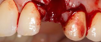
We make a cut - I prefer an L-shaped one along the transitional fold (learned from the Americans). We peel off the mucous membrane along with the periosteum - we almost always see bone perforation in the cyst area - especially with repeated exacerbations: In the right picture we also see the healing material “Metapex” brought into the cavity of the cyst - for a long time they tried to treat the cyst conservatively.
Using a cutter, we create access and visibility into the cyst cavity:
As a result, we see the following: On the right side of the surgical wound is the apex of the tooth root, which we have to remove. It's better to do this now to get a good overview.
Using a Lindeman cutter we remove the top of the tooth root. Ideally, we should clearly see the filling material and the stump itself - therefore, it is best to do the resection at an angle:
Now nothing prevents us from removing the entire cyst shell under visual control. You need to scrape the peri-root area especially carefully:
At this stage, if necessary, retrograde root canal filling is performed. The question of how best to fill the resulting bone cavity and whether it should be done at all remains a subject of debate. In my opinion, it is important to indicate what should never be put in there - these are all sorts of hemostatic sponges with iodine, iodoform turunds, antibiotics, etc. In our work we use xenografts Synthraft, Bioss, etc., which give good results result. But more often, just an FRP gel, which we made from the patient’s blood, is enough: Sometimes it’s worth adding bone chips, which can be collected with a scrubber around the bone defect.
Now we apply stitches: This is what L-cuts are useful for. They allow you to get by with a minimum of ligatures and maximum tightness.
Next, we prescribe the patient complex anti-inflammatory and antibacterial therapy (as part of the fact that we have already carried out antibiotic prophylaxis), and schedule examinations. We take control pictures once a month to track the dynamics of the healing processes. If you are interested in looking at radiographs, I can select them specifically for you.
As you can see, everything is quite simple. In terms of time, such an operation takes a maximum of twenty minutes with all the preparation:
and with proper planning it gives very good results.
That's it. If you have any questions, write in the comments.
Price
No practicing medical institution can give the exact cost of this procedure, since it depends on a number of factors:
- diagnostic measures;
- the volume of work performed during the operation;
- the cost of anesthetic and other costs of medicines;
- the cost of additional services and doctor’s consultations.
The region of residence of the patient and the status of the clinic where treatment is carried out play an important role.
On average, the cost of separation without taking into account additional costs will cost the patient from 5,500 rubles.
Forecast
When visiting a dentist, every patient wants to receive high-quality and professional treatment, as well as the opportunity to preserve and restore a problematic tooth using all available methods.
Premolarization is not yet widely used in dentistry. But in cases where, for some reason, this procedure remains the only one possible, it has a high success rate.
The dental fragments formed during separation retain their full functionality for an average of another 3-4 years.
Reviews
Despite the fact that this technique is still the subject of controversy among a number of practitioners, timely correction carried out in this way in most clinical cases justifies its use, especially when it comes to a child’s body.
If you or your loved ones have had personal experience with coronoradicular separation, you can leave your feedback on the operation and the course of the recovery period in the “comments” section.
If you find an error, please select a piece of text and press Ctrl+Enter.
Tags coronoradicular separation operations in dentistry
Did you like the article? stay tuned
Previous article
What can cause chipping of ceramics on a crown and how to eliminate the defect
Next article
Review and scope of nanocomposites in dentistry
Expected result and cost
The prognosis for premolarization can be considered favorable. If the operation is performed well, the separated tooth can be used for a long time.
Although the officially declared service life of a tooth subjected to coronoradicular separation is about 4 years, in reality it can last much longer.
The cost of the operation, if we mean one separation, is about 5,000 rubles. However, taking into account preparatory operations - sanitation, professional cleaning, endodontic treatment, etc. - the doctor’s bill can amount to a significantly higher amount.
The price of treatment is influenced by the status and location of the clinic. In general, only a doctor at a particular clinic can tell you the exact cost after drawing up a treatment plan.