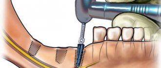
During treatment at the dentist, before performing various manipulations, additional hardware diagnostic procedures are often required. During a medical examination, 40% of dental tissues are accessible to external visualization, the rest are hidden. An instrumental examination is necessary to clarify the location of the pathological process; even visible dental tissues can hide pathological cavities, as well as to identify anatomical features.
You can understand which is better or dental x-rays by identifying indications for diagnosis using one or another method.
What does a dental x-ray show?
Implantation, prosthetics, sinus lifting, root canal treatment - all these operations are impossible without x-rays. It is necessary to take a photo of your teeth to assess the condition of the bone tissue, check the filling of the canals (photo of the roots of the teeth), and identify the development of hidden caries, which can appear under a filling, crown, or in the interdental spaces. An X-ray of the tooth also shows the condition of the periodontium (tissue around the root of the tooth) and periodontium (tissue around the entire tooth), cracks or inflammation of the canals. With the help of x-rays, the dentist can make an accurate diagnosis, especially if the patient's complaints and symptoms suggest several diseases or no obvious destruction. In this case, the dentist will not have to drill at random, which will save the patient from unnecessary painful manipulations.
When is a jaw x-ray taken?
Tell the patient exactly whether he needs and can have x-rays of his jaws
, should his attending physician. General indications for such a diagnostic procedure are:
- Suspicion of jaw development abnormalities (in childhood or adulthood). These may be congenital clefts of the hard/soft palate, impaired growth and proper development, or malocclusion.
- Suspicion of infectious-inflammatory and specific pathologies. These include osteomyelitis, periostitis, syphilis, actinomycosis, tuberculosis or necrosis of the jaw bone.
- To identify various acquired defects resulting from injury or removal of oncological formations.
- Suspicion of the presence of a false joint.
- Suspicion of a benign or malignant neoplasm.
What can you see in the photographs?
The following can be seen on x-rays of the upper and lower jaws:
- defects - cracks, fractures, presence or absence of bone fragments;
- pathological changes in bone tissue - areas of tissue thinning, compaction, thickening or thinning of the cortical plate;
- areas of necrosis - sequestra;
- sclerotic process;
- various periosteal layers, osteophytes (bone growths);
- tumor-like neoplasms.
What types of x-rays are there?
Depending on the task pursued by the dentist, he may prescribe one of the existing types of radiography: intraoral, or dental, panoramic, or orthopantomogram, computed tomography. Intraoral, or dental, x-rays are used if a targeted image of a specific tooth or several adjacent teeth (from one to four) is needed. If you need to get an idea of the jaw as a whole, identify foci of infection, assess the condition of the maxillary sinus, temporomandibular joint, periodontium, prepare for implantation, prosthetics or flap surgery, a panoramic photograph of the teeth or an orthopantomogram is prescribed. Computed tomography (CT) is indispensable when planning bone grafting, sinus lift or dental implantation. The tomograph takes a series of images of the patient’s jaw in different projections and then builds a 3D model of the patient’s jaw. This kind of 3D dental x-ray allows you to get the most complete, three-dimensional picture of the condition of the patient’s oral cavity. Contrary to popular belief, CT scans can be done in the presence of metal crowns and dental implants.
Contraindications to CT scan of the jaw
There are a number of contraindications for which the procedure is not recommended.
These include:
- weight more than 150 kg or girth more than 150 cm (the device is designed for a load of up to 150 kg);
- pregnancy (the risk of developing defects in the fetus increases);
- lactation period (during the procedure, rays can directly affect the composition of a nursing woman’s milk);
- severe mental illness (during the procedure the person must be at rest);
- the presence of metal objects in the area of interest, as they can reduce the information content of the results;
- children under 5 years old;
Are dental x-rays harmful?
Quite a lot of patients are concerned about whether it is harmful to take a dental x-ray. There is an opinion that dental x-rays are harmful. Today this is just a myth, fostered by panic fear of radiation sickness. As for the dose of X-ray radiation that a person receives during such an examination, we are talking about 0.002 - 0.003x mSv (millisievert is a unit used to indicate the amount of radiation), and according to SanPiN, up to 1 mSv can be received per year for research purposes , so you can take dental x-rays as often as you like, because the main goal of this research method is to improve the quality of diagnosis and treatment of oral diseases. It should also be taken into account that we naturally receive radiation of about 2-3 mSv per year from the environment, the same amount during a two- to three-hour flight on an airplane.
One should be afraid of mutations or radiation sickness leading to death only when radiation exposure exceeds 1000 mSv. According to the Scientific Committee on the Effects of Atomic Radiation at the UN, even with a single sharp irradiation of 500 mSv, a short-term change in the composition of the blood occurs, while the hematopoietic system is able to fully restore its functions, especially if not all cells of the body were exposed to irradiation.
Previously, only film similar to that used in a camera was used to obtain an X-ray image. It was on this that the photograph was projected. Modern digital technologies make it possible to replace traditional X-ray film with a sensor that converts X-rays into a digital signal. In this case, the emitting apparatus remains the same, only the receiving device changes. But a digital sensor requires a shutter speed five times faster than film, which means the radiation dose is reduced proportionally. It should be clarified that we are talking about only fractions of a second of irradiation: 0.05 - 0.3 s for a digital visiograph or 0.5 - 1.2 s for film. It is easy to calculate how many times a year you can safely take dental x-rays: about 300 - 500 times with a digital device or about 100 with a film one.
The issue of X-ray safety for children
Any X-ray radiation, including in the dental industry, carries a certain degree of danger to the patient’s health. Research in the field of medicine confirms the possibility of developing acute radiation sickness when exposed to increased radiation. However, X-ray equipment is not enough to provoke it.
X-raying of a child’s primary teeth can be safe if the examination schedule and radiation safety standards are followed.
The permissible annual exposure rate, according to regulatory documentation, is 5 mSV (millisievert). In the case of small patients, as well as pregnant and lactating women, this figure is halved.
Dental radiography is characterized by minimal mZV indicators. With digital X-rays, the amount of single exposure is 0.01-0.03 mSv. In this case, the permissible single dose is 0.1 mSV. The development of radiation sickness is discussed only when this figure increases by (0.7 ZV).
X-rays are often prescribed periodically, for example, as part of a long therapeutic course. How often can dental x-rays be taken? According to WHO, if 5-6 such procedures are carried out during the year, the radiation background of a small patient will not be disturbed. However, one should not lose sight of the factor of the presence of its own background radiation in the area in which the child lives. For example, for Moscow this figure is 20 µSV.
There is no official evidence that chronic doses of irradiation to the oral cavity (for example, long-term therapy) result in the development of cancer.
Is it possible to take dental x-rays during pregnancy?
You should prepare for pregnancy in advance, and your plan for such preparation must include a visit to the dentist. However, if it so happens that it is necessary to take a dental x-ray during pregnancy, you should be guided by specific indications and listen to the doctor’s instructions. It is not recommended to take an x-ray in the first trimester of pregnancy, but it is better to refrain from it until the fifth month. You also need to ensure that the total radiation dose during pregnancy does not exceed 1 mSv. As we have already calculated, this is approximately 300 dental photographs using a modern digital device, or 80 digital orthopantomograms, or 20 computer tomograms. Plus, you should use all possible types of protection: wear a special lead apron on your stomach and collar. It is worth remembering that the stress of going to the doctor and worry about the safety of a particular procedure, not to mention the pain, can cause much more harm to pregnant women than the x-ray itself.
What is the difference between computed tomography and magnetic resonance imaging?
CT, unlike MRI, is a radiological type of diagnosis, therefore it is not recommended for pregnant women and young children. One of the advantages of CT over MRI is the ability to conduct research in patients with claustrophobia, the presence of metal-containing implants, pacemakers, etc.
Regarding the diagnostic capabilities of computed tomography and magnetic resonance imaging, the first is especially effective in diagnosing hollow organs, for example, lungs and bronchi, and bones, the second - soft tissues, parenchymal organs, joints, and vertebral structures. In examining the abdominal cavity and pelvis, CT and MRI are often used interchangeably.
The contrast used in CT is more allergenic and has more contraindications and side effects than contrast agents in MRI.
The choice of MRI or CT analogue should be made only by the attending physician based on the indications.
Is a dental x-ray dangerous when breastfeeding?
X-ray radiation does not accumulate in the environment, but, like light waves, passes through objects, lingering a little longer in a dense environment, a little less in a more rarefied environment. So, all those who are concerned about the question of whether a nursing mother can have a dental x-ray done should answer: “Yes, you can!” Irradiation will not “spoil” milk. It can only linger in the mammary glands themselves. In dentistry in particular and in medicine in general, microdoses of radiation are used, several tens of times less than those recognized as dangerous and causing immediate dire consequences, so dental x-rays during breastfeeding, if there are indications and high-quality devices, are quite safe. For your own peace of mind after the procedure, you can skip one feeding - this will be enough.
Ultrasound or CT – which is better?
Ultrasound research methods are traditional, safe and more accessible than CT. Therefore, ultrasound can be used quite often, in contrast to CT screening.
Ultrasound can be performed on pregnant women to assess fetal development and the condition of the placenta. This method is suitable for diagnosing soft tissues, the heart, blood vessels in newborns, as well as the digestive organs, pelvis, mammary glands, some joints and parts of the spine. Ultrasound is usually prescribed as the primary method of examination at the beginning of treatment; it often requires clarification by other methods, in particular CT.
CT scans are used to diagnose not only internal structures, but also soft tissues, all blood vessels, the lymphatic system, and head structures. It is used in emergency cases in cases of suspected stroke, heart attack, or after extensive trauma. CT is more accurate than ultrasound and, unlike it, allows you to build a three-dimensional model of the anatomical area and verify formations as small as 1 mm.
Why take an X-ray of your child's teeth?
To adequately diagnose oral diseases in children and adolescents, the doctor needs to have as much information as possible. An x-ray of a child’s baby teeth can provide this information. More than 50% of the tooth is hidden, and the dentist simply does not see some areas where pathological processes may occur. In these cases, x-rays come to the aid of the doctor. An image of a child’s baby teeth will show whether there is caries between the teeth, whether the roots of the teeth are changed, and how the rudiments of permanent teeth are located. This way, the dentist can predict the timing of the loss of baby teeth and identify the presence of impacted, or crooked, rudiments. Such early diagnosis makes it possible to recognize a possible malocclusion and prevent it. At this stage, it is much easier to correct the bite, and there is a chance of avoiding wearing braces in the future.
The Ministry of Health has developed the timing of the examination, as well as safety rules under which neither the patient nor the clinic staff will be exposed to unnecessary radiation. According to his recommendations, children should have an X-ray of their children's teeth once every two years, and after the eruption of permanent teeth - once every 1.5 - 3 years.
How the research works
The examination does not require preparation, the only thing is that you will be asked to remove your jewelry. The procedure lasts 2-3 minutes, the results can be obtained in your hands after 10-15 minutes.
When x-raying the jaw joint, you need to take two pictures, asking the patient to first open and then close his mouth.
There are more than 30 methods of radiography of the joint. Since it is located in a hard-to-reach area, the radiologist uses special devices that differ in the method of placement to take the picture. Among them:
- Laying according to Schüller. The patient rests his lower jaw on a special stand, his head is fixed in a certain position opposite the X-ray machine using special clamps. This is the best way to determine damage to the mandibular joint, pathology of the temple bone, inflammation and neoplasm of the lower jaw, and trauma to the mastoid process.
- According to Mayer . The patient lies with the side of the head on a stand, the X-ray device is located on the side of the upper ear.
- According to Pordex. The patient sits in a chair and holds the film cassette on the side of the joint being examined. The beam of the device is directed from the side of the tragus, the projection magnification of the joint elements is clearly visible, without image superposition.
- General survey x-ray. This is an image taken using a dental apparatus.
Modern projections, in which images can be obtained quickly and with minimal radiation exposure to the patient:
- intraoral;
- extraoral;
- panoramic;
- orthopantomography;
- radiovisiography.
The image allows you to measure the width of the joint space, the position of the bones relative to each other, and suggest the development of pathological processes.
Price for dental x-ray
As for the cost of x-ray examination of patients, it all depends on the clinic, the equipment used for diagnosis, as well as the type of image required. On average, prices for a dental photograph in Moscow start at 250 rubles, and the cost of a panoramic photograph of the jaw ranges around 1,300 rubles. Where to take dental x-rays is up to you. Many clinics have adequate equipment and their patients do not need to go elsewhere, while others offer x-rays as a separate service.
Baby teeth and molars - what's the difference?
The differences between temporary and permanent are the following characteristics:
- The roots of baby teeth are shorter, widely spaced and angled. This is explained by the need for the formation of molars;
- the enamel of temporary teeth is bluish, it is much thinner, and therefore more susceptible to the development of caries;
- The child’s milk teeth are smaller than the molars;
- The number of some teeth is not equal to the number of others. There are fewer temporary ones, because some elements of the series immediately grow as radicals;
- the tissue of baby teeth is poorly mineralized;
- temporary teeth grow vertically, and the crowns of permanent teeth are directed outward (towards the lips and cheeks).
Where can I get a dental x-ray and how is it done?
Today, dental photographs can be taken in almost any clinic. As a rule, the best dental clinics in Moscow have equipment that allows you to take dental x-rays directly during the treatment process, without lifting the patient from the chair. These are so-called dental visiographs, the image from which is instantly transmitted to the screen, and the dentist can immediately make a diagnosis or assess the quality of treatment. In order for the most informative photograph of the teeth to be obtained, it is necessary to remove all jewelry. When performing an x-ray, be sure to wear a protective apron and collar. You will have to remain still for some time. As a rule, there is no need to take a photograph of your teeth at another dentistry, especially since many clinics refuse to issue photographs in person under various pretexts. However, according to the Russian law “on personal data”, they do not have the right to do this, and such a position of a dental clinic often negatively affects its reputation.
Publisher: Expert magazine about dentistry Startsmile.ru
Author of the material: Yulia Usacheva