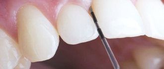
Hyaluronic acid is a type of polysaccharide that is part of connective, epithelial and nervous tissues and extracellular structures of the body. Hyaluronic acid is found in many biological fluids, that is, it is a substance produced in the body, its natural important component. Hyaluronic acid adds softness and elasticity to the skin and hair, and viscosity to the joint fluid. One molecule of this viscous substance attracts and holds up to a thousand water molecules, providing the necessary hydration.
If there is a deficiency of hyaluronic acid, it can be introduced into the body in the form of injections. Many have heard about injections of hyaluronic acid into the area of the lips and nasolabial folds - the lips become more voluminous and the folds are smoothed out. But, in addition to cosmetic use, hyaluronic acid injections are also used in dentistry. Injections of hyaluronic acid to dental patients are carried out in the aesthetic cosmetology department of the Family Dental Clinic.
Stages of occurrence
It happens that periodontal disease is divided into stages as it progresses. So, there are three of them in total:
- Mild - when there are no pronounced complaints yet, only sometimes a reaction to cold or hot appears.
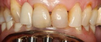
- Medium - the roots begin to become exposed. The gums can descend by 4-6 mm. Here the symptoms worsen - a burning sensation appears, the teeth react more sharply to the difference in food temperatures.
- Heavy - exposure of the roots is already measured at 8-10 mm, severe pain is felt when chewing.
How successful is periodontal disease treated? More details
Properties of hyaluronic acid used in dentistry:
- anti-inflammatory – used for gum inflammation, periodontitis, and dental implantation;
- bacteriostatic – suppresses the growth and development of bacteria in the area of inflammation, which is actively used in the treatment of periodontitis;
- regenerative – helps speed up the healing and recovery processes after surgery;
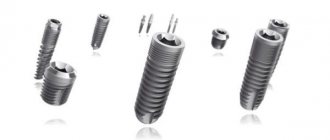
- improves the fixation of bone materials when used for dental implantation (bone grafting), maintaining the stability and volume of the bone graft;
- restores interdental papillae, reconstructing missing tissues;
- helps in the restoration of tissues inside and outside the joint (injections of hyaluronic acid are used for disorders of the temporomandibular joints), reducing pain, eliminating clicks in the joint and restoring mobility to it.
Let's take a closer look at some areas of application of hyaluronic acid in dentistry.
Reasons for development
To combat the problem, as well as to recognize it in a timely manner, you should first understand why periodontal disease develops. So, the list includes the following factors:
- Poor oral hygiene, when plaque accumulates due to insufficient brushing of teeth.
- Decreased immunity due to past illnesses.
- Problems with hormonal levels, for example during pregnancy, during menopause.
- Immunodeficiencies.
- Having bad habits, such as smoking, which negatively affects gum tissue.
- An unbalanced diet with a deficiency, when there are not enough fruits and vegetables, there are not enough vitamins and microelements in the diet, because when the body lacks all of this, it begins to take them from itself, including from the teeth.
- Taking medications for a long time that reduce the normal flow of saliva to the gums, the latter provides protection from negative factors.
So those who may notice such provoking factors should especially carefully monitor their dental health. Moreover, there are the first signs of a problem when it is not yet so difficult to cope with it.
Let's stop periodontal disease! Is it possible to slow down the progression of the disease Read more
Symptoms of gum inflammation
According to statistics, more than 70% of people have experienced inflammation of the gums between their front teeth at least once in their lives. The main signs of the problem include:
- redness of the gingival papilla,
- swelling of soft tissues,
- bleeding gums,
- bad breath,
- accumulation of pus in the interdental space,
- discomfort when brushing teeth and eating.
If you notice any of these signs, you should consult your doctor. These symptoms can indicate either injury to the gums from hard food or bones, or the development of serious diseases.
The first signs of periodontal disease
Gums are bleeding
“The very first sign of periodontal disease is bleeding gums. This is the first signal that under no circumstances should you miss or not react,” says dentist, chief physician of the dental clinic Ilya Antonov. As the doctor notes, bleeding develops against the background of an inflammatory process. Any inflammation, the specialist emphasizes, is a tissue reaction to the penetration of bacteria into the damaged area.
Article on the topic
Teeth as an indicator of health: what oral diseases will tell you about
The tissue around the tooth is injured
“The damage can be mechanical, usually resulting in injury to the circular ligament around the tooth. Such an injury can occur, for example, due to a poorly made crown or filling, when the edges of the restoration are not adjusted, there are no smooth transitions of materials at the junction with the tissue of the tooth,” notes the dentist. As Ilya Antonov says, in such places the gums are constantly injured by the protruding edges of the restoration due to the fact that food, passing the surface of the tooth, ends up on the gingival papilla protruded by the restoration. “In this way, a chronic injury appears, which leads to the formation of a pocket between the gum and tooth, where inflammation occurs, leading to periodontitis,” says Ilya Antonov.
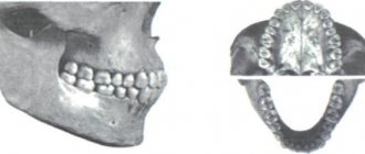
Exposing the roots of the teeth
The doctor notes that periodontal disease is, as a rule, a phenomenon that appears in chronic systemic diseases of the whole organism. “The first reason is the loss of bone tissue in the upper and lower jaw. There are two main types of such manifestations. One of them is when we simply see the roots of the teeth exposed. Such processes are accompanied by the appearance above the gum of the root part of the tooth, which was previously under it. The second type is accompanied by an active inflammatory process with swelling of the gums, the appearance of an unpleasant odor, and bleeding,” says Ilya Antonov.
Periodontitis or periodontal disease? Let's understand the differences between the two diseases Read more
New protocol for the formation of gingival papilla between dental implants
Restoring lost teeth using orthopedic structures supported by dental implants is a very common dental practice nowadays. However, aspects of osseointegration of supports, as well as the restoration of corresponding aesthetic parameters in the area of single and partial edentia, differ markedly.
An important aspect of rehabilitation remains the restoration of an adequate soft tissue contour and the architectonics of the interdental papilla, as extremely important components of the optimal smile profile. The absence of an interdental papilla compromises not only the patient’s appearance, but also provokes phonetic problems, as well as food getting stuck in the problem area.
Earlier studies have already proven the role of the distance from the apex of the interdental septum to the contact point between adjacent teeth as a factor influencing the amount of restoration of the papilla, at the same time, this parameter is variable for the papilla between adjacent natural teeth, between the implant and the own tooth, and also in the area of the overhanging part of the prosthesis. In cases where this distance between adjacent teeth is less than 5 mm, the papilla has the ability to completely fill the interdental space, while in the area between the implants the average height of the soft tissues, as a rule, does not exceed 3.4 mm, as a result of which in the implantation area often occurs deficiency in the height of the interdental papilla, which is critical in the rehabilitation of a patient with adentia in the frontal area.
There are many different approaches to restore the interdental papilla, but often due to compromised blood supply conditions and the formation of scar tissue, most known surgical techniques are not sufficiently predictive. Villareal in 2010 described a predictable approach to papillary regeneration using careful sequential soft tissue manipulation, including gentle incision and minimal flap separation. The main principle of the author's approach was to maintain adequate blood supply and the existing quality of the mucosa. This is why this approach recommended against suturing the intervention area, as this may cause additional trauma or inflammation, which ultimately will negatively affect the final result of treatment.
The purpose of this article is to present a series of clinical cases in which restoration of interdental papillae in the implantation area was performed using a modified surgical technique.
Materials and methods
Clinical data used in this study were obtained from the database of the Department of Periodontology and Implantology at New York University Kriser Dental Center. Data certification was carried out by the quality assurance department of the same university. The study was conducted in accordance with the Health Insurance and Identity Sharing Act and was approved by the University's Human Subjects Research Review Board.
Subjects studied
The study included ten clinical cases of restoration of the edentulous area of the central part of the upper jaw using dental implants. In the retrospective portion of the study, patients with existing restorations who had previously undergone interdental papilla augmentation between August 2011 and August 2012 were analyzed. The study group included 3 men and 7 women, whose average age was 45 years. During the study, the areas of the interdental papilla between two adjacent implants, between the implant and the natural tooth, as well as in the area of the intermediate part of the prosthesis in the area between the 13th and 23rd teeth were analyzed.
The inclusion criteria for the study group were as follows:
- The presence of an implant supporting the provisional restoration.
- Absence of interdental papilla (0 or 1 according to Jemt classification).
- Absence of a papilla in the anterior part of the upper jaw between two adjacent implants, an implant and a tooth, in the area of the intermediate part of the prosthesis.
To assess the severity of the interproximal papilla, the Jemt classification was used:
0 – absence of papilla; 1 – the presence of a papilla of only half its normal height; 2 – presence of more than half the height of the papilla; 3 – presence of a papilla of normal size; 4 – papillary hyperplasia.
The exclusion criteria from the study group were as follows:
- State of pregnancy or lactating women.
- Active periodontal disease in the area of remaining natural teeth.
- Having systemic diseases or taking medications that may affect the healing process of tissue around dental implants.
- Lack of motivation to carry out long-term maintenance therapy.
Measurements
Immediately after fixation of the provisional restorations, the distance from the contact areas of the superstructures to the papillary region of the gums was measured using a North Carolina periodontal probe (Hu-Friedy). After this, the results were interpreted in accordance with the Jemt classification. To improve the accuracy of the final results, measurements were carried out independently by two different examiners, but in no case did expert opinion differ and all papillae were scored as 0 or 1 according to the Jemt classification. During follow-up visits, measurements and classification of papillae were carried out according to the same scheme.
Surgical procedure
One hour before the intervention, patients took 2 g of amoxicillin orally, or 600 mg if allergic to penicillins. After local anesthesia with lidocaine with adrenaline at a concentration of 1: 100,000 (Henry Schein), the provisional structures were removed in order to visualize the area of the interdental papilla. Before surgery, patients underwent a procedure to expand the interdental space to ensure sufficient volume for future soft tissue (photo 1a).
Photo 1a. Clinical view of a provisional restoration with a missing papilla in the area of the implant at the site of the 12th tooth and an intermediate part in the area of the 11th tooth after augmentation.
Before modification of the provisional structures, each of the papillae was assessed according to the Jemt classification. After removing the temporary restorations from the vestibular mucosa apical to the papillary region, an oblique incision was made through the full thickness of the soft tissue (Figure 1b).
Photo 1b. An oblique incision of the mucosa from the vestibular side.
A similar incision was made on the palatal side (Figure 1c).
Photo 1c. Palatal incision.
The oblique direction of the incisions, as well as the formation of such at a certain distance from the existing papilla, was reasoned with the goal of maintaining an adequate level of blood supply in the recipient area. Using the interlingual (TLC) (Ebina), modified and double-angled (Figure 1d) curette, it was possible to provide tunnel access apical to the papilla without additional soft tissue trauma.
Photo 1d. Interlingual curette.
First, the working part of the instrument was placed in the area of the vestibular incision, after which the periosteum was carefully separated to form a subperiosteal tunnel to the alveolar ridge, located apical to the existing interdental papilla (photo 2).
Photo 2a-2c. Separation of the periosteum using an interlingual curette.
In this case, tissue separation was carried out so carefully that the area of the incision area was preserved in its original state. A similar manipulation was performed on the palatal side, which later helped to connect the two tunnel approaches.
The subepithelial connective tissue graft was collected from the palate after anesthesia. The procedure was carried out using Langer-Calagna and Hurzeler-Weng techniques. The wound area was sutured using 4/0 chrome catgut sutures (Ethicon). Two sutures were placed on the mesial and distal sides of the graft itself to facilitate its further positioning and stabilization in the defect area (Figure 3).
Photo 3. Stabilization suture on a connective tissue graft.
The graft was initially placed in the recipient area through the vestibular incision, after which it was able to be moved up to the palatal tunnel area (photo 4).
Photo 4. View of graft placement in the defect area.
After achieving the optimal position of the graft, it was fixed in the area of the previously formed vestibular and palatal incisions using catgut sutures (photo 5).
Photo 5a-5b. Schematic representation of the augmentation procedure.
In the postoperative period, patients were prescribed 500 mg amoxicillin or 150 mg clindamycin three to four times a day for 1 week, and ibuprofen (600 mg every 4-6 hours) was prescribed as painkillers. Patients were also advised to use 0.12% chlorhexidine solution as a mouth rinse twice daily, starting 24 hours after surgery for the next 2 weeks, as well as a bland diet while the wound healed. Cleaning the intervention area with a brush or dental floss was prohibited; for this purpose, it was recommended to use 0.9% saline solution 5 to 6 times a day, or the same chlorhexidine twice a day. Repeat examinations were carried out 7 and 14 days after the iatrogenic intervention (Figure 6).
Photo 6. View 7-14 days after augmentation.
3 months after augmentation, the final prosthetic restorations were fixed (photos 7a-7d), and the design of those in the mucosal area exactly matched the contour of the previously fitted provisional structures.
Photo 7a. Clinical appearance before fixation of the final prosthesis.
Photo 7b. Clinical view with the final prosthesis in place.
Photo 7c. Clinical appearance of the final superconstruction.
Photo 7d. X-ray of the implantation area at the site of the 12th tooth and the intermediate part in the area of the 11th tooth.
In some areas where it was not possible to completely restore the interdental papilla, a slight lengthening of the contact points was carried out directly on the final superstructures. For monitoring purposes, all patients re-visited the dentist every 3 months after fixation of the final restorations. The measurement of the height of the papillae, as well as the assessment of their parameters, according to the Jemt classification, was carried out during repeated examinations by two independent researchers. In one case report, a 55-year-old woman sought dental attention due to the presence of a “black space between implants” (Figure 8a).
Photo 8a. Papilla deficiency between installed implants.
In the edentulous area, in place of the left central and lateral incisors, she had two infrastructures installed, splinted with restorations. The papilla present was classified as class 0 according to the Jemt classification. Restoration of the papilla was carried out according to the method described above. After one year, the black space area was completely filled with soft gingival tissue (Jemt 3), after which the patient received a new prosthetic restoration (Figure 8b and 8c).
Photo 8b. View after 12 months: the new papilla has filled the defect area.
Photo 8c. X-ray of the implantation area to control the bone tissue between the titanium supports.
results
The mean follow-up period in the 10 case series was 16.3 months (range 11 to 30 months), with Jemt classification achieving papillary improvement of 0.8 to 2.4 (range 0 to 3) ). Moreover, in 2 clinical cases, augmentation was carried out in the area of the central incisors, and in 8 cases - between the central and lateral incisors. In only one patient the papilla was restored between the implant and the natural tooth, while in 5 patients it was restored between two implants, and in 4 patients it was restored in the area of the intermediate part of the prosthesis. During the study, zirconium abutments were used in 2 cases, and titanium abutments in 8 cases. In only one clinical case we were unable to improve the initial soft tissue parameters.
Discussion
In order to restore the area of the interdental papilla, several clinical approaches have been proposed. For example, Palacci et al used a full-tissue flap that was separated from the buccal and palatal sides and rotated 90 degrees to fill the space over dental implants. Adriaenssens proposed the so-called “palatal sliding flap” method for restoring the papilla between the installed implant and the natural tooth in the anterior region of the upper jaw. This approach consisted of moving the palatal mucosa in a vestibular direction. Nemcovsky et al suggested using a U-shaped incision to implement a similar approach. Arnoux developed several augmentation methods to restore aesthetic parameters around a single tooth, but later agreed that the proposed approaches were not sufficiently predictive due to impaired blood supply and the presence of scar tissue.
Chao developed a needle hole augmentation technique to restore the soft tissue covering of the tooth root area. This approach did not require any release incisions, sharp dissection, or even suturing. The Chao procedure is very similar to the technique described in this article, with the difference that the first one involves only a vestibular incision and the use of a bioresorbable membrane (Bio-Gide, Geistlich) or acellular dermal matrix (Alloderm, BioHorizons). Another peculiarity is that the Chao technique is also aimed at restoring the coverage of the recession area, and not reconstructing the interdental papilla.
This article presents a modified approach to interdental papilla restoration that provides predictable soft tissue regeneration results. According to the results obtained, it was possible to achieve an improvement in the papillary area from 0.8 to 2.4, according to the Jemt classification. Based on this, this method can be recommended for restoring the papilla in the area between adjacent implants, between the implant and the tooth, and also in areas of the intermediate part of the prosthetic superstructure. At the same time, analyzing the results of treatment, it was also possible to come to the conclusion that restoration of the papilla in the area between the implant and the tooth is more predictable than in the area between two implants. Based on the experience of the authors of the article, this is the first case of describing a technique for restoring the interdental papilla, which is quite predictable in the long term.
To adequately provide access and accurately form the mucoperiosteal tunnel, the use of specific dental instruments is required. Thus, the use of an anatomically shaped interlingual curette (TLC) significantly reduces the risk of soft tissue perforation, and also increases the predictability of the manipulation performed (photos 1d and 2). At the same time, complete restoration of the papillae was achieved in 6 out of 10 clinical cases, and only in 3 cases the doctor had to slightly lengthen the point of contact in the area of the final restorations. But this did not in any way affect the rate of patient satisfaction with the results of the treatment. In one clinical case, we were unable to restore soft tissue to the proper extent, which is why this patient underwent repeated surgery and is currently in the wound healing stage.
Further clinical studies are required to confirm the consistency of the results provided by this soft tissue reconstruction technique, but even based on the data obtained, it can be summarized that this technique is very predictable and effective for soft tissue restoration in the aesthetic zone.
Conclusion
Given the limitations of this study, the average Jemt papillary improvement score of 1.6 (range 0.8 to 2.4) was found to be acceptable for soft tissue restoration between two adjacent implants and between an implant and its own. tooth, as well as in the area of the intermediate part of the superstructure. The predicted treatment result is ensured by a precisely planned incision, an atraumatic approach and the provision of postoperative support at home. To confirm the effectiveness of the proposed technique, subsequent clinical studies are required.
Authors: Stuart Froum, DDS Miltiadis Lagoudis, DMD Giovanni Molina Rojas, DDS Takanori Suzuki, DDS, PhD Sang-Choon Cho, DDS
Dangerous time of year
The dentist notices that such manifestations worsen, as a rule, in spring and autumn. They can also worsen after an illness, which leads to a decrease in immunity and an exacerbation of other chronic pathologies present in the body, says Ilya Antonov.
“In the clinic, they undergo a series of manipulations that relieve the manifestations of the inflammatory process, preventing the situation from developing into irreversible forms, and also help maintain the oral cavity in a relatively healthy state,” says Ilya Antonov.
The specialist emphasizes that the main thing here is to understand that everything will not go away on its own and the problem will not resolve. The process, once established, will never stop. It may slow down for a fairly short time, but after that it will return again and with a vengeance. So, if bleeding occurs, and even more so if the gums begin to recede (recession occurs), you should not try to rub, smear, or brush your teeth in any special way. It is necessary to go to the doctor as quickly as possible and fight for your teeth.
Hyaluronic acid in the treatment of diseases of the temporomandibular joints
Disruption of the temporomandibular joints can be caused by their overload due to malocclusion, increased tone of the masticatory muscles, or other reasons.
Pain when opening the mouth, clicking in the TMJ - these are common complaints with which the patient consults a doctor. How can hyaluronic acid injections help?
Hyaluronic acid is the basis of synovial fluid - the fluid that fills the joint capsule and reduces friction of the articular surfaces. Joint dysfunction is usually accompanied by a decrease in the amount of hyaluronic acid. The depreciation in the joint decreases, the articular surfaces are less lubricated, which increases friction, and, as a result, there is pain when chewing and restrictions in opening the mouth.
Injections of a drug containing hyaluronic acid temporarily restore the functions and properties of the joint fluid, which leads to the elimination of pain and increases the range of motion in the joint. Hyaluronic acid helps restore cartilage tissue and intra-articular structures, stimulates the production of hyaluronic acid in joint tissues, and improves the properties of synovial fluid.
Indications for the use of hyaluronic acid for problems with the temporomandibular joints:
- osteoarthritis of I-III degree,
- post-traumatic changes in the temporomandibular joints,
- prevention of osteoarthritis with increased tone of the masticatory muscles and overloaded joints due to malocclusion.
The dose, frequency of use and duration of the drug course are determined by the doctor only after a face-to-face consultation.
Injections of hyaluronic acid can be recommended for patients who are allergic to ibuprofen, aspirin and other drugs that are usually prescribed for joint inflammation, as well as when physical therapy is ineffective. Minor discomfort after administration of the drug and the appearance of slight swelling are completely offset by the pronounced therapeutic effect.
Treatment methods
Treatment of inflamed interdental papillae is aimed at eliminating increased sensitivity and pain. This may include:
- rinsing the mouth with antiseptic solutions and chamomile decoction,
- applications of anti-inflammatory gels and ointments,
- taking antibacterial drugs,
- professional teeth cleaning,
- coagulation of overgrown tissues.
The treatment regimen is chosen by the doctor taking into account the diagnosis, clinical picture, and individual characteristics of the patient’s oral cavity.